Effects of experimental infections with larvae of
Eustrongylides ignotus
Jäegerskiold, 1909
and
Contracaecum multipapillatum
(Drasche, 1882) Baylis, 1920 in rabbits
[Efeitos de infecções experimentais em coelhos com larvas de Eustrongylides ignotus Jäegerskiold, 1909 e Contracaecum multipapillatum (Drasche, 1882) Baylis, 1920]
L.A. Barros1, R. Tortelly2, R.M. Pinto3, D.C. Gomes3*
1Faculdade de Agronomia e Medicina Veterinária da UFMT – Cuiabá, MT 2
Faculdade de Veterinária da UFF – Niterói, RJ
3Instituto Oswaldo Cruz
Avenida Brasil 4365 21045-900 - Rio de Janeiro, RJ
ABSTRACT
Rabbits were infected per os with 10 Eustrongylides ignotus L4 and with 50 Contracaecum
multipapillatum L3 per rabbit, recovered from naturally infected freshwater fishes (Hoplias malabaricus)
in order to evaluate the patogenicity of these two nematode species in mammalian host. Two rabbits (20%) infected with E. ignotus died before the fourth day post-inoculation (one after 51 and the other after 78 hours). Six rabbits (60%) were inappetent until the fifth day following experimental inoculation. No clinical signs in rabbits inoculated with C. multipapillatum were observed; nevertheless, eight (80%) animals were positive for this nematode species. Rabbits inoculated with E. ignotus, had gastric congestion with hematoma of the gastric wall in 60% of the cases. Peritoneum was congested in 20% of the animals with the presence of peritoneal abscess in 10% of the cases. All inoculated animals showed hyperemia of the gastric mucosa with hemorrhagic gastritis due to infections with E. ignotus. In C. multipapillatum inoculated animals, the hyperemia was followed by disruption of the epithelial mucosa in the sites of parasite attachment. In the gastric mucosa, miscellaneous leukocitary infiltrates, with multifocal necrosis reaching the submucosa in the infections with C. multipapillatum were observed under bright field microscopy. Perforating lesions in several organs, mainly in the gastric wall, pancreas and liver, always in the presence of a mixed inflammatory process, intensely fibrous, with hemorrhage and necrosis were observed in animals infected with E. ignotus.
Keywords: Eustrongilydes ignotus, Contracaecum multipapillatum, experimental infection, rabbits, pathology
RESUMO
Coelhos foram infectados experimentalmente per os com 10 larvas L4 de Eustrogylides ignotus (n= 10) e
50 L3 de Contracaecum multipapillatum (n= 50) coletados em traíras (Hoplias malabaricus) naturalmente
parasitadas a fim de se avaliar a patogenicidade induzida por essas espécies de nematóides em mamíferos. Dois coelhos (20%) infectados com E. ignotus morreram antes do quarto dia pós-infecção (um após 51 horas e outro após 78 horas). Seis coelhos (60%) mostraram-se inapetentes até o quinto dia após a infecção experimental. Não foram observados sinais clínicos nos coelhos infectados com C. multipapillatum. À necropsia, oito (80%) mostraram-se positivos para essa espécie de nematóide. Os coelhos infectados com E. ignotus apresentaram congestão gástrica, com formação de hematoma na
Bolsista do CNPq - Proc. no. 300374/80-1 e 302459/88 Recebido para publicação em 15 de janeiro de 2003
Recebido para publicação, após modificações, em 13 de maio de 2003 *Autor para correspondência (corresponding author)
parede gástrica em 60% dos casos. O peritônio estava congestionado em 20% dos animais, e em 10% dos casos foi observada a presença de abscesso. Todos os animais infectados apresentaram hiperemia da mucosa gástrica com gastrite hemorrágica provocada pela infecção com E. ignotus. Nas infecções com C. multipapillatum, a hiperemia foi seguida por ruptura da mucosa nos locais de fixação do parasito, e na mucosa gástrica observou-se infiltrado leucocitário, com necrose multifocais alcançando a submucosa. Nas infecções com E. ignotus foram constatadas lesões perfurantes em diversos órgãos, principalmente na parede gástrica, pâncreas e fígado, sempre na presença de processo inflamatório misto, intensamente fibrótico, com hemorragia e necrose.
Palavras-chave: Eustrongilydes ignotus, Contracaecum multipapillatum, infecção experimental, coelhos, patologia
INTRODUCTION
The nematodes Eustrongylides ignotus
Jäegerskiold, 1909 and Contracaecum
multipapillatum (Drasche, 1882) Baylis, 1920, of
evident zoonotic potential, are found parasitizing aquatic birds and are important due to the lesions they induce in these hosts. Human infections with these nematodes occur after ingestion of raw or poorly cooked fish meat, since fishes act as intermediate hosts in the development of their life-cycles. The pathogenicity to humans can be different and, most of times, more conspicuous than that observed in birds, the natural definitive hosts for both species.
The possibility of human infection determines a more effective investigation of the helminth fauna of aquatic birds, increasing the interest for results of experimental inoculation of mammals with helminth larvae recovered from fishes (Barros, Amato 1995a, 1995b, 1996; Conroy, Perez 1985; Guerin et al., 1982; Shirazian et al., 1984; Vidal-Martinez et al., 1994). Guerin et al. (1982) were the first to report a natural (accidental) human infection with E. ignotus. Other clinical reports of natural human infection due to Eustrongylides spp. were made by Eberhard et al. (1989) and Wittner et al. (1989). Shirazian et al. (1984) inoculated six New Zealand rabbits with 7–10 larvae / rabbit of
Eustrogylides sp. Vidal-Martinez et al. (1994),
utilizing C. multipapillatum larvae, experimentally inoculated rats (7–8 larvae) and cats (number not established). The other above referred authors utilized larval forms of the digenetic Ascocotyle
(Phagicola) longa Ramson, 1920. Thus,
experimental infections of rabbits with nematode larvae recovered from fishes were performed in order to better characterize the pathological effects of nematodes with possible zoonotic importance in Brazil.
MATERIALS AND METHODS
Twenty-two male New Zealand rabbits
(Oryctolagus cuniculi), 60 day- old, weighting 2 kg,
were used. Ten animals were experimentally infected per os with 10 E. ignotus fourth stage larvae/rabbit. Other 10 rabbits were inoculated with
50 C. multipapillatum third stage larvae/rabbit. Two
non-inoculated rabbits were maintained as controls. Larval stages of the two nematode species were recovered from the guts and muscles of 202
specimens of the freshwater fish Hoplias
malabaricus (Bloch), fished in the Cuiabá River, at
haematoxylin and eosin. Micrographs of the histological sections were obtained in a Zeiss Axiophot system. The development of this protocol has been authorized by the Committee of Ethics for the Use of Animals (CEUA-Fiocruz nº P00095-01).
RESULTS
Two rabbits inoculated with E. ignatus died before the fourth day post-inoculation, one after 51 hours, and another after 78 hours (Tab. 1). Six
rabbits were inappetent until the fifth day after inoculation, and animals did not show clinical signs. Gross alterations in the feces were not observed and animals were also negative for nematode eggs. By ultrasonography, 48 and 72 hours after infections, more echogenic hepatic images were observed in the infected animals compared to those observed in control rabbits, suggesting the occurrence of fibrosis with variable intensities. In animals inoculated with C. multipapillatum, no alterations were detected.
Table 1. Recovery of nematode larvae after experimental infection per os of rabbits with Eustrongylides ignotus and Contracaecum multipapillatum
E. ignotus (L4) C. multipapillatum (L3)
Rabbit
Number* Recovered Period*** Number Recovered Period***
C 01 10 04 24 50 25 24
C 02 10 03 48 50 15 48
C 03 10** 02 51 50 30 72
C 04 10 06 72 50 08 120
C 05 10** 01 78 50 06 168
C 06 10 02 120 50 09 216
C 07 10 01 168 50 08 288
C 08 10 0 240 50 02 360
C 09 10 0 360 50 0 480
C 10 10 0 600 50 0 600
*Number of larvae; **Died; ***Post-infection period in hours.
Post-mortem examination of gross lesions in the infections by E. ignotus revealed congestion of the gastric serosa, with hematoma in the deepest layer of the gastric wall, visible through the gastric serosa in 60% of the infected rabbits. The gastric mucosa showed hyperemia, edema, and ulcerated lesions with elevated margins and necrotic center. The peritoneum was congested and had abscesses of 0.5×1.0cm in diameter, that appeared as single lesions in 20% of the cases. Hepatic perforations, followed by hemorrhagic processes, with larvae in different sites appeared in 50% of the cases.
In eight rabbits (80%) parasitized with C. multipapillatum, hyperemia of the gastric mucosa with low or high intensities was observed with disruption of the epithelium, formation of ulcerative lesions of variable sizes with elevated margins, in the sites of larval attachment. Perforation of gastric wall, migration of the larvae in the peritoneal cavity and death of animals did not occur.
E. ignotus larvae were alive up to the seventh day whereas those of C. multipapillatum lived up to the 15th day (Tab. 1).
Under brightfield microscopy, in the infections with
E. ignotus an intense inflammatory reaction in the
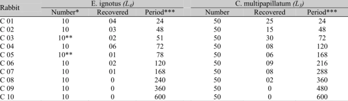
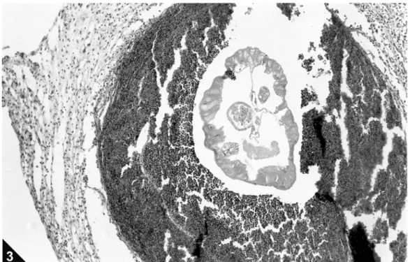
gastric wall, with predominance of eosinophils, disruption of the mucosa and submucosa hemorrhagy were observed. Mixed inflammatory reaction, necrosis, abscesses in the body cavity, mixed inflammatory reaction in the peritoneum, with predominance of mononuclear cells and with pancreatitis were observed (Fig. 1-4).
Figure 1. Hemorrhagic gastric submucosa of rabbits 48 hours after experimental infection wit E. ignotus L4 larvae. Bar = 0.10mm. (Bar in Fig. 2 is common to Fig. 1).
Figure 2. Gastric mucosa of rabbits 51 hours after experimental infection wit E. ignotus L4 larvae,
Figure 3. Peritoneal abscess surrounding the parasite, in rabbits 78 hours after experimental infection with E. ignotus L4 larvae. Bar = 0.10mm. (Bar in Fig. 4 is common to Fig. 3).
Figure 5. Gastric mucosa of rabbits 120 hours after experimental infection with C. multipapillatum L3
larvae, showing disruption, with parasite debris, necrosis and rare eosinophils. Bar = 0.05mm. (Bar in Fig. 6 is common to Fig. 5).
DISCUSSION
Experimental inoculation with 7-10 E. ignotus larvae in rabitts have been previously reported by Shirazian et al. (1984). Clinical signs were absent and deaths did not occur in the post-infection period. Nevertheless, during necropsies, gastritis and lesions due to the migration of larvae in the body cavity were observed. In the post-inoculation period, during tomographic exams, images compatible with abdominal abscesses were observed. These findings were further confirmed in necropsies but differ from previous results, considering that the rabbits investigated herein had clinical signs with the occurrence of deaths. During the post-mortem examination, there were evidences of a larval migratory process, always originating in the gastric wall and not compromising the thoracic cavity. Images by ultrasonography indicated a thickening of the hepatic parenchyma but they were not specific for abscesses.
There are no reports of experimental inoculation with C. multipapillatum in rabbits. In a domestic cat experimentally inoculated with a non established number of larvae of this nematode, necropsy findings revealed ulcerated lesions and hyperemia of the intestinal mucosa (Vidal-Martinez et al., 1994). Nevertheless, no intestinal lesions due to C. multipapillatum were found in the experimentally infected rabbits presently investigated.
The association of allergenic reactions with infections of anisakid worms was analyzed in humans by Alonso et al. (1977), Cuende et al. (1998), Del Pozo et al. (1996), Fernández de Corres et al. (1996) and Montoro et al. (1997), when clinical signs of urticaria, anaphylaxis and arthritis, after the ingestion of infected fish were reported. In the present study the hypersensitivity symptoms were revealed by a hyperemic reaction restricted to the gastric mucosa.
The high prevalence of Contracaecum sp. larvae, associated to the wide range of fishes that can serve as potential hosts for these helminths are, in fact, the most important factor in the transmission to humans; nevertheless, E. ignotus seems to be of major relevance, considering the severity of the lesions and, consequently, the possibility of sudden deaths due to peritonitis.
Ratifying data after Okumura et al. (1999) and Chieffi et al. (1992), the recommendation to avoid consumption of raw or poorly cooked fish is still the best preventive procedure.
The application of a simple device with a lamp under a glass wooden-framed board was effective for the visualization of either E. ignotus or C. multipapillatum cysts in the musculature of the examined fishes. This technique can be easily applied in industries for inspection purposes.
Data herein presented suggest that the species E. ignotus and C. multipapillatum might have zoonotic importance, since larval forms of these nematodes are potentially harmful to mammalian hosts.
REFERENCES
ALONSO, A.; DASCHNER, A.; MORENO, A. Anaphylaxis with Anisakis simplex in the gastric mucosa. N. Engl. J. Med., 337, p.350-351, 1977.
AMATO, J.F.R.; BOEGER, W.A.P.; AMATO, S.B. Protocolos para laboratório: coleta e processamento de parasitos de pescado. Impressa Universitária, Universidade Federal do Rio de Janeiro, Seropédica, RJ, Brasil, 1991. 81p.
APA. Código de ética experimental com animais. Sociedade Zoófila Educativa, Rio de Janeiro, RJ, Brasil, 1989. 8p.
BARROS, L.A.; AMATO, S.B. Aspectos patológicos observados em hamsters (Mesocricetus auritus) infectados experimentalmente com metacercárias de Phagicola longus (Ranson, 1920) Price, 1932 (Digenea, Heterophyidae). Rev. Bras. Parasitol. Vet., v.4, p.43-48, 1995a.
BARROS, L.A.; AMATO, S.B. Infecções experimentais em cães com metacercárias de Phagicola longus (Ranson, 1920) Price, 1932. Rev. Bras. Parasitol. Vet., v.5, p.61-64, 1996.
BARROS, L.A.; AMATO, S.B. Infecções experimentais em gatos, Felis domestica, com metacercárias de Phagicola longus. Rev. Univ. Rural. Sér. Ciên. Vida, v.17, p.49-54, 1995b.
Phagicola sp. (Trematoda-Heterophyidae) in the municipality of Registro, São Paulo State, Brazil. Rev. Inst. Med. Trop. São Paulo, v.32, p.285-288, 1992.
CONROY, G.; PEREZ, K. A report on the experimental infection of a smooth-headed capuchin monkey (Cebus apella) with metacercariae of Phagicola longa obtained from silver mullet. Riv. Ital. Pisc. Ittiopatol., v.4, p.154-155, 1985.
CUENDE, E.; ALDICANA, M.T.; GARCIA, M. et al. Rheumatic manifestations in the course of anaphylaxix caused by Anisakis simplex. Clin. Exp. Rheumatol., v.16, p.303-304, 1998.
DEL POZO, M.D.; ALDICANA, M.; DIEZ, J.M. et al. Anisakis simplex a relevante etiologic factor in acute urticaria. Allergy, v.52, p.576-579, 1996.
EBERHARD, M.L.; HURWITZ, H.; SUN, A.M. et al. Intestinal perforation caused by larval Eustrongylides (Nematoda: Dioctophymatoide) in New Jersey. Am. J. Trop. Med. Hyg., v.40: p.648-650, 1989.
FERNANDEZ DE CORRES, L.; ALDICANA, M.; DIEZ, J.M. et al. Anisakis simplex induces not only anisakiosis: report of 28 cases of allergy caused by this nematode. J. Investig. Allergol. Clin. Immunol., v.6, p.315-319, 1996.
GUERIN, P.F.; MARAPENDI, S.; MC GRAIL, L. et al. Intestinal perforation caused by larval Eustrongylides. Morb. Mort. Week. Rep., v.31, p.383-389, 1982.
MONTORO, A.; PERTERGUER, M.J.; CHIVATO, T. et al. Recidivuous acute urticaria caused by Anisakis simplex. Allergy, v.52, p.985-991, 1997.
OKOMURA, M.P.M.; DÉREZ, A.C.A.; ESPINDOLA, A. Principais zoonoses parasitárias transmitidas por pescado – revisão. Rev. Ed. Cont. CRMV-SP, v.2, p.66-80, 1999.
RÊGO, A.A.; VICENTE, J.J.; SANTOS, C.P. et al. Parasitas de anchovas, Pomatomus saltatrix (L.) do Rio de Janeiro. Ciên. Cultura, v.35, p.1329-1336, 1983.
SHIRAZIAN, D.; SCHILLER, E.L.; GLASER, C.A. et al. Pathology of larval Eustrongylides in rabbits. J. Parasitol., v.70, p.803-806, 1984.
VIDAL-MARTINEZ, V.M.; OSORIO-SARABIA, D.; OVERSTREET, R.M. Experimental infection of Contracaecum multipapillatum (Nematoda: Anisakinae) from Mexico in the domestic cat. J. Parasitol., v. 80, p.576-579, 1994.