Suspicion of Obstructive Sleep Apnea by Berlin Questionnaire
Predicts Events in Patients with Acute Coronary Syndrome
Eryca Vanessa S. de Jesus
1, Euvaldo B. Dias-Filho
1, Bethania de M. Mota
1, Luiz de Souza
3, Celi Marques-Santos
2,
João Bosco G. Rocha
2, Joselina L. M. Oliveira
1,2, Antônio C. S. Sousa
1,2, José Augusto Barreto-Filho
1,2Departamento de Cardiologia da Universidade Federal de Sergipe1, Aracaju, SE; Serviço de Cardiologia da Clínica e Hospital São Lucas2; Departamento de Estatística da USP - Ribeirão Preto3, Ribeirão Preto, SP - Brazil
Mailing address: José Augusto Barreto-Filho •
Núcleo de Pós-Graduação em Medicina - Universidade Federal de Sergipe Hospital Universitário - Campus da Saúde Prof. João Cardoso Nascimento Jr Rua Cláudio Batista, S/N - prédio CCBS/HU - Bairro Sanatório - 49060-100 - Aracaju, SE - Brazil
E-mail: [email protected]
Manuscript received August 11, 2009; revised manuscript received November 01, 2009; accepted November 24, 2009.
Abstract
Background: From a mechanistic standpoint, obstructive sleep apnea (OSA) may further disturb cardiovascular homeostasis in the setting of acute coronary syndrome (ACS).
Objective: We sought to investigate if a standardized clinical diagnosis of OSA, in acute coronary syndrome patients, predicts the risk of cardiovascular events during hospitalization.
Methods: In a prospective cohort study, a group of 200 patients diagnosed with ACS between September 2005 and November 2007 were stratified by the Berlin Questionnaire (BQ) regarding the risk for OSA (high or low risk). We tested if the subgroup of high risk for OSA was prone to a higher frequency of cardiovascular events. The primary endpoint evaluated was a composite outcome of cardiovascular death, recurrent cardiac ischemic events, acute pulmonary edema and stroke during hospitalization.
Results: Ninety four (47%) patients assessed by the BQ were likely to have OSA. High risk for OSA was associated with a non-significant higher mortality (4.25% vs 0.94%; p=0.189), but a significant higher incidence of composite cardiovascular events (18.08% vs 6.6%; p=0.016). In the logistic regression model, multivariate predictors of composite cardiovascular events were age (OR = 1.048; 95% CI 1.008 to 1.090; p=0.019), left ventricular ejection fraction (OR = 0.954; 95% CI 0.920 to 0.989; p=0.010), and higher risk for OSA (OR= 3.657; 95% CI 1.216 to 10.996; p=0.021).
Conclusion: The use of a simple and validated questionnaire (BQ) to identify patients with higher risk for OSA may help in the prediction of cardiovascular outcome during hospitalization. Moreover, our data suggests that OSA is very common in patients with ACS. (Arq Bras Cardiol 2010; 95(3): 313-320)
Key words: Sleep apnea, obstructive/complications; questionnaires; acute coronary syndrome.
the impact of OSA adding risk to patients with ACS. Polysomnography is the gold standard method to diagnose sleep apnea syndrome; nevertheless it has potential limitations to its widespread use in clinical practice6,13. The Berlin
Questionnaire (BQ) is a simple and validated method to diagnose OSA in the general population13 and, recently, it has
also been validated in cardiovascular patients6. Therefore, we
tested the impact of the clinical diagnosis of OSA suspected by BQ in predicting cardiovascular events during hospitalization of ACS patients.
Methods
Study design
In this prospective, observational study, all patients referred to the Chest Pain Unit in São Lucas Hospital, state of Sergipe, Brazil, from September 2005 to November 2007, and diagnosed with ACS were invited to respond the BQ and were followed until hospital discharge. The protocol was approved by the local ethics review board, and all subjects provided written informed consent before study enrollment.
Introduction
Obstructive sleep apnea (OSA) is an emerging cardiovascular risk factor. From an epidemiological standpoint, ample evidence supports that OSA may cause hypertension1,2, stroke3, heart failure4,5 and may increase the
risk of atrial fibrillation6,7. Moreover, OSA is associated with
signs of surrogate markers of premature atherosclerosis8. In
addition, the hallmarks of OSA pathophysiology in regard to the imbalance of oxygen delivery and demand suggest that apnea-related hypoxemia may impose an additional risk to patients in the setting of acute coronary syndromes (ACS)9-12.
Subjects
Inclusion criteria included a diagnosis of ACS defined by medical history and at least one of the following findings: electrocardiographic changes consistent with ACS, serial increases in serum markers of cardiac necrosis and documentation of coronary artery disease (coronary angiography or detection of myocardial ischemia by noninvasive method). Patients were diagnosed with ST-segment elevation myocardial infarction (STEMI), non-STEMI or unstable angina according to current guidelines14,15. Patients
who refused to be enrolled in the present investigation, who was transfered to other hospital or who were not capable of answering the questionnaire were excluded.
Baseline assessment
Data on demographic characteristics, prior medical history and life habits were obtained with the use of a standardized questionnaire administered by a trained medical resident (EVSJ) and corroborated by the data in the medical files. Risk-factor data included history of hypertension, diabetes mellitus, dyslipidemia, smoking or prior cardiovascular disease, either reported by the patient on the baseline medical record or based on the current therapy used by the patient. Renal failure was defined by creatinine at hospital admission above the upper limit of normality.
The patients’ height and weight were recorded and used to calculate the body-mass index (BMI). Obesity was defined as a BMI > 30 kg/m2. Biochemical and electrocardiographic
findings, treatment practices and hospital outcome data were collected. Echocardiography was performed at the discretion of the patient’s physician and left ventricular ejection fraction (LVEF) was analyzed according to the current guidelines.
Patients were considered to have clinical signs of acute congestive heart failure (CHF) at admission in the presence of rales or crackles in the lungs, an S3 gallop, and elevated jugular venous pressure, frank acute pulmonary edema (APE) or cardiogenic shock (defined as hypotension, measured as systolic blood pressure lower than 90 mmHg, and evidence of peripheral vasoconstriction).
Risk of OSA (Berlin questionnaire)
Risk of OSA was assessed by the Berlin questionnaire. Patients were divided in high risk and low risk of OSA based on responses to symptom questions grouped in three categories. A patient was considered to be at high risk for sleep apnea if 2 of the 3 following criteria were met: 1) snoring with two of the following features: louder than talking, at least 3 to 4 times a week, complaints by others about snoring, witnessed breathing pauses at least 3 to 4 times a week; 2) early morning and daytime fatigue exceeding 3 to 4 times a week or having fallen asleep while driving; and 3) presence of hypertension or obesity13.
Outcome
Patients were followed for clinical endpoints and adverse events during their index hospitalization. The cardiovascular events of interest were cardiovascular death, recurrent
ischemic events (angina, infarction or reinfarction), APE and stroke. A diagnosis of recurrent myocardial ischemia was based on recurrent ischemic symptoms, new electrocardiographic changes compared to the index ECG that were not considered an evolution of the index recording and/or a subsequent increase in CKMB levels after a decrease from a previous peak value14,15. APE was classified as a relevant cardiovascular
outcome due to the epidemiological and pathophysiological association of OSA and heart failure4. APE was defined
as the presence of clinical signs of left ventricular failure, manifestation of dyspnea, signs of hypoxia and fluid in the lungs (i.e. chest auscultation revealingrales and a chest radiography showing bilateralpulmonary infiltrates consistent with congestion).Stroke was defined as rapidly developing clinical signs of focal (or global) disturbance of cerebral function lasting for more than 24h with no apparent cause other than a vascular origin.
Statistical analysis
Baseline characteristics of patients in the 2 groups were expressed as means (with standard deviations) and counts (with percentages). Patients with BQ suggestive of OSA were compared to patients with low probability of OSA by the 2-tailed T test and X2 test or Fisher exact test.
To ascertain the independent effect of high risk of OSA on primary endpoints, we identified potential confounding variables and covariates by performing univariate analysis, comparing patients with high and low risk of OSA and patients with and without cardiovascular composite events. Significant confounders (p < 0.10) identified at the univariate analysis were included in the model for the multivariate logistic regression. A stepwise logistic regression was used to develop a multivariate model that identified the best set of variables associated with composite cardiovascular events. The multivariate model was constructed based on the Hosmer-Lemeshow approach. A forward stepwise procedure was used to identify the final model. Statistical significance was established by p < 0.05. All analyses were performed using SPSS 16.0 for Windows (SPSS Inc., Chicago, Illinois).
Results
Characteristics of the study population
Two hundred and seventy-three patients were selected for the study. Of these, 50 declined participation, 19 were not capable of answering the questionnaire and 4 were transferred to another hospital, resulting in a final study population of 200 patients. The baseline characteristics of the excluded patients were similar to those of the participants in the study (data not shown).
cardiac necrosis were not different. Ninety-one percent of the patients (181 patients) underwent coronary angiography. There were nosignificant differences between the groups with respect to prevalence of multivessel and left main coronary
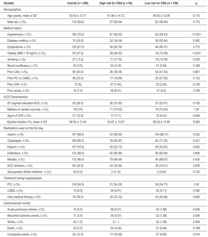
Table 1 -Baseline characteristics of patients in relation to suspicion of OSA
Variable Overall (n = 200) High risk for OSA (n = 94) Low risk for OSA (n = 106) p
Demographics
Age (years), mean ± SD 63.54 ± 13.11 61.96 ± 14.12 64.92 ± 12.06 0.115
Male sex, n (%) 119 (59.5) 57 (60.64) 62 (58.49) 0.775
Medical history
Hypertension, n (%) 150 (75.0) 87 (92.55) 63 (59.43) < 0.001
Diabetes mellitus, n (%) 70 (35.0) 32 (34.04) 38 (35.84) 0.882
Dyslipidemia, n (%) 123 (61.5) 59 (62.76) 64 (60.37) 0.772
Obesity (BMI > 30 kg/m2), n (%) 54 (27.0) 38 (40.42) 16 (15.09) < 0.001
Smoking, n (%) 27 (13.5) 11 (11.70) 16 (15.09) 0.538
Renal insuficiency, n (%) 05 (2.5) 04 (4.25) 01 (0.94) 0.189
Prior CAD, n (%) 80 (40.0) 36 (38.29) 44 (41.50) 0.667
Prior PCI or CABG, n (%) 46 (23.0) 17 (18.08) 29 (27.35) 0.132
Prior CHF, n (%) 10 (5) 07 (7.44) 03 (2.83) 0.195
Prior stroke, n (%) 15 (7.5) 08 (8.51) 07 (6.6) 0.789
ACS Characteristics
ST segment elevation ACS, n (%) 53 (26.5) 26 (27.65) 27 (25.47) 0.750
Markers of cardiac necrosis, n (%) 150 (75) 71 (75.53) 79 (74.53) 1.00
Signs of CHF, n (%) 21 (10.5) 11 (11.7) 10 (9.43) 0.649
Ejection fraction (%), mean ± SD 56.54 ± 13.44 54.67 ± 13.67 58.29 ± 13.06 0.083
Medications used on the irst day
Aspirin, n (%) 197 (98.5) 93 (98.93) 104 (98.11) 0.632
Clopidogrel, n (%) 160 (80.0) 78 (82.97) 82 (77.35) 0.321
Heparin, n (%) 107 (53.5) 49 (52.12) 56 (52.83) 0.920
β-Blockers, n (%) 132 (66.0) 62 (65.95) 69 (65.09) 0.898
Nitrates, n (%) 172 (86.0) 79 (84.04) 94 (88.67) 0.435
ACE inhibitors, n (%) 60 (30.0) 32 (34.04) 35 (33.01) 0.878
Glycoprotein IIb/IIIa inhibitors , n (%) 05 (2.5) 2 (2.12) 3 (2.83) 0.732
Treatment during hospitalization
PCI, n (%) 109 (54.5) 51 (54.25) 58 (54.71) 1.00
CABG, n (%) 13 (6.5) 08 (8.51) 05 (4.71) 0.390
Only medical therapy, n (%) 78 (39.0) 35 (37.23) 43 (40.56) 0.665
Cardiovascular events
Acute pulmonary edema, n (%) 10 (5.0) 08 (8.51) 02 (1.88) 0.048
Recurrent ischemic events, n (%) 11 (5.5) 09 (9.57) 02 (1.88) 0.048
Stroke, n (%) 02 (1.0) 0 ( - ) 02 (1.88) 0.499
Death, n (%) 05 (2.5) 04 (4.25) 01 (0.94) 0.189
Composite events, n (%) 24 (12.0) 17 (18.08) 07 (6.60) 0.016
Markers of cardiac necrosis indicate positive CKMB and/or Troponin-I. Only 166 patients were submitted to echocardiography: 80 from the group with high risk and 86 from the group with low risk, p=0.455. Only the irst event was considered in composite events.
artery disease (CAD). In addition, no differences were observed in regard to evidence-based treatments between the two groups (Table 1).
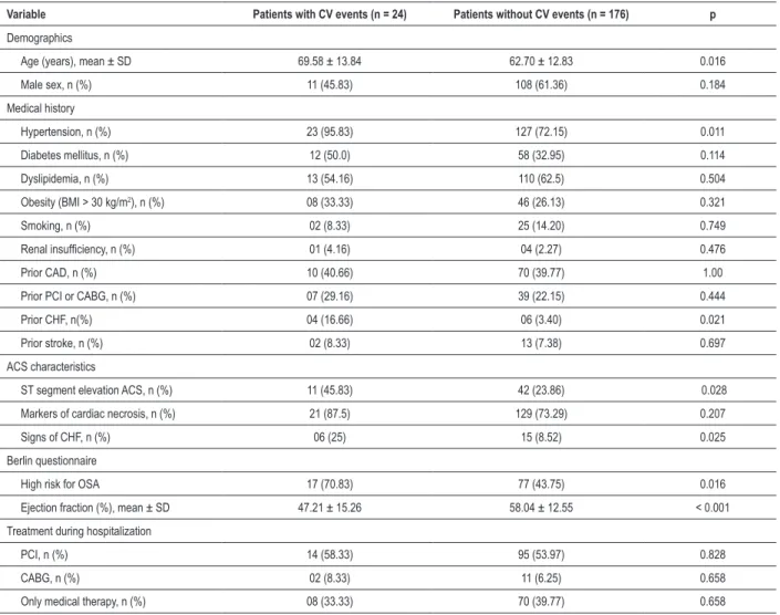
Of the total number of patients, 24 (12%) presented cardiovascular events during hospitalization (Table 2). The total mortality of the population was 2.5% (05 patients). The group with higher risk of OSA presented 4.25% (04 patients) of mortality compared with 0.94% (01 patient) in the group with low clinical probability of OSA (p=0.189). The OSA group presented 18.08% (n=17) of composite cardiovascular events compared to 6.6% (n=07) in the group without suspicion of OSA (p=0.016) (Table 1). Patients with cardiovascular events presented higher prevalence of hypertension (p=0.011), higher prevalence of medical history of prior CHF (p=0.021) and signs of CHF upon hospital admission (p=0.025), lower ejection fraction (p<0.001), higher prevalence of ST elevation ACS (p=0.028) and older age (p=0.016). There were nosignificant differences between patients with and without cardiovascular events with respect to the prevalence of multivessel and left main coronary artery disease. No differences were observed in treatment strategies when comparing the two groups (Table 2).
Association of suspicion of OSA with cardiovascular events
A higher risk of OSA by the BQ persisted significantly associated with composite cardiovascular events during hospitalization after adjustment (Table 3). The variables age and ejection fraction were also independently associated with CV events in the multivariate model.
Discussion
The novel finding of this study is that the subgroup of patients with ACS and high clinical suspicion of OSA by the BQ are at higher risk of composite cardiovascular events during
Table 2 -Comparison of patients in relation to cardiovascular events
Variable Patients with CV events (n = 24) Patients without CV events (n = 176) p
Demographics
Age (years), mean ± SD 69.58 ± 13.84 62.70 ± 12.83 0.016
Male sex, n (%) 11 (45.83) 108 (61.36) 0.184
Medical history
Hypertension, n (%) 23 (95.83) 127 (72.15) 0.011
Diabetes mellitus, n (%) 12 (50.0) 58 (32.95) 0.114
Dyslipidemia, n (%) 13 (54.16) 110 (62.5) 0.504
Obesity (BMI > 30 kg/m2), n (%) 08 (33.33) 46 (26.13) 0.321
Smoking, n (%) 02 (8.33) 25 (14.20) 0.749
Renal insuficiency, n (%) 01 (4.16) 04 (2.27) 0.476
Prior CAD, n (%) 10 (40.66) 70 (39.77) 1.00
Prior PCI or CABG, n (%) 07 (29.16) 39 (22.15) 0.444
Prior CHF, n(%) 04 (16.66) 06 (3.40) 0.021
Prior stroke, n (%) 02 (8.33) 13 (7.38) 0.697
ACS characteristics
ST segment elevation ACS, n (%) 11 (45.83) 42 (23.86) 0.028
Markers of cardiac necrosis, n (%) 21 (87.5) 129 (73.29) 0.207
Signs of CHF, n (%) 06 (25) 15 (8.52) 0.025
Berlin questionnaire
High risk for OSA 17 (70.83) 77 (43.75) 0.016
Ejection fraction (%), mean ± SD 47.21 ± 15.26 58.04 ± 12.55 < 0.001
Treatment during hospitalization
PCI, n (%) 14 (58.33) 95 (53.97) 0.828
CABG, n (%) 02 (8.33) 11 (6.25) 0.658
Only medical therapy, n (%) 08 (33.33) 70 (39.77) 0.658
Table 3 -Multivariate predictors of cardiovascular events
Variable Odds ratio 95% CI p
Age 1.048 1.008 - 1.090 0.019
Left ventricular
ejection fraction 0.954 0.920 - 0.989 0.010
hospitalization. Moreover, the study also confirmed that the incidence of OSA in the setting of ACS is much higher than that in the general population6,16,17.
It is reasonable to hypothesize that OSA is an emerging cardiovascular factor1,3,4,6-10, 18,19. Nevertheless, this hypothesis
is not universally accepted16. In addition, very limited
information is known about the role of OSA as an aggravating factor to established cardiovascular disease.
ACS is generally caused by reduced myocardial perfusion as a consequence of a thrombus that develops on a disrupted plaque. The hallmark of OSA pathophysiology is intermittent episodes of hypoxia elicited by the mechanical interruption of upper airway flow10,17. The cardiovascular response during
hypoxia in order to compensate systemic oxygen desaturation is complex and may be altered in pathological conditions20,21.
In patients with ACS and OSA, the association of a) severe reduction in coronary blood flow, b) acute oxygen desaturation during apneic episodes, c) impairment of coronary defense response during hypoxia caused by endothelial dysfunction and atherosclerosis and d) blood pressure elevation during hypoxemia increasing myocardial oxygen demand may, altogether, aggravate myocardial ischemia and further impair cardiac function. Although it is tempting to suppose that OSA may be a risk factor for atherothrombosis and aggravate the ACS prognosis, the concept is yet to be proven.
OSA is associated with surrogate pathways and markers of premature atherosclerosis8,22. CAD is more prevalent in
OSA patients18,19 and the respiratory disturbance index is a
predictor of cardiovascular mortality in small cohorts9. Sleep
apnea has been implicated in patients with nocturnal angina pectoris. Nocturnal angina and ST depression are diminished during treatment of sleep apnea by CPAP10, 23. Similarly, Hanly
et al24 observed that the ST depression occurred in about a
third of patients with severe OSA. ST depression was markedly attenuated during nasal CPAP. However, these patients did not have proven coronary artery disease, and artifactual ST changes related to breathing patterns may have contributed. Araújo et al25, studying a group of 53 patients with stable
coronary disease, did not observe significant difference in the number or duration of ischemic episodes during sleep among patients with OSA and controls. Moreover, in this study there were no alterations in the circadian pattern of myocardial ischemia in OSA or related to sleep apnea and heart rate variability or arrhythmias.
In longer-term studies, OSA in patients with CAD was associated with a significant increase in the composite endpoint of death, myocardial infarction (MI), and cerebrovascular events at a 5-year median follow-up interval26. However,
neither oxygen desaturation index nor AHI was independently predictive of single endpoints of MI or death. In a case-control study, there was a graded increase in the odds of acute MI with increased sleep apnea severity, even after adjustment for possible confounding factors27.
In a large cohort study, OSA was associated with risk of death and stroke, even when adjusted by hypertension or other confounders3. It was observed that OSA patients
presented a higher risk of having sudden cardiac death during sleep time28. The same group also showed that patients with
nocturnal onset of acute myocardial infarction had a higher likelihood of having OSA29. Collectively, the data point out to
OSA as a condition that may be a trigger for cardiovascular death, stroke and ACS. Although much is known about the association between OSA and CAD, myocardial ischemia and infarction, the data provide mostly observational, albeit valuable, insights.
We studied patients with ACS who were admitted to a single cardiovascular center. The BQ identified that 47% of the overall population presented high probability of having OSA. This is in agreement with recent studies showing the extremely high prevalence of OSA in this scenario (57% and 66.6%)16,18,19 . Taken together, these numbers underscore the
high frequency of OSA in patients with ACS and the necessity of further research to understand the OSA-ACS interaction.
In addition, modeling the association of OSA and cardiovascular events to covariates that generally are accepted as confounder factors demonstrated that clinical suspicion of OSA was independently associated to cardiovascular events during hospitalization. In our population, differences in treatment strategies did not explain the higher prevalence of cardiovascular events in OSA patients (Table 1).
When compared to patients with low risk of OSA, it was demonstrated that high risk of OSA was associated with an increased risk of composite adverse cardiovascular events (i.e. cardiovascular mortality, recurrent myocardial ischemic events, APE and stroke). It should be stressed that the sample size was relatively small to detect differences in cardiovascular mortality between OSA and non-OSA patients. Nevertheless, recurrent cardiac ischemic events and APE (Table 1) were more prevalent in OSA patients. In addition to the potential role of hypoxic episodes precipitating ischemic cardiac events, the association of OSA, heart failure and impairment in cardiac function has also been documented4,30.Moreover, hypoxia
may increase pulmonary vascular permeability through an endothelin-related mechanism increasing the risk of pulmonary edema31. This evidence was the base of our choice
to include APE as a cardiovascular endpoint in this prospective study and may explain our finding of a higher prevalence of APE in OSA patients.
In a prospective cohort study, patients with and without OSA were compared regarding the frequency of MI during different intervals of the day based on the time of onset of chest pain29. The odds of having OSA in patients whose MI
occurred between 12 AM and 6 AM was 6-fold higher than in cases whose MI occurred during the remaining 18 h of the day (95% confidence interval: 1.3 to 27.3, p > 0.01). Of all patients having an MI between 12 AM and 6 AM, 91% had OSA. These findings suggest that OSA may be a trigger for MI.
Cassar et al19 have reported, in a retrospective and
who were submitted to PCI during the course of ACS. They demonstrated that the incidence of MACE (cardiac death, reinfarction and target vessel revascularization) was higher in OSA patients (23.5 vs 5.3%; p=0.02). In addition, they documented that OSA was an independent predictor of MACE, greater angiographic late loss and a higher binary restenosis rate.
A second prospective observational study assessed the incidence of fatal and nonfatal cardiovascular events in healthy men, snorers, and patients with treated and untreated OSA. In men with severe untreated OSA (AHI > 30), both fatal and nonfatal cardiovascular events were markedly increased. In contrast, fatal and nonfatal cardiovascular events in treated OSA patients approached levels seen in simple snorers32.
A recent study reported different conclusions from ours. Mehra et al16 analyzed 104 patients with ACS and complete
sleep studies performed within 72 h of hospital admission. Similar to our data, they observed a very high prevalence (66.4%) of OSA in the setting of ACS. However, that did not demonstrate OSA as a predictive factor for cardiovascular events during a 6-month follow-up. In spite of the merits of their study for having performed polysomnography to diagnose OSA, the absence of an objective definition of relevant cardiovascular events as potential limitations may have masked the real impact of OSA in the setting of ACS.
One major limitation of our study was the fact that it was not possible to use polysomnography to confirm our data as the gold standard method. In the study of Mehra et al16, neither
the pre-test probability for sleep apnea through the Cleveland questionnaire, an expanded Berlin Questionnaire, nor the degree of subjective sleepiness predicted OSA. Araújo et al25
observed that 23 patients (43.4%) of the 53 classified as having
high risk for OSA by the BQ had an AHI ≤ 15. Nevertheless,
the BQ has been validated in several clinical scenarios and has been used in the identification of patients with suspicion of OSA with reasonable sensitivity and specificity6,13,33. Gami et
al6 reported that the risk of AF was increased in OSA patients
identified by the BQ. Recently, these data were confirmed, by the same group, in a larger study using polysomnography, supporting the role of the BQ in the clinical scenario7. In
addition, because of the epidemiological importance of ACS and the major economic and logistical obstacles to the widespread use of sleep studies in patients with ACS, new simple and inexpensive methods to identify patients with OSA
are urgently desirable.
One strength of the current study includes the high prevalence of ACS, the growing epidemiological importance of OSA and the major economic and logistical obstacles to the widespread use of sleep studies in all patients with ACS.
Conclusions and clinical implications
The first major finding of this study is that almost half of patients with ACS may also have OSA. The second major finding is that our data suggest that OSA is an aggravating cardiovascular risk factor for patients in the setting of ACS. Therefore, further research is necessary to confirm our data and, more importantly, to test the effects of treating OSA on cardiovascular endpoints during the course of ACS. A randomized trial addressing this issue will be essential to answer this fundamental question, especially if considering the potential harmful effect of continuous positive airway pressure (CPAP) at aggravating myocardial ischemia34. Finally, our data
disclosures the possibility of improving our prediction model of cardiovascular events during ACS hospitalization by using a simple and inexpensive method.
Acknowledgments
The authors gratefully acknowledge the help of the cardiologists from the Chest Pain Unit and the Coronary Care Unit of São Lucas Hospital whose cooperation made this study possible.
Potential Conflict of Interest
No potential conflict of interest relevant to this article was reported.
Sources of Funding
This study was partially funded by Projeto PIBIC do CNPq/ UFS.
Study Association
This study is not associated with any post-graduation program.
References
1. Peppard PE, Young T, Palta M, Skatrud J. Prospective study of the association between sleep-disordered breathing and hypertension. N Engl J Med. 2000; 342 (19): 1378-84.
2. Dart RA, Gregoire JR, Gutterman DD, Woolf SH. The association of hypertension and secondary cardiovascular disease with sleep-disordered breathing. Chest. 2003; 123 (1): 244-60.
3. Yaggi HK, Concato J, Kernan WN, Lichtman JH, Brass LM, Mohsenin V. Obstructive sleep apnea as a risk factor for stroke and death. N Engl J Med. 2005; 353 (19): 2034-41.
4. Wang H, Parker JD, Newton GE, Floras JS, Mak S, Chiu KL, et al. Influence of
obstructive sleep apnea on mortality in patients with heart failure. J Am Coll Cardiol. 2007; 49 (15): 1625-31.
5. Bradley T, Hall M, Ando S, Floras J. Hemodynamic effects of simulated obstructive apneas in humans with and without heart failure. Chest. 2001; 119 (6): 1827-35.
6. Gami AS, Pressman G, Caples SM, Kanagla R, Gard JJ, Davison DE, et al. Association of atrial fibrillation and obstructive sleep apnea. Circulation. 2004; 110 (4): 364-7.
Cardiol. 2007; 49 (5): 565-71.
8. Drager LF, Bortolotto LA, Lorenzi MCl, Figueiredo AC, Krieger EM, Lorenzi-Filho G. Early signs of atherosclerosis in obstructive sleep apnea. Am J Respir Crit Care Med. 2005; 172 (5): 613-8.
9. Peker Y, Hedner J, Kraiczi H, Löth S. Respiratory disturbance index: an independent predictor of mortality in coronary artery disease. Am J Respir Crit Care Med. 2000; 162 (1): 81-6.
10. Peled N, Abinader EG, Pillar G, Sharif D, Lavie P. Nocturnal ischemic events in patients with obstructive sleep apnea syndrome and ischemic heart disease: effects of continuous positive air pressure treatment. J Am Coll Cardiol. 1999; 34 (6): 1744-9.
11. Yamauchi M, Nakano H, Maekawa J, Okamoto Y, Ohnishi Y, Suzuki T, et al. Oxidative stress in obstructive sleep apnea. Chest. 2005; 127 (5): 1674-9.
12. Alonso-Fernández A, García-Río F, Racionero MA, Pino JM, Ortuno E, Martinez I, et al. Cardiac rhythm disturbances and ST-segment depression episodes in patients with obstructive sleep apnea-hypopnea syndrome and its mechanisms. Chest. 2005; 127 (1): 15-22.
13. Netzer NC, Stoohs RA, Netzer CM, Clark K, Strohl KP. Using the Berlin Questionnaire to identify patients at risk for the sleep apnea syndrome. Ann Intern Med. 1999; 131 (7): 485-91.
14. Anderson JL, Adams CD, Antman EM, Bridges CR, Califf RM, Casey DE Jr, et al. ACC/AHA 2007 Guidelines for the management of patients with unstable angina/non-ST-elevation myocardial infarction-executive summary: a summary of the ACC/AHA/Task Force on practice guidelines development in collaboration with American Emergency Physicians. J Am Coll Cardiol. 2007; 50 (7): e1-e157.
15. Antman EM, Anbe DT, Armstrong PW, Batesx ER, Green LA, Hand MH, et al. ACC/AHA guidelines for the management of patients with ST-elevation myocardial infarction: a report of the American College of Cardiology/ American Heart Association Task Force on Practice Guidelines (Committee to Revise the 1999 Guidelines for the Management of Patients with Acute Myocardial Infarction). Circulation. 2004; 110 (9): e82-e292.
16. Mehra R, Principe-Rodriguez K, Kirchner L, Strohl KP. Sleep apnea in acute coronary syndrome: high prevalence but low impact on 6-month outcome. Sleep Medicine. 2006; 7 (6): 521-8.
17. Young T, Palta M, Dempsey J, Skatrud J, Weber S, Badr S. The occurrence of sleep-disordered breathing among middle-aged adults. N Engl J Med. 1993; 328 (17): 1230-5.
18. Yumino D, Tsurumi Y, Takagi A, Suzuki K, Kasanuki H. Impact of obstructive sleep apnea on clinical and angiographic outcomes following percutaneous coronary intervention in patients with acute coronary syndrome. Am J Cardiol. 2007; 99 (1) : 26-30.
19. Cassar A, Morgenthaler T, Lennon R, Rihal C, Lerman A. Treatment of obstructive sleep apnea is associated with decreased cardiac death after percutaneous coronary intervention. J Am Coll Cardiol. 2007; 50: 1310-4.
20. Barreto-Filho JAS, Consolim-Colombo FM, Lopes HF, Sobrinho CRM, Guerra-Riccio GM, Krieger EM. Dysregulation of peripheral and central chemoreflex
responses in Chagas’ heart disease patients without heart failure. Circulation. 2001; 104: 1792-8.
21. Barreto-Filho JAS, Consolim-Colombo FM, Guerra-Riccio GM, Santos RD, Chacra AP,Lopes HF, et al. Hypercholesterolemia blunts forearm vasorelaxation and enhances the pressor response during acute systemic hypoxia. Arterioscler Thromb Vasc Biol. 2003; 23 (9): 1660-6.
22. Drager LF, Bortolotto LA, Figueiredo AC, Krieger EM, Lorenzi-Filho G. Effects of continuous positive airway pressure on early signs of atherosclerosis in obstructive sleep apnea. Am J Respir Crit Care Med. 2007; 176 (7): 706-12.
23. Franklin KA NJ, Sahlin C, Naslund U. Sleep apnoea and nocturnal angina. Lancet. 1995; 345 (8957): 1085-7.
24. Hanly P SZ, Zuberi N, Lunn K. ST-segment depression during sleep in obstructive sleep apnea. Am J Cardiol. 1993; 71 (15): 1341-5.
25. Araújo CM, Solimene MC, Grupi CJ, Genta PR, Lorenzi-Filho G, Da Luz PL. Evidence that the degree of obstructive sleep apnea may not increase myocardial ischemia and arrhythmias in patients with stable coronary artery disease. Clinics. 2009; 64 (3): 223-30.
26. Mooe T, Franklin KA, Holmström K, Rabben T, Wiklund U. Sleep-disordered breathing and coronary artery disease: long-term prognosis. Am J Respir Crit Care Med. 2001; 164 (10 Pt 1): 1910-3.
27. Hung J, Whitford EG, Parsons RW, Hillman DR. Association of sleep apnoea with myocardial infarction in men. Lancet. 1990; 336 (8710): 261-4.
28. Gami AS, Howard DE, Olson EJ, Somers VK. Day–night pattern of sudden death in obstructive sleep apnea. N Engl J Med. 2005; 352 (12): 1206-14.
29.Kuniyoshi FHS, Garcia-Touchard A, Gami AS, Romero-Corral A, Van der Walt C, Pasalavidijasagon, et al. Day night variation of acute myocardial infarctation in obstructive sleep apnea. J Am Coll Cardiol.2008;52(5): 343-6.
30. Javaheri S, Parker TJ, Wexler L, Michaels SE, Stamberry E, Nishyama H, et al. Occult sleep-disordered breathing in stable congestive heart failure. Ann Intern Med. 1995; 122 (7): 487-92.
31. Carpenter T, Schomberg S, Steudel W, Ozimek J, Colvin K, Stenmark K, et al. Endothelin B receptor deficiency predisposes to pulmonary edema formation via increased lung vascular endothelial cell growth factor expression. Circ Res. 2003; 93 (5): 456-63.
32. Marin JM CS, Vicente E, Agusti AG. Long-term cardiovascular outcomes in men with obstructive sleep apnoea-hypopnoea with or without treatment with continuous positive airway pressure: an observational study. Lancet. 2005; 365 (9464): 1046-53.
33. Tasali E, Cauter EV, Ehrmann DA. Relationships between sleep disordered breathing and glucose metabolism in polycystic ovary syndrome. J Clin Endocrinol Metab. 2006; 91 (1): 36-42.