Complete depigmentation of a small aperture corneal inlay implanted for
compensation of presbyopia
Despigmentação completa de um implante corneano de pequena abertura para correção da presbiopia
Mauro CaMpos1, sandra Beer1, eliane MayuMi nakano1, Cristina MuCCioli1, ruBens Belfort Jr.1, WallaCe ChaMon1,2
Submitted for publication: October 31, 2016 Accepted for publication: December 6, 2016
1 Department of Ophthalmology and Visual Sciences, Escola Paulista de Medicina, Universidade Federal de São Paulo (UNIFESP), São Paulo, SP, Brazil.
2 Department of Ophthalmology & Visual Sciences, College of Medicine, University of Illinois at Chicago (UIC), Chicago, IL, USA.
Funding: No specific financial support was available for this study.
Disclosure of potential conflicts of interest: Wallace Chamon is an advisor for Abbott Vision and holds patents on Biomechanics, Crosslinking, and Wavefront.
Corresponding author: Mauro Campos. Rua Monsanto, 166 - São Paulo, SP - 05412-030 - Brazil - E-mail: [email protected]
ABSTRACT
We describe a case of late-onset remarkable depigmentation of a small aper-ture corneal inlay implanted for presbyopia compensation. The patient was a participant in a clinical trial designed to evaluate the safety and efficacy of the AcuFocus™ ACU-10R160, which is a 10 µm-thick polyimide film tinted with an organic dye. Inlay implantation occurred under mechanical microkeratome Lasik flaps set for a depth of 120 µm. The patient returned to the clinic 11 years after surgery and reported loss of near-vision acuity. Clinical examination showed the complete absence of pigments in the device and the total loss of the initial effect on near vision, despite normal distance vision. Manifest refraction remained stable during the follow-up period. Scheimpflug images characterized the loss of the small aperture effect on incoming light. Confocal analysis revealed small hyper-reflective round images on the endothelium and no signs of inflammation.
Keywords: Corneal transplantation; Corneal stroma; Presbiopia/surgery; Polyvinyls; Visual acuity; Case report
RESUMO
Descrevemos um caso de importante despigmentação de início tardio de im plante corneano de pequena abertura implantada para compensação de presbiopia. O paciente foi um dos participantes de ensaio clínico destinado a avaliar a segurança e eficácia do AcuFocus™ ACU-10R160, uma película de po liimida de 10 microns de espessura, tingida com um corante orgânico. A implantação ocorreu sob um flap de Lasik criado por microcerátomo mecânico ajustado para profundidade de 120 μm. O caso aqui descrito foi avaliado 11 anos após a cirurgia, relatando diminuição de acuidade de visão para perto. O exame clínico mostrou ausência total de pigmen-tos no dispositivo e perda total do efeito inicial na visão de perto, apesar da visão normal para distância. A refração manifesta permaneceu estável durante o período de seguimento. As imagens de Scheimpflug caracterizaram a perda do efeito da abertura pequena na luz entrante. A análise de microscopia confocal revelou peque nas imagens hiper-reflexivas redondas sobre o endotélio, sem sinais de inflamação.
Descritores: Transplante de córnea; Substância própria; Presbiopia/cirurgia; Po livinil; Acuidade visual; Relatos de casos
INTRODUCTION
Presbyopia develops with aging and currently affects millions of people around the world(1). Near-vision impairment can significantly affect daily activities, and its surgical correction remains a challenge(2). Small-aperture corneal inlays increase the depth of focus and have the potential advantage of modifying the refractive status of the eye while preserving corneal tissue and allowing possible reversibility(3-8).
During the last 4 decades, inventors have filed a series of patents based on small apertures optical devices to enhance vision. These inventions include masked contact lenses and intraocular lenses(9). A system and method for increasing the depth of focus of the human eye was assigned to AcuFocus Inc. (Irvine, USA) and, recently, the Food and Drug Administration (FDA) approved the use of the Kamra™ Inlay for compensation of presbyopia(6).
The case reported herein involves a patient who underwent im-plantation of a small aperture corneal inlay and returned for eva lua-tion 11 years later presenting marked depigmentalua-tion of the device and loss of the achieved near vision improvement.
CASE REPORT
A 67-year-old female returned to our university-based refractive surgery clinic for evaluation with a complaint of progressive near-vi sion disturbance in her left eye following surgery for near-near-vision
improvement. According to her medical records, the patient had un-dergone monocular implantation of a small aperture corneal inlay 11 years earlier, under an approved Institutional Review Board protocol. Five patients included in the protocol had normal eye exami nation results, except for presbyopia. Full preoperative evaluation was performed. The non-dominant eye was selected for surgery, which consisted of creation of a corneal flap by using a mechanical microke-ratome set for a depth of 120 µm. After flap lifting, the device was placed in the center of the visualized pupil, and the flap was replaced. The routine postoperative medical regimen included a combination of topical antibiotic and a steroid for 14 days. All surgeries were per-formed in 2004.
The implanted device was an AcuFocus™ ACU-10K160, a 10 µm-thick opaque polyimide inlay with an overall outer diameter of 3.8 mm and an inner diameter of 1.6 mm. The polyimide material was a liquid formulation designed for thin-film casting, supplied by Dupont®. The radius of curvature of the inlay was 7.5 mm. An organic dye (Sudan Black B) was incorporated in the inlay to provide opacity. The inlay had visible light transmission of 6.0% to 7.5%. This device had laser-drilled 25 µm diameter holes (1,600 pores) designed to facilitate nutrient and particle flow to the cornea. The outer and inner edges were free of these porosity holes(10).
during the first 24 months after implantation because of persistent epithelial defects or progressive topographic changes that affected satisfaction.
The patient reported herein was the only with an eye having the implant in place. She had been followed up for 3 years but did not return until recently. Data obtained under the original clinical proto-col regarding her first year of follow-up visits are included in table 1. The patient showed improvement in UNVA after implantation, with no other remarkable clinical findings.
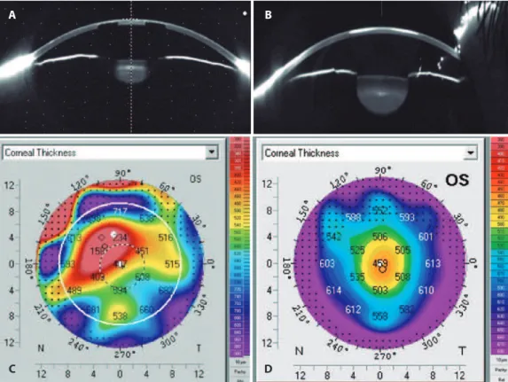
At the most recent evaluation, provocative questioning revealed that she had experienced glare and halos besides decreased UNVA. Figure 1 presents slit-lamp photos of the patient at 7 days and 1, 3, and 11 years after surgery. Until 3 years, the cornea and inlay had a normal appearance. At 11 years, marked depigmentation of the inlay had occurred. Careful observation of the clinical images revealed a reticular rounded appearance of an annular structure in the center of the clear cornea, with rare dots of dark pigments. No signs of in-flammation or edema were present. A slight amount of corneal haze surrounding the inlay was observed. The remaining stroma and en-dothelium were unremarkable and the anterior chamber structures were easily seen throughout the inlay. No signs of pigment dispersion were seen at the anterior chamber angle. Applanation tonometry was 18 mmHg. On optical coherence tomography angiographic ima-ges, the inlay was not markedly distinct from the presumed stromal opacities and had an estimated depth of 120 µm from the corneal surface and a total thickness of approximately 500 µm (Figure 2). Fi gures 3A and 3B show corneal Scheimpflug images taken at the 1-year and 11-year follow-up visits, respectively. Irregularities increa-sed during this period but were not followed by worsening of visual acuity or change in refraction.
Figure 4 represents a Scheimpflug sagittal view of the anterior segment. The initial image clearly shows the shadowing effect of the inlay on internal structures, which blocked the incoming light (Figure 4A). A dark zone observed between the borders of the crystalline lens and the border of the iris illustrates the pinhole effect. At the most recent examination, light clearly passed through the entire inlay area of the cornea and homogeneously illuminated the lens (Figure 4B). A mistaken pachymetry of 158 µm, measured under the inlay opacity annulus (Figure 4C), was corrected after the loss of inlay opacity to achieve more accurate pachymetry of 456 µm (Figure 4D).
Figure 5, obtained using confocal microscopy (Nidek models CS 2 and 4) 30 days after the implantation (Figure 5A), revealed round, well-limited, randomly distributed hypo-reflective structures (pores) with almost no signs of stromal edema. Current confocal microscopy images (Figures 5B-5D) showed a hazy, moderate reflectance of the inlay surface and small hyper-reflective dots (arrow) observed over its surface, and hypo-reflective round images (doubled arrow) deno-ted the pores of the inlay (Figure 5B). The inner border of the inlay
appeared intact with no signs of material deterioration or cracking (Figure 5C). No inflammation was noticed, and endothelium images demonstrated small, hyper-reflective, round images (arrow), similar to those seen on the inlay surface, but that were larger in number (Fi-gure 5D). These hyper-reflective dot images are thought to represent pigment aggregates. The endothelial density was 2,400 cells/mm2.
DISCUSSION
The implant presented herein is not the same as that approved recently by the FDA (KAMRA® Model ACI 7000). Their main differen-ces are related to their thicknesses (10 µm for the older version and 6 µm for the approved one), material composition (polyimide vs. polyvinylidene fluoride - PVDF), method of inlay pigmentation (Su-dan black vs. Carbon black), and pore configuration. Presently, under the current FDA approved technique, a femtosecond-guided pocket with a 200 µm-depth should be used for implantation(10).
The present case illustrates several concepts that drive current knowledge about inlay use in the cornea. On the basis of finite ele-ment model analysis, the company concluded that a 10 µm-device, with pores >15 µm in diameter and covering >5% of its area, would not produce significant glucose depletion(10). The examination did not show any signs of corneal inflammation or cell depletion. The biocompatibility of the current PVDF inlay material on rabbit corneas has been reported in the literature, with a limited amount of tissue inflammation in the first weeks after surgery(7).
A recent paper about long-term outcomes on the use of a more recent generation of the inlay indicated increased UNVA and subtle changes on distance corrected visual acuity (DCVA) in 32 emmetropic eyes(4). Of the 32 operated eyes, 21 lost one or more lines of DCVA, and one eye had the inlay removed because of patient dissatisfaction. The authors mentioned topographic changes and elevation over the area of the implant with a central divot and correlated these findings with the presence of spot-like iron deposits that were described in 56.3% of their cases.
Another long-term study in the literature reported the experien-ce with this technique on 39 (31%) non-previously operated em-metropic eyes that had undergone previous hyperopic Lasik correc-tion (69%)(8). Twenty-two eyes were followed for 4 years, and 93% achieved UNVA of at least J3; all eyes presented uncorrected distance visual acuity of 20/40 or better. Four eyes had their implants removed; one due to a button hole that was not noticed during surgery, two that presented refractive shifts, and one with a flap of 58 µm. Despite these reported complications, no healing defects or iron deposits were mentioned in the study.
The experience by these last two authors with a similar inlay concept, but a deeper implantation depth (170 µm vs. 120 µm) to that reported here, is different from ours. We included five eyes in
Table 1. Summary of clinical indings
Preop 1 month 3 months 6 months 9 months 12 months 11 years
Uncorrected distance VA 20/16 20/16 20/16 20/20 20/16 20/20 20/20 Uncorrected near VA 20/63 20/32 20/40 20/25 20/25 20/40 20/60 Corrected distance VA 20/16 20/16 20/16 20/20 20/16 20/20 20/20 Distance-corrected near VA 20/63 20/32 20/40 20/20 20/20 20/20 20/60 Near-corrected near VA 20/32 20/12 20/16 20/16 20/16 20/16 20/20 Manifest refraction +0.25 0.00 0.00 +0.25 0.00 -0.50+0.50@110° -0.25+0.50@165° K1 45.10 44.23 43.80 45.30 44.94 44.06 40.70 K2 45.50 44.46 44.80 45.73 45.18 44.88 43.40 Steep axis 063º 072º 088º 068º 068° 099° 089°
our series and had to explant four because of persistent epithelial defects and topographic irregularities within the first 2 years after implantation. The other two studies did not report epithelial healing problems or relevant topographic changes. Careful observation of the topographic maps obtained from our patient reveals increased corneal irregularity. Although a considerable amount of keratometric flattening was detected, refraction and uncorrected vision remained unchanged. The current standard of care with an implantation depth of ≥200 µm of a 6 µm-thick inlay will most likely not produce surface changes.
The most significant finding in the present reported case is de-pigmentation of the inlay, which occurred between 3 and 11 years of follow-up. Unfortunately, because the patient did not return to our institution between years 3 and 11 postoperatively, we were unable to estimate the pattern or the onset of depigmentation. The material used in this case, polyimide, is similar to Kapton® HN polyimide film, which has been approved by the FDA for IOL haptics (10). The material approved by the FDA in the current AcuFocus inlay model, PVDF, has proved to be biocompatible(4,7-8). In our case, polyimide was con sidered to be biocompatible by means of the slit-lamp and con-focal appearance. Depigmentation was the only detected structural change. The drilled pores were difficult to detect and appeared to be clogged with collagen deposits. None of the long-term studies cited here addressed the issue of depigmentation. To our knowledge, this is the first report of this occurrence in an inlay designed to have a pinhole effect.
Carbon black nanoparticles, the dye currently used for tation in the FDA-approved Kamra device, is widely used for pigmen-tation. In 2013, the European Scientific Committee on Consumer Safety, evaluated the use of carbon black and found no evidence of photo toxicity(11). Carbon black particles in other tissues, such as the alveolar region of lungs, can produce oxidation and have been considered to be risk factors for lung cancer in rats(12).
One concern raised in our case is that there may be hazards related to the migration of smaller particles used for pigmentation. Determining if the primary mechanism of clearance is by macropha-ge phagocytosis or simple drainamacropha-ge trough conventional aqueous humor routes may be helpful because they can be present as aggre-gates or agglomerates with particle sizes varying from 100 to 800 nm. Considering the size on our confocal images, the structures seen at the endothelial level are very likely to represent Sudan black particle agglomerates.
We had partial access to technical details of the manufacturing process of the inlay used here. The correct size of the dye particle,
Figure 2. Optical coherence tomographic image of the inlay after 11 years. Horizontal measurements show the limits of the outer border of the inlay from the center of the corneal image in millimeters. The vertical measurements show the evaluated depths on both sides (118 µm and 120 µm). The residual stroma is approximately 370 µm. Figure 1. Slit-lamp images of the inlay implanted in the patient eye. (A-E) show images taken during the postoperative period: at 7 days (A), 1 year (B), 3 years (C), and 11 years (D and E).
A
D E
B C
Figure 3. Topographic analysis obtained by using a Scheimplug analysis system at 1 (A) and 11 years (B). Surface irregularity is noticed in both examinations, and increased multifocality is observed in the latter examination results.
Figure 5. Confocal evaluation obtained during the follow-up. (A) 1 month after implantation showing the pores of the inlay; (B-D) show recent images; the arrow in (5B) shows remaining pigment, and the double arrows point to clogged holes; (5C) the curved dense image shows the border limits of the inlay; and (5D) shows presumed in lay-originated pigments on the endothelium level.
A B
D C
Figure 4. Scheimplug sagittal view of the implanted eye clearly shows the efect on the light intensity passing through the cornea at one (A) and 11 years (B). Mis-read pachymetric results (C) were not as prevalent (more reliable) at 11 years (D).
A B
amount used to tint a single inlay, form (liquid or powder), and the potential hazards of systemic migration of the particle from a single inlay, could not be evaluated by us. When addressing the differences between the inlay presented here and the current FDA approved implant, the authors were informed that during the product deve-lopment, the manufacturer detected a presumed UV sensitivity of the polyimide material and instability of the material and dye composite, but this information was not made publicly available(10).
REFERENCES
1. Holden BA, Fricke TR, Ho SM, Wong R, Schlenther G, Cronjé S, et al. Global vision im-pairment due to uncorrected presbyopia. Arch Ophthalmol. 2008;126(12):1731-9. 2. McDonnell PJ, Lee P, Spritzer K, Lindblad AS, Hays RD. Associations of presbyopia with
vision-targeted health-related quality of life. Arch Ophthalmol. 2003;121(11):1577-81. 3. Waring GO IV. Correction of presbyopia with a small aperture corneal inlay. J Refract Surg.
2011;27(11):842-5.
4. Dexl AK, Jell G, Strohmaier C, Seyeddain O, Riha w, Rückl T, et al. Long term outco-mes of a monocular inlay implantation for the surgical compensation of presbyopia. J Cataract Refract Surg. 2015;41(3):566-75.
5. Limnopoulou AN, Bouzoukis DI, Kymionis GD, Panagopoulou SI, Plainis S, Pallikaris AI, et al. Visual outcomes and safety of a refractive corneal inlay for presbyopia using femtosecond laser. J Refract Surg. 2013;29(1):12-28.
6. Food and Drug Administration. Department of Health & Human Services. KAMRA inlay. Device regulation and guidance document [Internet]. Irvine, California: AcuFocus Inc.; 17 april 2015. [cited 2015 June 11]. Available from: http://www.accessdata.fda.gov/ cdrh_docs/pdf12/P120023a.pdf
7. Yilmaz OF, Alagoz N, Pekel G, Azman E, Aksoy EF, Cakır H, et al. A. Intracorneal inlay to correct presbyopia: long-term results. J Cataract Refract Surg. 2011;37(7):1275-81. 8. Kanno M, Kawakami H, Nagaoka S, Kubota S. Biocompatibility of fluorinated
polyi-mide. J Biomed Mater Res. 2002;60(1):53-60.
9. Miller D, Blanco E. System and method for increasing the depth of focus of the human eye. Boston: Boston Innovative Optices; 2003. Patent US 6554424 B1, 29 abr 2003. Avaliable at: http://www.google.com/patents/EP1173790A2?hl=pt-BR
10. Ziemer Ophthalmology. The presbyopia solution. Femto LDV and KAMRA inlay lead the way in presbyopia correction [Internet]. Irvine,California: Acufocus Inc.; 2012. [cited 2015 Nov 21]. Available from: http://www.domedics.ch/fileadmin/_migrated/ content_uploads/Ziemer_Femto_LDV_and_Kamra_Inlay_Brochure.pdf
11. European Commission. Scientific Committee on Consumer Safety. Opinion on Car-bon Black (nano-form). 2a revision. Luxembourg: SCCS; 2015. (SSCS/1515/13 revision
of December 15, 2015). [cited 2015 June 11]. Available from: http://ec.europa.eu/ health/scientific_committees/consumer_safety/docs/sccs_o_144.pdf
12. Nolte T, Thiedemann KU, Dungworth DL, Ernst H, Paulini I, Heinrich U, et al. Morpho-logy and histogenesis of squamous cell metaplasia of the rat lung after chronic ex posure to a pyrolized pitch condensate and/or carbon black, or to combinations of pyrolized pitch condensate, carbon black and irritant gases. Exp Toxicol Pathol. 1993;45(2-3):135-44.