https://doi.org/10.1590/0004-282X20170053 ARTICLE
Decompressive craniotomy for the treatment
of malignant infarction of the middle cerebral
artery: mortality and outcome
Craniotomia descompressiva para tratamento do infarto maligno da artéria cerebral
media: mortalidade e desfecho
Gianise Toboliski Bongiorni1, Marjeane Cristina Jaques Hockmuller1, Cristini Klein2, Ápio Cláudio Martins Antunes3
1Universidade Federal do Rio Grande do Sul, Programa de Pós-Graduação em Ciências Médicas - Ciência Cirúrgica, Porto Alegre RS, Brasil; 2Universidade Federal do Rio Grande do Sul, Programa de Pós-Graduação em Ciências Médicas, Porto Alegre RS, Brasil;
3Universidade Federal do Rio Grande do Sul, Hospital de Clínicas de Porto Alegre, Serviço de Neurocirurgia, Porto Alegre RS, Brasil.
Correspondence: Gianise Toboliski Bongiorni; Hospital de Clínicas de Porto Alegre;
Rua Ramiro Barcelos, 2350; 90035-007 Porto Alegre RS, Brasil; E-mail: gianise1@hotmail.com
Conflict of interest: There is no conlict of interest to declare. Received 30 November 2016; Accepted 14 February 2017.
ABSTRACT
Objective: To assess, by Rankin scale, the functional disability of patients who had a malignant middle cerebral artery (MCA) ischemic stroke, who underwent decompressive craniotomy (DC) within the irst 30 days. Methods: A cross-sectional study in a University hospital. Between June 2007 and December 2014, we retrospectively analyzed the records of all patients submitted to DC due to a malignant MCA infarction. The mortality rate was deined during the hospitalization period. The modiied outcome Rankin score (mRS) was measured 30 days after the procedure, for stratiication of the quality of life. Results: The DC mortality rate was 30% (95% CI 14.5 to 51.9) for the 20 patients reported. The mRS 30 days postoperatively was ≥ 4 [3.3 to 6] for all patients thereafter. Conclusion: DC is to be considered a real alternative for the treatment of patients with a malignant ischemic MCA infarction.
Keywords: craniotomy; cerebral infarction; intracranial hypertension.
RESUMO
Objetivo: Avaliar a capacidade funcional de pacientes com acidente vascular cerebral isquêmico no território da artéria cerebral média (ACM) submetidos à craniotomia descompressiva (CD) no período de 30 dias pela escala de Rankin. Métodos: Estudo transversal em um hospital universitário. Entre junho de 2007 e dezembro de 2014, analisados retrospectivamente os registros de todos os pacientes submetidos a CD devido a enfarte maligno na ACM. A taxa de mortalidade foi deinida durante o período de internação. O resultado da estratiicação da qualidade de vida foi através da escala Rankin modiicado (mRS) mensurado em 30 dias após o procedimento. Resultados: A taxa de mortalidade CD foi de 30% (IC 95% 14,5-51,9) para os 20 pacientes relatados. A mRS 30 dias de pós-operatório foi => 4 [3,3-6] para todos os pacientes.
Conclusão: CD deve ser considerada uma alternativa real para o tratamento de pacientes com enfarte isquêmico no território da ACM.
Palavras-chave: craniotomia; infarto cerebral; hipertensão intracraniana.
A decompressive craniotomy (DC) has been a therapeutic option for an infarction of the middle cerebral artery (MCA)
region. he goal of the surgical treatment is the reduction of the intracranial hypertension. he procedure is based on a
fronto-parieto-temporal craniotomy, ipsilateral to the lesion, followed by plastic reconstruction of the dura mater, allowing
immediate decompression of the brain1,2,3,4,5.
Some complications, such as infection of the surgical area, temporal muscle hematoma with cerebral compression, intrace-rebral hemorrhage, or worsening of the neurological condition
may follow DC2,3,4. In order to clearly deine the indication for the
surgical treatment and to decrease complications derived from
DC in a university hospital in southern Brazil, a speciic proto -col for DC in the treatment of intracranial hypertension derived from ischemia of the MCA region has been implemented since 2007, in conjunction with the neurology department. Even
though studies have demonstrated the eicacy of DC in patients
presenting with malignant MCA infarction5,6,7,8,9,10, some
resis-tance to its application still exists, particularly regarding the functionality of the patients who survive the acute event.
with intracranial hypertension due to an ischemic infarction of the MCA region. A secondary objective was to assess the quality of life of the patients discharged from hospital after
the procedure, by means of the modiied Rankin scale (mRS)
score, 30 days after discharge from the hospital.
METHODS
A retrospective cohort study was performed in a single center setting, in a public teaching hospital. All patients sub-jected to DC for the treatment of intracranial hypertension in the MCA region, between June 2007 and December 2014, were included. Data were extracted from electronic records, including general patient characteristics, clinical and surgi-cal variables: time elapsed from the ictus to admission in the Hospital de Clínicas de Porto Alegre emergency unit, National
Institutes of Health Stroke Scale (NIHSS) score upon admis
-sion, time elapsed between the irst neurosurgery evaluation
and the therapeutic decision to proceed with DC, interval in hours between the primary event and the surgery, clinical
worsening after the irst neurosurgery evaluation, number
of CT scans necessary for the surgical decision, time
inter-val between the last CT and the DC, mRS score at the time
of hospital discharge and after 30 days, the outcome after DC (discharge/death); and the in-hospital time interval.
he sample consisted of 20 patients, corresponding to
the total number of patients submitted to DC in the referred period. For the surgical indication, the institutional protocol is detailed in Table 1.
Surgical technique
he following were the stages of the surgery. Patient was positioned in dorsal decubitus. Head pin-ixed, elevated 60º and rotated approximately 90º, to make it parallel to the loor.
Elevation of the ipsilateral shoulder. Upper and lower limbs were stretched out along the body. Surgical asepsis was done with chlorhexidine, followed by alcohol chlorhexidine. Demarcation of the surgical incision with a dermographic pen, in a curved line, extending from the frontal region (about 1 to 2 cm away from the midline), involving the pari-etal region and ending at the base of the ipsilateral temporal
bone. Placement of surgical towels. Skin incision, following the previous delimited marking. Hemostasis and divulsion of
the planes (some authors advocate the use of a single plane), with preservation of the ipsilateral temporal artery, as well
as the frontal branch of the facial nerve. Identiication and
incision the temporal muscle fascia: opening in a single plane or not. Detachment of the temporal muscle from the bone plate and traction. Bone wax hemostasis. Craniotomy, preferably with a craniotome, with previous demarcation of the main points, such as the inion, with preservation of the frontal sinuses. Detachment of the bone plate. Hemostasis of the bone edges with bone wax. Dural opening in arc, with
Table 1. Institutional decompressive craniotomy protocol.
Protocol inclusion criteria
National Institutes of Health Stroke Scale (NIHSS) > 15, clinical evidence of MCA extensive infarction (complete hemiparesis contralateral to the lesion, homonymous hemianopsia contralateral to the lesion, tendency for oculocephalic deviation towards the lesion, language alteration, heminegligence);
Age ≤ 60 years (in patients older than 60 years, the decision must be individualized);
Tomographic evidence of early hypodensity involving cortico-subcortical topography > 50% of the MCA territory.
Protocol exclusion criteria
Other previous incapacitating diseases (skeletal muscle, neurologic or clinical);
Signs of severe neurological deterioration at the time of surgical indication (unilateral or bilateral pupil dilatation, with mydriasis or absence of photo-reaction, and/or signs of pathological extension or lexion);
Severe clinical complications;
Terminal illness; mRS ≥ 3;
Clotting disorders;
Unavailability of the intensive care unit (ICU) beds;
Hemodynamic instability at the time of surgical indication.
Patient initial follow-up
Second CT between 6–12 hours after patient arrival, or less, in the case of neurological deterioration – early detection of the malignant evolution;
After the second CT: new CT every six to eight hours.
Indication for decompressive surgery
Any neurological deterioration in relation to the admission or history of neurological deterioration since the beginning of the symptoms;
Any tomographic worsening (extension of the infarction or worsening of the space-occupying effect).
Levels of evidence of the recommendations
Early DC is recommended for the treatment of MCA space occupying cerebral infarction in patients up to 60 years of age, before the manifestation of clinical or radiological signs of cerebral herniation, within the time span of 48 hours (evidence level 1, recommendation grade A - Systematic Review (with homogeneity) of controlled and randomized clinical trials).
The decision of DC for patients older than 60 years of age or after 48 hours must be individualized. (evidence level 4, recommendation grade C - case reports (including cohort or case control of lesser quality).
The independent variables studied were: age, sex, marital status, time elapsed between ictus and surgery, irst CT and surgery and the score of the National Institutes of Health Stroke Scale (NIHSS) upon admission.
The mRS evaluates the functionality of the individual after a CVA (either ischemic or hemorrhagic) and it became an important clinical tool capable of estimating the prognosis and the outcome. It comprises seven items, with a score of zero being considered without symptoms, and a score of 6 being death (Addendum 1).
the base towards the parietal lobe, or in a cruciform shape. Hemostasis and dural anchoring to the bone edges. Brain irri-gation with warm saline solution. Dural opening in a cruci-ate fashion, followed by duraplasty, preferably with
aponeu-rotic galea previously prepared or fascia lata, taken from the
lateral vastus muscle, with continuous stitches and sealed with biological glue. Placement of a closed draining
sys-tem. Subcutaneous tissues and skin closing. Head dressing
with surgical gauze, bandage and adhesive tape, with a label: “WITHOUT BONE FLAP”. Straight incision in the upper quadrant of the ipsilateral hemiabdomen. Hemostasis and
identiication of the subcutaneous tissue. Identiication, hemostasis and detachment of the fat tissue. Identiication of the muscular fascia. Insertion of the bone lap under the
muscular fascia. Careful review of the hemostasis. Suture by planes. Simple abdominal dressing.
Statistical analysis
he data analysis was done using the Statistical Package for Social Sciences (SPSS) 18.0. he categorical variables
were expressed as absolute value and percentage; the continuous variables as average ± standard deviation or median and interquartile ranges, according to data
distri-bution. he Kaplan-Meier curve was created to report the
in-hospital mortality.
RESULTS
In the period of the study, a total of 20 patients were sub-jected to DC for the treatment of intracranial hypertension
due to malignant MCA ischemia. he socio-demographic
characteristics of the patients included are shown in Table 2.
he time elapsed between the ictus and the admission
to the emergency unit of the Hospital de Clínicas de Porto Alegre was an average of 13.2 ± 1.4 hours: in two patients, it was not possible to accurately establish the time of the pri-mary event. Of the total of patients evaluated by neurosur-gery, 65% needed a second evaluation due to clinical
wors-ening, when the procedure was indicated. he number of
CTs before surgery was 2 [1.2–3], until an eventual deinitive hypodense area was established. he time interval between
the last CT and the surgical procedure was 7.7 [2.3–14.8] hours, considering the time elapsed from the neurosurgi-cal evaluation and the availability of the operating room for
the performance of the operation. he median mRS 30 days
after the procedure was 4 [3.3–6] for all patients undergoing
DC. hose who were discharged with neurological deicits had a mRS score 4 [3–4].
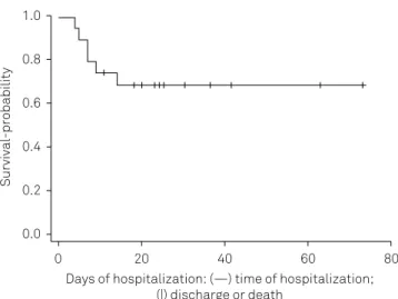
he clinical and surgical characteristics are shown in Table 3. he DC mortality rate evaluated 30 days postoperatively
(Figure) was 30% (95%CI 14.5–51.9).
Table 2. Socio-demographic characteristics of the patients included in the study.
Demographic variables n = 20
Age (years)* 52.7 ± 7.4
Sex (%)**
Male 11 (55)
Female 9 (45)
Skin color (%)**
White 15 (75)
Black 5 (25)
*Continuous variable expressed as average ± standard deviation; **Categorical variables expressed as n (%).
Table 3. Clinical and surgical characteristics of the patients included in the study.
Variables n = 20
Ictus time x admission (hours)* 13.2 ± 1.4
Hospitalization NIHSS (score)* 17.6 ± 3.2
NCI evaluation x surgery (hours)** 2.9 [1.9–4.4]
Ictus time x surgery (hours)** 28.1 [21.2–50.6]
Clinical worsening after 1st NCI evaluation (%)*** 13 (65)
Nº of CTs before surgery** 2 [1.2–3]
Time interval between surgery and last CT (hours)** 7.7 [2.3–14.8]
Rankin (30 days score)** 4 [3.3–6]
Rankin (discharge score)** 4 [3–4]
Discharge rate (%)*** 14 (70)
Death rate (%)*** 6 (30)
Hospitalization time (days)** 21.5 [9.5–34.5]
NIHSS: National Institutes of Health Stroke Scale; NCI: neurosurgery; CT: computerized tomography. *Continuous variable expressed as average ± standard deviation; **Asymmetrical variables expressed as median and interquartile interval; ***Categorical variables expressed as n (%).
Figure. Kaplan-Meier curve of in-hospital mortality of patients included in the study.
Survival-probability
Days of hospitalization: (—) time of hospitalization; (|) discharge or death
1.0
0.8
0.6
0.4
0.2
0.0
DISCUSSION
To the best of our knowledge, this is the first study
that assessed the DC outcome in malignant MCA infarc-tion in an academic hospital in Brazil, considered a
ref-erence for the treatment of stroke patients nationwide.
As a main finding, there was a 30% mortality rate among patients subjected to DC. Another Brazilian study, involv-ing 34 patients, showed similar data, with a surgical
mor-tality rate of 26%11.
Previous studies12,13 have recommended performing DC
for the treatment of malignant cerebral infarction in patients aged 60 years or less, before clinical or radiological signs of brain herniation, within a time frame of 48 hours. In the pres-ent study, we had an average age of 52.7 ± 7.4 years,
includ-ing three patients over 60 years of age. he average age of patients in the randomized controlled trials HeADDFIRST14,
DECIMAL9, DESTINY8 and HAMLET7 was, respectively, 54.6;
43.4; 44.6 and 48.2 years.
Several studies have reported that age should be con-sidered an important factor in patients to be submitted
to the surgical procedure15,16,17. he decision for patients
over 60 years of age must be individualized: there is great
controversy regarding this cut-of point, as some authors describe beneits beyond this age-group12,15,18,19, while
oth-ers report unfavorable outcomes6,7,8,9. In the present study,
three patients over 60 years of age were included. A Chinese
study evaluated the efectiveness of DC in patients over
80 years of age, concluding that DC, even in patients in this age group, may increase the survival rate without severe
compromise of functionality18. In a recent American study,
involving more than 1,600 patients, the authors reported the results according to age as uni- and multivariate
analy-sis, not taking into account this variable as a signiicant pre
-dictor of clinical outcome20.
In the present study, an average of 3,5 points in the
mRS score was found among survivors, a value considered
acceptable (mild to moderately severe disability), as those patients probably would have died without surgical treat-ment. A recent cross-sectional and multicenter study aimed at identifying the opinion of more than 1,800 phy-sicians, with clinical experience in MCA treatment, about what they considered an acceptable disability. The
major-ity of respondents (79.3%) considered acceptable mRS
scores of 3 or greater21.
Individuals afected by a malignant ischemic event are seldom discharged without severe clinical deicits; this data
is in accordance with a recent study of meta-analysis carried out in China, involving more than 700 patients, that
consid-ered an mRS of 4 a good functional result10.
In two out of the 20 patients in the sample, it was not possible to clearly determine the approximate time
lapse from stroke to admission in the emergency unit of
the hospital. In those where this information could be
obtained, the average time was 13.2 ± 1.4 hours. Such data has a potential clinical significance, since the longer the time between the ictus, the diagnosis and the opera-tion procedure, the smaller the chances of positive out-comes are.
The median time between the ictus and the opera-tion was 28.1 [21.2–50.6] hours, different from another Brazilian study with 34 cases that presented an average
time of 2.05 days11. We highlight that, among the
stud-ied patients, three were submitted to the procedure late,
due to the lack of a clear definition of the MCA territory
hypodensity > 2/3. This was confirmed by the number of CTs before the operation (2.2 [1–5]), data that suggests the caution with which the surgical procedures were indicated. In the same manner, the time elapsed from the last CT to the surgical procedure was 8.7 [2–23] hours, somewhat large, but justified by the logistical difficulties, since this hospital is a teaching hospital with a very busy emergency department requiring surgery, and a wait-ing list for the available operatwait-ing rooms. Even without immediate availability of an operating room, the neuro-surgery team remained in constant contact, evaluating and handling of these patients, which can be underlined by the time-elapsed hours between the last neurosur-gery evaluation and the surgical procedure: up to 50% of the evaluated patients were subjected to surgery within 2,9 hours.
he median time of hospitalization, including the ICU and inirmary stay, was 21.5 [9.5–34.5] days. In a retrospec -tive study carried out in France, where ten patients sub-jected to DC were evaluated, the average ICU stay was
22 days (3–58)22. It is important to highlight that the time
of the present study was measured from the time of
hospi-talization to the inal outcome, including one (5%) who was hospitalized for an elective tracheoplasty and ifteen days later sufered a stroke.
An average NIHSS score of 17.6 ± 3.2 was found upon hospitalization, which demonstrates the clinical sever-ity of the patients. In a recent Japanese study involving 355 patients, an average NIHSS score of 18 points (mini-mum 0 – maxi(mini-mum 38) was shown at admission, and in
the DC group it was 215. These results suggest that the
patients in our study followed strict selection criteria for the surgical procedure.
A real limitation of this study is the type of outlining used, since the group of patients subjected to the DC proce-dure was not compared to any other patients subjected to conservative treatment, as reported in the major
random-ized clinical trials7,8,9,12.
References
1. Musabelliu E, Kato Y, Imizu S, Oda J, Sano H. Surgical treatment of patients with ischemic stroke decompressive craniectomy. In: Rodríguez JCG, editor. Acute ischemic stroke. Rijeka: InTech; 2012. p. 165-86.
2. Mellado TP, Castillo FL, Campos PM, Bugedo TG, Dougnac LA, Andresen HM. [Decompressive hemicraniectomy for malignant middle cerebral artery infarction: report of two cases]. Rev Med Chil. 2005;133(4):447-52. Spanish.
3. Neugebauer H, Heuschmann PU, Jüttler E. DEcompressive surgery for the Treatment of malignant INfarction of the middle cerebral arterY - Registry (DESTINY-R): design and protocols. BMC Neurol. 2012;12(1):1-6. https://doi.org/10.1186/1471-2377-12-115
4. Ropper AH. Hemicraniectomy: to halve or halve not. N Engl J Med. 2014;370(12):1159-60. https://doi.org/10.1056/NEJMe1315721
5. Suyama K, Horie N, Hayashi K, Nagata I. Nationwide survey of decompressive hemicraniectomy for malignant middle cerebral artery infarction in Japan. World Neurosurg. 2014;82(6):1158-63. https://doi.org/10.1016/j.wneu.2014.07.015
6. Chen CC, Cho DY, Tsai SC. Outcome of and prognostic factors for decompressive hemicraniectomy in malignant middle cerebral artery infarction. J Clin Neurosci. 2007;14(4):317-21. https://doi.org/10.1016/j.jocn.2005.05.024
7. Hofmeijer J, Kappelle LJ, Algra A, Amelink GJ, Gijn J, Worp HB et al. Surgical decompression for space-occupying cerebral infarction (the Hemicraniectomy After Middle Cerebral Artery infarction with Life-threatening Edema Trial [HAMLET]): a multicentre, open, randomised trial. Lancet Neurol. 2009;8(4):326-33. https://doi.org/10.1016/S1474-4422(09)70047-X
8. Jüttler E, Schwab S, Schmiedek P, Unterberg A, Hennerici M, Woitzik J et al. Decompressive surgery for the treatment of malignant infarction of the middle cerebral artery (DESTINY): a randomized, controlled trial. Stroke. 2007;38(9):2518-25. https://doi.org/10.1161/STROKEAHA.107.485649
9. Vahedi K, Vicaut E, Mateo J, Kurtz A, Orabi M, Guichard JP et al. Sequential-design, multicenter, randomized, controlled trial of early decompressive craniectomy in malignant middle cerebral artery infarction (DECIMAL Trial). Stroke. 2007;38(9):2506-17. https://doi.org/10.1161/STROKEAHA.107.485235
10. Lu X, Huang B, Zheng J, Tao Y, Yu W, Tang L et al. Decompressive craniectomy for the treatment of malignant infarction of the middle cerebral artery. Sci Rep. 2014;4(7070):7070. https://doi.org/10.1038/srep07070
11. Nobre MC, Monteiro M, Albuquerque AC, Veloso AT, Mendes VA, Silveira MF et al. [Decompressive craniectomy for treatment of intracranial hypertension secondary to large ischemic cerebral infarction: analysis of 34 cases]. Arq Neuropsiquiatr. 2007;65(1):107-13. Portuguese. https://doi.org/10.1590/S0004-282X2007000100022
12. Jüttler E, Unterberg A, Woitzik J, Bösel J, Amiri H, Sakowitz OW et al. Hemicraniectomy in older patients with extensive middle-cerebral artery stroke. N Engl J Med. 2014;370(12):1091-100.
https://doi.org/10.1056/NEJMoa1311367
13. Huttner HB, Schwab S. Malignant middle cerebral artery infarction: clinical characteristics, treatment strategies, and future perspectives. Lancet Neurol. 2009;8(10):949-58. https://doi.org/10.1016/S1474-4422(09)70224-8
14. Frank JI, Schumm LP, Wroblewski K, Chyatte D,
Rosengart AJ, Kordeck C et al. Hemicraniectomy and durotomy upon deterioration from infarction-related swelling trial: randomized pilot clinical trial. Stroke. 2014;45(3):781-7. https://doi.org/10.1161/STROKEAHA.113.003200
15. Fiorot Junior JA, Silva GS, Cavalheiro S, Massaro AR. Use of decompressive craniectomy in the treatment of hemispheric infarction. Arq. Neuropsiquiatr. 2008;66(2A):204-8. https://doi.org/10.1590/S0004-282X2008000200012
16. Gupta R, Connolly ES, Mayer S, Elkind MS. Hemicraniectomy for massive middle cerebral artery territory infarction: a systematic review. Stroke. 2004;35(2):539-43. https://doi.org/10.1161/01.STR.0000109772.64650.18
17. Antuña-Ramos A, Alvarez-Vega MA, Seijo-Fernández F, Calleja-Puerta S, González-Delgado M, Torres-Campa JM et al. [Surgical treatment of the stroke in the middle cerebral artery]. Rev Neurol. 2009;49(7):354-8. Spanish.
18. Zhao J, Su YY, Zhang Y, Zhang YZ, Zhao R, Wang L et al. Decompressive hemicraniectomy in malignant middle cerebral artery infarct: a randomized controlled trial enrolling patients up to 80 years old. Neurocrit Care. 2012;17(2):161-71. https://doi.org/10.1007/s12028-012-9703-3
19. Inamasu J, Kaito T, Watabe T, Ganaha T, Yamada Y, Tanaka T et al. Decompressive hemicraniectomy for malignant hemispheric stroke in the elderly: comparison of outcomes between individuals 61-70 and >70 years of age. J Stroke Cerebrovasc Dis. 2013;22(8):1350-4. https://doi.org/10.1016/j.jstrokecerebrovasdis.2013.02.008
20. Daou B, Kent AP, Montano M, Chalouhi N, Starke RM,
Tjoumakaris S et al. Decompressive hemicraniectomy: predictors of functional outcome in patients with ischemic stroke. J Neurosurg. 2016;124(6):1773-9. https://doi.org/10.3171/2015.6.JNS15729
21. Neugebauer H, Creutzfeldt CJ, Hemphill JC 3rd, Heuschmann PU, Jüttler E. DESTINY-S: attitudes of physicians toward disability and treatment in malignant MCA infarction. Neurocrit Care. 2014;21(1):27-34. https://doi.org/10.1007/s12028-014-9956-0