ARTICLE DOI: 10.1590/0004-282X20130037
Multiparametric multidetector computed
tomography scanning on suspicion of
hyperacute ischemic stroke: validating a
standardized protocol
Avaliação multiparamétrica por tomografia computadorizada multidetectores na suspeita
de isquemia cerebral hiperaguda: validando um protocolo padronizado
Felipe Torres Pacheco1, Antônio José da Rocha1, Ingrid Aguiar Littig1, Antonio Carlos Martins Maia Júnior1,
Rubens José Gagliardi2
Within the last few years, brain imaging has revolution-ized the approach for patients with acute ischemic stroke, conidently diferentiating hemorrhagic from ischemic stroke, depicting the extent of early parenchymal efects and most recently deining the parameters to guide thrombolytic therapy1.
Magnetic resonance imaging (MRI) protocols are reliable for the detection of early cytotoxic brain edema, mismatch areas, and arterial low abnormalities2. However, the
acquisi-tion is not as feasible in the daily routine due to the required time and inherent limitations of the procedure. Noncontrast computed tomography (NCCT) has become a useful tool in
1Division of Neuroradiology, Santa Casa de Misericórdia de São Paulo, São Paulo SP, Brazil; 2Division of Neurology, Santa Casa de Misericórdia de São Paulo, São Paulo SP, Brazil.
Correspondence: Antonio Jose da Rocha; Rua Doutor Cesário Motta Junior 112; 01221-020 São Paulo SP - Brasil; E-mail: [email protected] Conflict of interest: There is no conflict of interest to declare.
Received 10 November 2012; Received in final form 22 November 2012; Accepted 29 November 2012. ABSTRACT
Multidetector computed tomography (MDCT) scanning has enabled the early diagnosis of hyperacute brain ischemia. We aimed at validating a standardized protocol to read and report MDCT techniques in a series of adult patients. The inter-observer agreement among the trained examiners was tested, and their results were compared with a standard reading. No false positives were observed, and an almost perfect agreement (Kappa>0.81) was documented when the CT angiography (CTA) and cerebral perfusion CT (CPCT) map data were added to the noncontrast CT (NCCT) analysis. The inter-observer agreement was higher for highly trained readers, corroborating the need for specific train-ing to interpret these modern techniques. The authors recommend addtrain-ing CTA and CPCT to the NCCT analysis in order to clarify the global analysis of structural and hemodynamic brain abnormalities. Our structured report is suitable as a script for the reproducible analysis of the MDCT of patients on suspicion of ischemic stroke.
Key words: tomography, perfusion, angiography, stroke, cerebral infarction.
RESUMO
A tomografia computadorizada multidetectores (TCMD) permitiu o diagnóstico precoce de isquemia cerebral hiperaguda. O presente estudo objetivou validar a interpretação e a descrição padronizada de um protocolo de TCMD multiparamétrica em uma série de pacientes adultos. A concordância entre os examinadores foi testada, e seus resultados confrontados com uma leitura padrão. Não foram observados resultados falso-positivos, e foi documentado um elevado grau de concordância (Kappa>0,81) quando os dados da angiotomografia (ATC) e dos mapas de perfusão cerebral por TC (PCTC) foram adicionados à análise da TC sem contraste (TCSC). A concordância interobservador foi superior para os leitores melhor treinados, corroborando a necessidade de formação específica para a interpretação dos exames. Os autores recomendam acrescer a interpretação da ATC e da PCTC à análise da TCSC, visando à análise global das anormalidades cerebrais estruturais e hemodi-nâmicas. O presente protocolo é adequado como um roteiro reprodutível para a análise da TCMD de pacientes com suspeita de acidente vascular cerebral isquêmico.
the evaluation of acute stroke due to its wide availability, fast acquisition, and high accuracy for ruling out intracra-nial hemorrhage3-5. More recently, multidetector CT (MDCT)
scanning has enabled the development of advanced tech-niques, including CT angiography (CTA) and cerebral perfu-sion computed tomography (CPCT)6-9.
Although patient selection could be based primarily on NCCT10, recent reports have been gathering evidence about
the safe use of multiparametric MDCT to include patients for either intravenous or intra-arterial thrombolysis from early or later ictus in diferent protocols11-13.
In order to determine which patients would most beneit with the available protocols for thrombolytic therapies, some objective queries must be efectively understood in an emer-gency setting8,14:
• Could the imaging results indicate something other than
ischemia?
• Is there intracranial hemorrhage?
• Is there a parenchymal abnormality? What is its extent? • Is there a mismatch area in the brain perfusion? • Is there a large vessel obstruction?
A comprehensive training program must comprise the theoretical basis of stroke, and include a practical scenario to enable specialists to solve these inquiries. he sequential steps to read and report multiparametric MDCT in hyper-acute brain ischemia are the focus of this study.
METHODS
his is part of a larger study using MDCT techniques to evaluate hyperacute stroke. he protocol has been reviewed and approved by the Institutional Review Board and the lo-cal ethics committee. A cohort of individuals with previous-ly known outcomes was selected to include a range of im-aging indings in the scenario of hyperacute brain ischemia. Multiparametric MDCT examinations from the digital ar-chives of the Radiology Division of Santa Casa de Misericórdia de São Paulo were analyzed, with the aim of testing the re-producibility of a standardized protocol for their analysis and description.
Adult patients (≥18 years-old) with hyperacute symp-toms (<six hours) suspected of focal acute ischemia in the middle cerebral arteries (MCA) and who had been submit-ted to multiparametric brain MDCT were considered eligible. he informed consent was signed personally by all subjects or their guardians. We excluded patients with contraindica-tions to ionizing radiation exposure or to the intravenous io-dine contrast agent, those who refused to participate in the study, and those whose examinations included images with inadequate quality or technical artifacts.
All exams were conducted using a previously deined imag-ing acquisition protocol with a minimum dose of both ionizimag-ing
radiation and intravenous iodinated contrast in a 64-slice CT scanner (Brilliance CT 64 Channel, Philips Medical, Eindhoven, The Netherlands), including NCCT, CPCT maps, and CTA, which could take up to seven minutes.
In all cases, a dual-head power injector (Medrad, Warrendale, USA) with an 18-G i.v. access, usually located in the cubital vein, was used for injection. Brain perfu-sion was performed after the administration of 50 mL of a nonionic contrast agent at an injection rate from 4 to 5 mL/s. After 5 seconds from the start of contrast injection, continuous images in the region of interest were acquired (120 kV, 80 mA, rotation of 0.75 second, and duration of 20 seconds). A total of 640 images, axial through 16 and each 2.5 mm thick, were acquired, resulting in a total area of 3.75 cm2 of the brain studied in the MCA territory.
hereafter, the CTA was obtained by the intravenous ad-ministration of an additional 50 mL of nonionic iodinated contrast agent. he region of interest (ROI) was previously placed in the aortic arch for determining the self-timing of the apparatus, when the attenuation in this region reached 160 Hounsield units (HU), initiating the acquisition of the sections from the aortic arch to the vertex of the skull.
The perfusion analysis was post-processed from the source images to obtain semiautomated maps of the CPCT, including cerebral blood volume (CBV), ce-rebral blood flow (CBF), and mean transit time (MTT). The CTA analysis included the maximum intensity pro-jection (MIP) and tridimensional (3D) views of the in-tracranial and exin-tracranial arteries. All data were post-processed using a commercially available software on a workstation (Extended Brilliance Workspace v3.5.0.2250, Philips Medical Systems Nederland B.V., PC Best, The Netherlands).
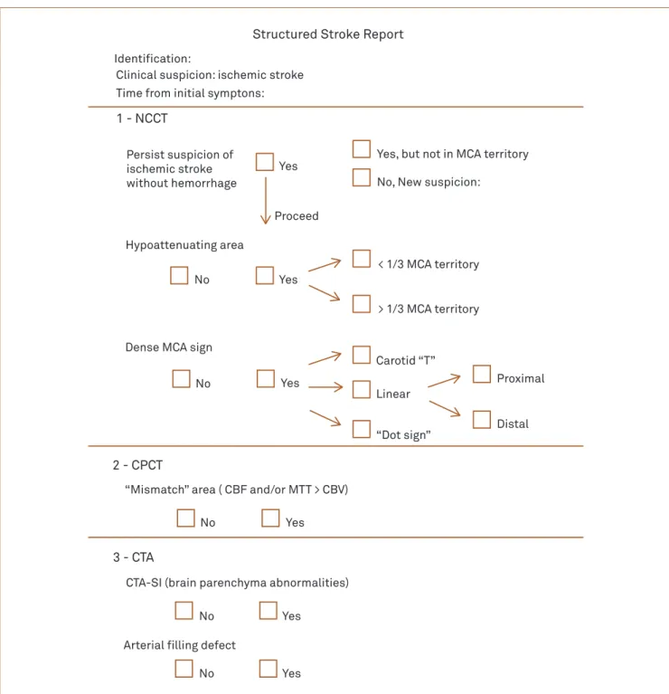
A structured form was adapted from previous reports considering the relevant imaging findings to build an ef-fective script for the step-by-step interpretation and to objectively answer those queries to guide the clinical de-cision-making process concerning patients with suspect-ed ischemic stroke (Fig 1)8.
All studies were anonymous, and the standard read-ing was based on the authors’ interpretation of all exams, which were read by consensus and considered the imag-ing follow-up results. Subsequently, in order to consider only the background knowledge of the examiners, we de-cided to include 22 residents from our current program of radiology (11 second- and 11 third-year residents) and 6 neuroradiology fellows. The purpose of the study was de-scribed, and doubts about how to fill out the report were clarified. Neither specific training nor an image review was offered to prepare them for the study.
ribbon sign or loss of basal ganglia definition) was graded according to the one third rule15. On the CTA
post-pro-cessed images, cervical and intracranial arteries from the carotid system were mainly examined to detect wall ab-normalities, intraluminal plaques, or thrombus ( filling defects). Intraluminal fresh thrombi (carotid-MCA) were defined as indicated in previous reports, according to their form and location, as a T-carotid thrombus, linear M1 thrombus or “dot sign” distal thrombus8,16,17.
Source images from the CTA (CTA-SI) acquisition were analyzed to detect hypoattenuating brain areas and
identify either an arterial obstruction or intraluminal thrombus. Mismatch areas were defined (present or ab-sent) on the CPCT post-processed images (CBF / MTT > CBV), and they were summarized on an automatic post-processed map (“summary map”)8,18.
All examiners sequentially analyzed every parameter from the same patient to create an individual structured report for each person. The inter-observer agreement was assessed by obtaining Kappa’s coefficient for the evalua-tion of the proposed stroke report.
Fig 1. Structured stroke report.
NCCT: noncontrast computed tomography; MCA: middle cerebral arteries; CPCT: cerebral perfusion computed tomography; CTA: tomography angiography.
Structured Stroke Report
Identification:
Clinical suspicion: ischemic stroke Time from initial symptons:
1 - NCCT
Persist suspicion of ischemic stroke without hemorrhage
Yes
Yes Yes
Yes
Yes
Yes Proceed
Yes, but not in MCA territory
No, New suspicion:
Hypoattenuating area
No
No
No
No No
< 1/3 MCA territory
> 1/3 MCA territory
Dense MCA sign
Carotid “T”
Linear
“Dot sign”
Proximal
Distal
2 - CPCT
“Mismatch” area ( CBF and/or MTT > CBV)
3 - CTA
CTA-SI (brain parenchyma abnormalities)
RESULTS
According to the proposed criteria, 70 consecutive sub-jects were submitted to the established protocol, with 11 be-ing excluded later.
he standard reading (consensus from authors and im-aging follow-up analysis) included the following paragraphs.
Normal results of the multiparametric MDCT studies were documented in 33 subjects (33/59 – 55.9%). All unre-markable studies were properly identiied, and none of them depicted brain ischemia on the imaging follow-up (no false positives). he remaining 26 ones (26/59 – 44.1%) had at least one abnormal parameter on the MDCT, and their im-aging follow-up conirmed the variable MCA territory brain ischemia.
Hypoattenuating areas were documented on the NCCT scans in 14 subjects (14/26 – 53.8%), which were associated or not with the dense middle cerebral ar-tery sign. After additional analysis of the CTA-SI, the sensitivity significantly increased to greater than 80%. Furthermore, the NCCT analysis, compared with both the
CTA and CPCT, failed to detect these abnormal areas in 12 subjects (12/26 – 46.1%).
The detection rate of the dense middle cerebral ar-tery sign on NCCT was 38.4 % (10/26). It was labeled as the linear type (7/10), carotid “T” type (2/10), and “dot sign” (1/10). After the analysis of the arterial circulation by CTA, we found that 65.3% (17/26) of the patients had an intravascular filling defect (Fig 2).
According to the CPCT parameters, penumbra areas and ischemic core were detected in all 26 scans (Fig 3), which confirmed brain ischemia on the imaging follow-up (no false negatives). The concurrent analysis of the CPCT maps and CTA-SI allowed the detection of brain ischemia in seven additional subjects for whom the NCCT had un-remarkable results (7/26 – 27.0%). CPCT was the only technique that enabled ischemia detection in another five subjects (5/26 – 19.0%).
The results of the examiners’ analysis ( filled imaging reports, not considering the imaging follow-up) were the following parts herein.
All unremarkable studies were also correctly identi-fied by examiners (no false positive reports). Considering
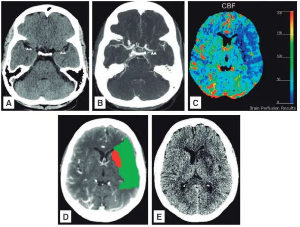
Fig 2. Intra-luminal thrombus undetected on the noncontrast computed tomography obtained one hour after the onset of ischemic symptoms in a 68-year-old woman. Note the absence of hyperdense artery signs on the M1 segments (arrowheads) in this noncontrast computed tomography axial scan (A). The computed tomography angiography axial post-processed image (B) depicted a filling defect in the left M1 segment (arrowheads). The perfusion computed tomography map of the carebral blood flow (C) and “summary map”(D) demonstrated a large mismatch (green-colored area) with a small infarcted core (red-colored area) on the left brain hemisphere. The comparative noncontrast computed tomography axial scan for the imaging follow-up (E) confirmed the final infarct extension to the left striatum (larger than the previous core).
CBF: cerebral blood flow.
A
B
C
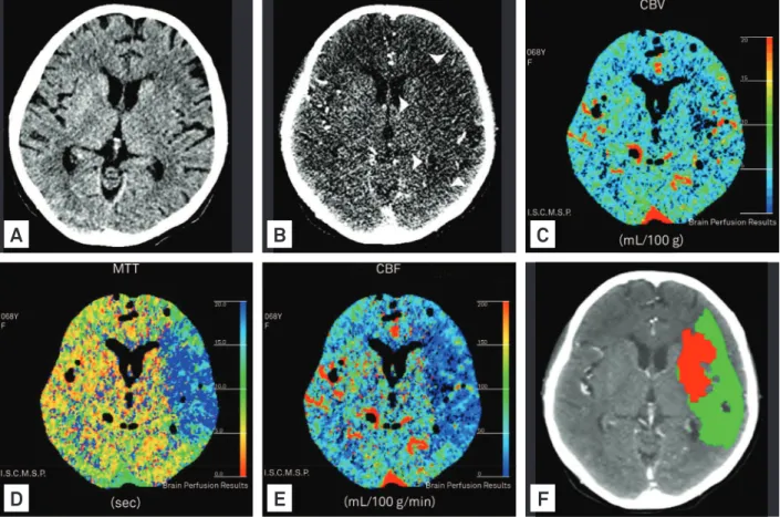
Fig 3. Multiparametric multidetector computed tomography on hyperacute ischemic stroke obtained four hours after the onset of ischemic symptoms in a 68-year-old woman. The noncontrast computed tomography axial scan at the basal ganglia level (A) exhibited the loss of putaminal defi nition and an insular ribbon sign on the left brain hemisphere. The source images from the computed tomography angiography (B) depicted a larger hypoattenuation in the middle cerebral arteries territory (arrowheads). The perfusion computed tomography map of the cerebral blood volume (C), mean transit time (D), and cerebral blood fl ow (E) confi rmed left middle cerebral arteries hypoperfusion (blue-colored area), and the “summary map” (F) shared information showing the mismatch (green-colored area) with a infarcted core (red-colored area).
CBV: cerebral blood volume; MTT: mean transit time; CBF: cerebral blood fl ow.
A
B
C
D
E
F
the abnormal examinations, 72.0% of the reports were correctly filled out by the second-year residents, when they analyzed the NCCT images, reaching 81.8% after the inclusion of the CTA and CPCT maps. These results char-acterized an almost perfect agreement (Kappa>0.81). The third-year residents and Neuroradiology fellows detected abnormalities in 84.1 and 90.3%, respectively, when eval-uating the NCCT images, and in 90.1 and 95.8%, respec-tively, after the inclusion of the CPCT maps. These results also showed an almost perfect agreement (Kappa>0.81). These data are summarized on Tables 1 and 2.
DISCUSSION
The emergency evaluation of hyperacute stroke pa-tients requires team harmony to quickly detect abnormal-ities and correctly determine the appropriate treatment. Conversely, traditional Brazilian radiologic reports de-scribe normal and abnormal findings in an inappropriate form for this scenario. Structured protocols for imaging analysis have been proposed abroad, but the validation of
their use in Brazilian practice and for training our special-ists remains to be performed8.
Fundamental queries concerning imaging diagnosis must be answered before starting therapeutics8. NCCT
analysis has proven to be an accurate method for rul-ing out intracranial hemorrhage and identifyrul-ing subtle early signs of brain ischemia16,19. However, several
stud-ies have demonstrated only poor-to-moderate inter-ob-server agreement in the detection of infarcts by NCCT in acute settings20,21. NCCT findings might indicate brain
swelling as a relatively late phenomenon characterized by the loss of insular ribbon sign and obscuration of the len-tiform nucleus sign, both detected at a lower rate in the first six hours than the results from the decreased density of brain tissue often associated with an increased density of arteries22.
Successful thrombolytic therapy depends on the se-lection of patients, and MDCT parameters have proven to be useful for this purpose3,12,23,24. To attend the need of
NCCT CTA CPCT
Patient
Hypoa
tt
enua
ting ar
ea
(< or > 1
/3 MC
A t
er
rit
ory)
Insular rib
bon sign
L
oss o
f de
finition o
f BG
Dense car
o
tid/MC
A ar
tery
Hypoa
tt
enua
ting ar
ea (small
er/
lar
g
er/
similar t
o NCC
T)
Intr
aluminal thr
ombus /
s
tenosis
Abnor
mal perf
usion (small
er/lar
g
er/
similar t
o C
TA)
1 - - - - Similar - Larger
2 + (< 1/3) - + - Larger + Larger
3 - + + Linear Larger + Larger
4 + (> 1/3) + + - Similar + Larger
5 - + - “Dot sign” Larger + Larger
6 + (> 1/3) + + “T” Similar + Larger
7 - - - - Similar + Larger
8 + (> 1/3) + + “T” Larger + Similar
9 - - - - Similar - Larger
10 + (< 1/3) - - - Similar - Larger
11 + (< 1/3) + - - Larger - Larger
12 + (> 1/3) + + - Larger + Similar
13 - - - - Similar - Larger
14 - + + Linear Larger + Larger
15 - + + - Larger + Larger
16 + (> 1/3) + + Linear Similar + Similar
17 - - - - Larger - Larger
18 + (< 1/3) + + Linear Larger + Similar
19 + (< 1/3) + + Linear Larger + Similar
20 + (< 1/3) + - - Similar - Larger
21 + (> 1/3) + + - Similar + Larger
22 - + + Linear Larger + Similar
23 + (< 1/3) - - - Similar - Larger
24 + (< 1/3) + + - Similar + Larger
25 - + + Linear Larger + Similar
26 + (< 1/3) - - - Similar - Larger
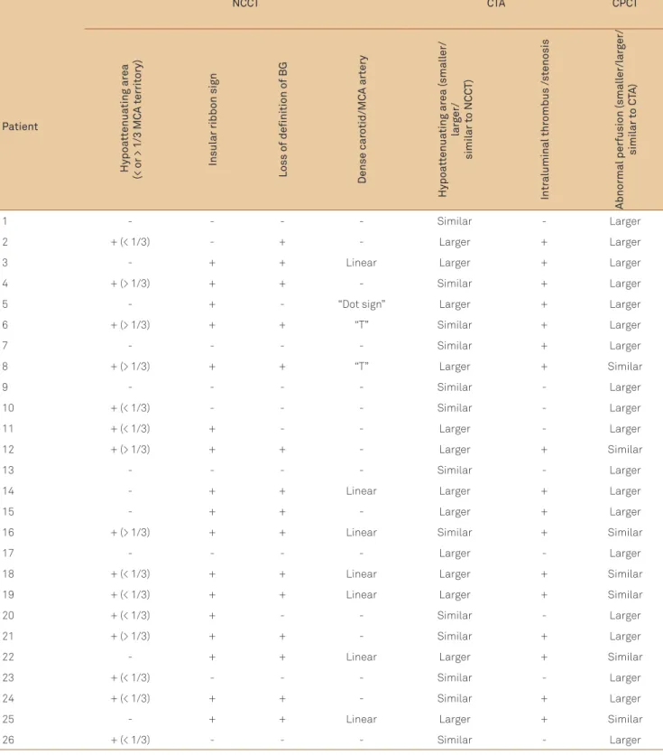
Table 1. Data of images.
MCA: middle cerebral arteries; NCCT: noncontrast computed tomography; CTA: tomography angiography; CPCT: cerebral perfusion computed tomography.
NCCT: noncontrast computed tomography; CTA: tomography angiography; CPCT: cerebral perfusion computed tomography.
Inter-observer agreement NCCT CTA CPCT
R2 72.0% 80.3% 81.8%
R3 84.1% 87.1% 90.1%
Neuroradiology fellows 90.3% 95.9% 95.8%
the clinical treatment. No false positives were derived from the NCCT analysis in our series of patients since all normal examinations were properly identified by the examiners.
After the NCCT evaluation, our proposed imaging re-port expressed the CPCT study, assessing the occurrence of penumbra and ischemic core areas. Some trials around the world have confirmed the relevance of patient’s selec-tion according to the demonstraselec-tion of “viable ischemic brain tissue”, as defined by perfusion mismatch8,14,24-26. Our
results are in line with these previous studies, confirming the relevance of CPCT in detecting abnormal brain perfu-sion even without parenchymal NCCT abnormalities with an almost perfect agreement among the examiners6,20,21,23.
Understanding CPCT maps added physiologic informa-tion to the anatomic and structural analyses of the NCCT and CTA. No false positives occurred in the analysis of the CPCT maps in our series of patients.
The last stage of our stroke protocol assessed the CTA information. On the NCCT scans, the occurrence of a hy-perdense artery sign predicted an intravascular filling defect in the CTA images17. A dense middle cerebral
ar-tery was detected in 38.4% of the patients from our series, which increased to 65.3% after the CTA analysis (vessel obstruction). Similarly, parenchymal abnormalities were detected on the NCCT of our patients in agreement with the detection rate reported in the current literature17.
After the additional analysis of the CTA-SI, the sensitivi-ty significantly was superior among our examiners. These results confirmed previous reports that CTA images add sensitivity and specificity to the interpretation of NCCT for detecting neglected brain abnormalities and intravas-cular filling defects6,17,24,25.
he increased red blood cell content in the thrombus enables the detection of potential fresh red thrombi (hyper-dense artery sign on the carotid or MCA) on NCCT, which inluences the therapeutic decision-making process25.
However, contrast injection on CTA enables the detection of illing defects even in the absence of red thrombus, avoiding the false-positive detection of hyperdense artery signs on the NCCT, as previously reported in cases of dehydration or ad-vanced atherosclerosis8. In acute ischemic stroke, CTA has
proven to be an accurate method for the detection of intra-cranial proximal arterial occlusions and stenoses that predict functional outcome, inal infarct size, and response to intra-venous thrombolysis, thereby facilitating the decision-mak-ing for the intra-arterial rescue procedures5,25,26. Hence, future
decisions about the optimal therapy for acute strokes should consider the detection of vascular thrombus17.
he inter-observer agreement among the Neuroradiology fellows was higher than among the younger residents, presum-ably as a result of not neglecting relevant details during the
interpretation. Although our normal results were conirmed to be accurate for all examiners, the abnormal ones exhibited a progressive learning curve according to the radiology train-ing program. his observation reinforces the previous sugges-tion that inter-observer agreement might be higher for highly-trained readers in the hyperacute ischemic stroke setting4. We
argue that our results corroborate the need for speciic train-ing to interpret multiparametric examinations.
The absence of false positives was most likely derived from the interference among the judged MDCT param-eters, suggesting that the sequential analysis of multipa-rametric complementary data facilitates the interpreta-tion and optimistically reinforces the confidence in the diagnosis and inter-rater reliability. The addition of CPCT semiautomated maps to CTA-SI more clearly depicted the hypoattenuating areas and their size, contributing to the ability to delineate the mismatch areas.
Our study design is in line with the current guidelines reinforcing the need for the correct global analysis of data to detect parenchymal, vascular, or hemodynamic abnor-malities. The development and application of multi-pro-fessional, integrated written protocols for the treatment of any suspected stroke cases are also addressed herein by this proposed stroke report10.
The limitations of the MDCT technology to evalu-ate ischemic stroke patients have been reported9,17.
Furthermore, our study has some limitations inherited from the proposed design, including the small patient sample and absence of intraobserver analysis. However, our study design is acceptable for testing the inter-observ-er agreement among the training radiologists. It is a pre-liminary attempt to develop an auditable tool that docu-ments specific skills to analyze multiparametric MDCT in the hyperacute stroke scenario. Related Brazilian societ-ies and medical services could use this objective script for training and certifying specialists to work in stroke units.
In conclusion, the proposed protocol for the multipara-metric MDCT study is a fast and uncomplicated tool that will help to objectively answer the most important issues of decision-making in the emergency scenario. Normal ex-ams were correctly identified (no false positives), minimiz-ing the risk of inappropriate intervention. Furthermore, to ensure a confident interpretation and avoid false negative results, the authors recommend adding CTA and CPCT to the NCCT analysis, clarifying the global evaluation of the structural and hemodynamic brain abnormalities.
1. Gonzalez RG. Imaging-Guided acute ischemic stroke therapy: from “time is brain” to “physiology is brain”. AJNR 2006;27:7.
2. Mullins ME. CT and conventional and diffusion-weighted MR imaging in acute stroke: study in 691 patients at presentation to the emergency department. Radiology 2002;224:253-260.
3. Latchaw RE. Recommendations for imaging of acute ischemic stroke. Stroke 2009;40:3646-3658.
4. Vu D, Lev MH. Noncontrast CT in acute stroke. Semin Ultrasound CT MR 2005;26:380-386.
5. Saqqur M, Uchino K, Demchuk AM, et al. Site of arterial occlusion identified by transcranial Doppler predicts the response to intravenous thrombolysis for stroke. Stroke 2007;38:948-954.
6. Camargo ECS. Acute brain infarct: detection and delineation with CT angiographic source images versus nonenhaced CT scans. Radiology 2007;244:542-548.
7. Tan JC. Systematic comparison of perfusion-CT and CT-angiography in acute stroke patients. Ann Neurol 2007;61:533-543.
8. de Lucas EM, Sanchez E, Gutierrez A, et al. CT protocol for acute stroke: tips and tricks for general radiologists. Radiographics 2008;28:1673-1687.
9. Suzuki K, Morita S, Masukawa A, Machida H, Ueno E. Utility of CT perfusion with 64-row multi-detector CT for acute ischemic brain stroke. Emerg Radiol 2011;18:95-101.
10. Oliveira Filho J. Guidelines for acute ischemic stroke treatment - Part I. Arq Neuropsiquiatr 2012;70:621-629.
11. Wintermark M. Imaging of acute ischemic brain injury: the return of computed tomography. Curr Opin Neurol 2003;16:59-63.
12. Lovblad KO, Baird AE. Actual diagnostic approach to the acute stroke patient. Euro Radiol 2006;16:1253-1269.
13. Leiva-Salinas C, Wintermark M. Imaging of acute ischemic stroke. Neuroimaging Clin N Am 2010;20:455-468.
14. Konstas AA. CT Perfusion Imaging in acute stroke. Neuroimaging Clin N Am 2011;21:215-238.
15. Kalafut MA. Detection of early CT signs of > 1/3 middle cerebral artery infarctions: interrater reliability and sensitivity of CT interpretation by physicians involved in acute stroke care. Stroke 2000;31:1667-1671.
16. Wardlaw JM, Mielke O. Early signs of brain infarction at CT: observer reliability and outcome after thrombolytic treatment--systematic review. Radiology 2005;235:444-453.
17. Romero JM. CT angiography source image evaluation for stroke. Semin Ultrasound CT MR 2005;26:387-393.
18. Wintermarck M. Perfusion-CT assessment of infarct core and penumbra. Stroke 2006;37:979-985.
19. Kucinski T. Unenhanced CT and acute stroke physiology. Neuroimaging Clin N Am 2005;15:397-407.
20. Schriger DL, Kalafut M, Starkman S, Krueger M, Saver JL. Cranial computed tomography interpretation in acute stroke: physician accuracy in determining eligibility for thrombolytic therapy. JAMA 1998;279:1293-1297.
21. Shinar D, Gross CR, Hier DB, et al. Interobserver reliability in the interpretation of computed tomographic scans of stroke patients. Arch Neurol 1987;44:149-155.
22. Sarikaya B, Provenzale J. Frequency of various brain parenchymal findings of early cerebral ischemia on unenhanced CT scans. Emerg Radiol 2010;17:381-390.
23. Paciaroni M. Systematic thrombolysis in patients with acute ischemic stroke and internal carotid artery occlusion. Stroke 2011;43:125-130.
24. Lev MH, Farkas J, Rodriguez VR, et al. CT angiography in the rapid triage of patients with hyperacute stroke to intraarterial thrombolysis: accuracy in the detection of large vessel thrombus. J Comp Assist Tomogr 2001;25:520-528.
25. Barlinn K, Alexandrov AV. Vascular imaging in stroke: comparative analysis. Neurotherapeutics 2011;8:340-348.
26. Puetz V, Dzialowski I, Hill MD, et al. Intracranial thrombus extent predicts clinical outcome, final infarct size and hemorrhagic transformation in ischemic stroke: the clot burden score. Int J Stroke 2008;3:230-236.