UNIVERSIDADE FEDERAL DO CEARÁ
FACULDADE DE FARMÁCIA, ODONTOLOGIA E ENFERMAGEM PROGRAMA DE PÓS-GRADUAÇÃO EM ODONTOLOGIA
IRACEMA MATOS DE MELO
EFEITOS DO ANASTROZOL NA PERIODONTITE INDUZIDA POR LIGADURA EM RATAS OVARIECTOMIZADAS
Dados Internacionais de Catalogação na Publicação Universidade Federal do Ceará
Biblioteca de Ciências da Saúde
M485e Melo, Iracema Matos de
Efeitos do anastrozol na periodontite induzida por ligadura em ratas ovariectomizadas / Iracema Matos de Melo. – 2012.
66 f. : il.
Dissertação (Mestrado) – Universidade Federal do Ceará, Faculdade de Farmácia, Odontologia e Enfermagem, Programa de Pós-Graduação em Odontologia, Fortaleza, 2012.
Orientação: Profa. Dra.Vilma de Lima
1. Periodontite 2. Ovariectomia 2. Estrogênios 3. Inflamação I. Título.
IRACEMA MATOS DE MELO
EFEITOS DO ANASTROZOL NA PERIODONTITE INDUZIDA POR LIGADURA EM RATAS OVARIECTOMIZADAS
Dissertação apresentada ao Programa de Pós-Graduação em Odontologia, da Universidade Federal do Ceará, como requisito parcial para a obtenção do título de Mestre em Odontologia.
Orientadora: Profª Dra Vilma de Lima
IRACEMA MATOS DE MELO
EFEITOS DO ANASTROZOL NA PERIODONTITE INDUZIDA POR LIGADURA EM RATAS OVARIECTOMIZADAS
Dissertação apresentada ao Programa de Pós-Graduação em Odontologia, da Universidade Federal do Ceará, como requisito parcial para a obtenção do título de
Mestre em Odontologia.
Aprovada em 16/02/2012
BANCA EXAMINADORA
Profª Drª Vilma de Lima (Orientadora)
Universidade Federal do Ceará - UFC
Profª Drª Renata Ferreira Carvalho Leitão
Universidade Federal do Ceará - UFC
Prof Dr Sérgio Luis da Silva Pereira
AGRADECIMENTOS
À minha orientadora, profª Dra Vilma de Lima, por todos esses anos de dedicação,
disponibilidade, atenção e paciência. Pela imensa contribuição na minha formação científica
e profissional,
A todos os professores do Programa e Pós-graduação em Odontologia e do
Programa de Pós-graduação em Farmacologia que contribuíram em minha formação
científica,
Aos professores Gerly Anne de Castro Brito e Ronaldo de Albuquerque Ribeiro pela pronta cessão de seus espaços laboratoriais para realização de algumas fases desse
estudo,
Aos professores Renata Ferreira Carvalho Leitão, Sérgio Luis da Silva Pereira e
Rodrigo Otávio Cito César César Rego, por terem aceitado participar da comissão
avaliadora desse estudo,
A todos os meus colegas do Laboratório de Farmacologia Oral, Ana Patrícia Lima,
Paula Goes, Larice Monteiro, Aline Dantas, Ana Cristina Fiallos e Karinn Soares pela
contribuição em vários experimentos e companheirismo,
Aos alunos de iniciação científica, Mariana Guimarães, Pedro Henrique Acioly,
Luciana Cândido, Vilana Araújo, Thayanne Brasil, Camila Carvalho pela participação em
diversas fases desse estudo,
Às turmas de monitoria da disciplina de Farmacologia Geral para Odontologia pelo
convívio e contribuição no meu processo de aprendizagem,
À minha turma de Pós-graduação, pelos bons momentos que passamos juntos,
Aos funcionários da Universidade Federal do Ceará, pelos serviços prestados,
Ao Conselho Nacional de Desenvolvimento Científico e Tecnológico (CNPq) e à
Fundação Cearense de Apoio ao Desenvolvimento Científico e Tecnológico (FUNCAP), pelo
suporte financeiro a esse estudo,
À Coordenação de Aperfeiçoamento de Pessoal de Nível Superior (CAPES) através
do Programa de Reestruturação e Expansão das Universidades Federais (REUNI), pela
AGRADECIMENTOS ESPECIAIS
A Deus, a razão da minha existência e a fonte da minha maior alegria, pela força e capacitação para o desenvolvimento deste estudo,
Aos meus pais, Ducelino Gomes de Melo e Dorotéa Mª Santos Matos de Melo, que mesmo distantes, estão sempre presentes na minha vida, apoiando-me nas minhas decisões e alegrando-se com minhas conquistas,
Ao meu irmão, Ivens Matos de Melo, pelo carinho e pelos momentos de descontração que me proporciona,
À minha tia, Dalila Mª dos Santos Matos, e às minhas primas, Diana Alves dos Santos e Aryane Santos de Oliveira, pela pronta disposição a ajudarem-me,
A toda à minha família, pela compreensão, amor e ajuda dispensados a mim,
“O temor do Senhor é o princípio da ciência; os loucos desprezam a sabedoria e a instrução.”
RESUMO
A deficiência de estrógeno tem mostrado aumentar a remodelação óssea, podendo
afetar algumas doenças ósseas como a periodontite. A biossíntese desse hormônio
é catalisada pela enzima aromatase e sua inibição é importante para terapia do
câncer de mama. Os pacientes que usam inibidores da aromatase como o
Anastrozol (ANA) mostram maior número de fraturas e menor densidade óssea.
Considerando, então, o papel do estrógeno na resposta inflamatória e no
metabolismo ósseo, além dos efeitos adversos do ANA e da periodontite ser
caracterizada por um processo inflamatório que resulta em perda óssea alveolar, o
objetivo desse estudo foi de investigar se o ANA afeta a periodontite em ratas
ovariectomizadas. Para isto, o ANA (0,02, 0,1 e 0,5 mg/kg-v.o.) foi avaliado em um
modelo de periodontite induzida por ligadura em ratos durante 11 dias. A
periodontite foi analisada através de macroscopia, de histologia e da atividade de
mieloperoxidase (MPO). As dosagens séricas de estrógeno, o leucograma, a
variação de massa corpórea e as funções hepática, renal e esplênica também foram
consideradas. A ovariectomia reduziu os níveis séricos de estrógeno após 14 dias,
mas não exacerbou a periodontite ou aumentou a atividade de mieloperoxidase
(MPO) quando comparada aos animais falso ovariectomizados (F-OVX). Embora o
ANA não tenha aumentado a perda óssea alveolar (POA), a administração deste
fármaco por 11 dias aumentou a atividade de MPO. A ovariectomia combinada ou
não ao ANA não promoveu nenhuma mudança nas funções hepática, renal ou
esplênica, mas causou uma leucocitose promovida por células mononucleares no
11º dia. Os animais ovariectomizados que receberam salina (SAL) também
mostraram maior ganho de massa corpórea e a curva de peso do ANA foi similar à
curva do SAL. Em conclusão, embora a redução de estrógeno por 25 dias e a
administração de ANA por 11 dias não tenha aumentado a POA, o ANA aumentou a
atividade de MPO.
ABSTRACT
Estrogen deficiency was shown to increase bone remodeling, may affect some bone
diseases as periodontitis. Biosynthesis of this hormone is catalyzed by aromatase
enzyme and her inhibition is important for breast cancer therapy. The patients who
use aromatase inhibitors as Anastrozole (ANA) show greater number of fractures and
lower bone density. Since estrogen role in the inflammatory response and the bone
metabolism, besides those adverse effects of ANA and periodontitis is characterized
by an inflammatory process that results in alveolar bone loss, the aim of this study
was to investigate whether ANA affects the periodontitis in ovariectomized rats. For
this, ANA (0.02, 0.1 and 0.5 mg/kg-v.o.) was evaluated in ligature-induced
periodontits for 11 days in ovariectomized rats. Periodontitis was analyzed through
macroscopy, histology and by myeloperoxidase (MPO) activity. Serum dosage of
estrogen, leukogram, corporal mass variation and liver, renal and spleen functions
were also analyzed. The ovariectomy reduced the serum levels of estrogen after 14
days, but did not worsen the periodontitis nor increased MPO activty when compared
to sham ovariectomized (S-OVX) animals. Although, ANA did not increase alveolar
bone loss (ABL), administration of this drug for 11 days increased the MPO activity.
The ovariectomy combined or not with ANA no promoved any changes in liver, renal
or spleen functions, but caused leukocytosis promoved by mononuclear cells at 11th
day. Ovaricetomized animals that received saline (SAL) also showed more gain of
corporal mass and ANA weight curve were similar to SAL curve. In conclusion,
although the estrogen reduction for 25 days and the administration of anastrozole for
11 days did not increase the ABL, the ANA increased the MPO activity.
LISTA DE ABREVIATURAS
MPO Mieloperoxidase
TNF Fator de Necrose Tumoral
IL- Interleucina
α Alfa
β Beta
PG Prostaglandina
RANK Receptor Ativador de Fator Nuclear kappa B
RANKL Ligante do Receptor Ativador de Fator Nuclear kappa B
OPG Osteoprotegerina
DNA Ácido desoxirribonucleico
RNAm Ácido ribonucleico mensageiro
ATAC The Anastrozole or Tamoxifen Alone or in Combination
ANA Anastrozol ou Anastrozole
S-OVX Sham ovariectomized
OVX Ovariectomizado ou Ovariectomized
SAL Saline ou Salina
ABL Alveolar Bone Loss
ANOVA Análise de Variância
POA Perda Óssea Alveolar
IgG Imunoglobulina G
AST Aspartato aminotransferase
SUMÁRIO
1 INTRODUÇÃO 12
2 PROPOSIÇÃO 20
3 CAPÍTULO 21
3.1 Capítulo 1 21
4 CONSIDERAÇÕES FINAIS 44
5 CONCLUSÃO 46
REFERÊNCIAS BIBLIOGRÁFICAS 47
APÊNDICE 55
Apêndice A - Metodologia 55
ANEXOS 65
1 INTRODUÇÃO Periodontite
A periodontite crônica é uma doença oral prevalente (BURT, 2005), onde a
porcentagem de brasileiros adultos entre 35 e 44 anos que apresentaram algum
problema periodontal, tais como sangramento ou presença de bolsas periodontais,
foi de 78,1% no ano de 2003 (SB Brasil, 2004). É uma condição caracterizada pela
destruição dos tecidos de sustentação do dente, tais como o ligamento periodontal e
o osso alveolar (PAGE; KORNMAN, 1997), sendo o desenvolvimento da gengivite o
primeiro passo para a instalação desta doença (LORENCINI et al., 2009). Durante
as décadas de 1970 e 1980 grandes avanços foram feitos para elucidar a natureza
infecciosa da periodontite, no entanto, na década de 1990 percebeu-se que embora
as bactérias sejam essenciais, elas são insuficientes para que a doença ocorra.
Assim, os diversos fatores de defesa do hospedeiro, tais como recrutamento de
neutrófilos e liberação de citocinas, ativados devido a esse desafio microbiano são
responsáveis pela maior destruição observada na periodontite (PAGE; KORNMAN,
1997; KINANE et al., 2011).
Com o objetivo de destruir os patógenos periodontais e em decorrência da
interação micro-organismo-hospedeiro, um processo inflamatório é instalado e
consiste inicialmente pelo recrutamento de células inflamatórias (NUSSBAUM;
SHAPIRA, 2011) com consequente aumento dos níveis de mieloperoxidase (MPO)
(MENEZES et al., 2005). Tal evento está associado ao aumento da síntese e
liberação de diversos mediadores inflamatórios, tais como o de diferentes citocinas
como fator de necrose tumoral-α (TNF-α), interleucina-1β (IL-1β) (ASSUMA et al., 1998; GRAVES; COCHRAN, 2003), IL-6 (JOHANNSEN et al., 2007), IL-18
(OROZCO et al., 2006), bem como da produção de prostaglandinas (PGs)
(AIRILA-MANSSON et al., 2006) e de óxido nítrico (LEITÃO et al., 2005).
Na periodontite, as citocinas e produtos liberados pelas células inflamatórias,
como espécies reativas de oxigênio liberadas por neutrófilos, influenciam a
diferenciação e função osteoclásticas, provando a relação entre o estabelecimento
de uma resposta inflamatória e o processo de destruição óssea observado na
periodontite (NUSSBAUM; SHAPIRA, 2011). A regulação do metabolismo ósseo
de Fator Nuclear kappa B, RANKL, o seu ligante (Ligante de Receptor Ativador de
Fator Nuclear kappa B) e OPG, osteoprotegerina (COCHRAN, 2008). A literatura
relata que algumas citocinas, incluindo aquelas envolvidas na progressão da
periodontite como IL-1β, IL-6, e TNF-α aumentam a expressão de RANKL e diminuem a expressão de OPG, o que resultaria, então, em reabsorção aumentada
do tecido ósseo (NAKASHIMA et al., 2000). De fato, os níveis de RANKL estão
aumentados em pacientes com doença periodontal (CROTTI et al., 2003; KAWAI et
al., 2006; WARA-ASWAPATI et al., 2007), enquanto os níveis de OPG encontram-se reduzidos nesses pacientes (CROTTI et al., 2003). Foi demonstrado também que a
expressão de RANKL pode ser diretamente aumentada pela presença do patógeno
periodontal Porphyromonas gingivalis (REDDI et al., 2011).
Estrógeno e periodontite
Tem sido relatado que algumas doenças ou condições sistêmicas específicas
podem afetar o início, a progressão e a gravidade da doença periodontal, tais como
diabetes (CHEN et al., 2010), artrite reumatoide (MIRRIELEES et al., 2010) e menopausa (HAAS et al., 2009). Os efeitos prejudiciais da menopausa na saúde
periodontal estão associados às mudanças hormonais típicas desse período,
sobretudo à deficiência estrogênica, já que esta se relaciona com o aumento da
remodelação óssea (AMES et al., 2010), bem como afeta as respostas inflamatória
(KOH et al., 2001) e imune (TYAGI et al., 2011). Especificamente sobre o metabolismo ósseo, a redução nos níveis de estrógeno aumenta o processo de
reabsorção óssea pelo o aumento numérico da formação osteoclástica e o tempo de
vida dessas células (NAKAMURA et al., 2007; MARTIN-MILLAN et al., 2010). Uma
importante citocina relacionada ao aumento da osteoclastogênese durante a
deficiência estrogênica é o TNF, principalmente pelo aumento do número de células
T produtoras de TNF promovido por esta condição (ROGGIA et al., 2001).
Além do TNF, outras citocinas pró-reabsortivas também podem ser
influenciadas pelos níveis de estrógeno. Cheung et al. (2003) mostraram uma
redução significante na produção de IL-6, IL-1α e IL-1β após o tratamento de osteoblastos com estradiol. Ainda, o estrógeno está relacionado com o aumento da
supressão de RANKL (SHEVDE et al., 2000), de forma que durante a supressão
estrogênica, a síntese de OPG encontra-se diminuída e a de RANKL aumentada, o
que favorece a diferenciação osteoclástica (D'AMELIO et al., 2008). Especificamente
sobre a expressão de OPG e RANKL nos tecidos periodontais, Liang et al. (2008) mostraram que quando células do ligamento periodontal são tratadas com estradiol
há um aumento de OPG e diminuição de RANKL, indicando que esse hormônio
possui propriedade antirreabsortiva no osso alveolar, pela regulação do eixo
RANKL/OPG nas células do ligamento periodontal.
Considerando tais efeitos do estrógeno e a etiopatogenia da periodontite
crônica compreende-se a relação que é sugerida entre ambas. Justificando ainda
essa relação, a presença de receptores de estrógeno nos tecidos periodontais
sugere algum efeito desse hormônio no periodonto. Existem dois tipos de receptores de estrógeno, tipo α e tipo β, codificados por genes dos cromossomos 6 e 14, respectivamente, podendo ser expressados em níveis semelhantes ou com
predominância de um subtipo em diferentes tecidos e órgãos (NILSSON;
GUSTAFSSON, 2011). Jonsson et al. (2004) demonstraram que células do
ligamento periodontal possuem receptores de estrógeno, mais especificamente o receptor tipo β. Embora o tratamento dessas células com estradiol não tenha causado aumento na síntese de colágeno ou de DNA por essas células, isto é, não
tenha interferido na proliferação das células do ligamento periodontal (JONSSON et
al., 2005), tal condição esteve relacionada com a formação aumentada de nódulos mineralizados e com o consequente aumento nos níveis de fosfatase alcalina, um
marcador de formação óssea (MORISHITA et al., 1999). Resultados semelhantes
também foram observados por Tang et al. (2008), em que apesar de detectarem a presença de RNAm do receptor de estrógeno tipo α, o receptor tipo β foi o subtipo predominante nas células do ligamento periodontal e o único cujos níveis de
expressão aumentaram diante de um estímulo osteogênico. Além disso, esses
autores também mostraram a formação de nódulos mineralizados após o tratamento
combinado das células do ligamento periodontal com um estímulo osteogênico e
com estradiol, acompanhada por uma marcação significante para fosfatase alcalina
nessas células (TANG et al., 2008). Receptores de estrógeno do tipo β também são encontrados nas células do tecido ósseo alveolar, tanto em osteoblastos e
de osteoclastos em apoptose no grupo de células tratadas com estradiol, sugerindo
que o estrógeno pode afetar diretamente na homeostase do osso alveolar, incluindo
o processo de reabsorção óssea (CRUZOÉ-SOUZA et al., 2009).
De fato, tem sido sugerido que mulheres pós-menopáusicas que não fazem
reposição hormonal possuem risco aproximadamente 2 vezes maior de
apresentarem periodontite, possuindo um estado periodontal pior que mulheres
pré-menopáusicas (HAAS et al., 2009), como também mulheres sob terapia de
reposição hormonal parecem ter algum benefício no controle da infecção por
periodontopatógenos, tais como Porphyromonas gingivalis e Tannerella forsythia (TARKKILA et al., 2010). O efeito da redução de estrógeno na periodontite também
tem sido extensamente pesquisado em animais, cuja condição correspondente à
menopausa em humanos é obtida por modelos de ovariectomia. Alguns desses
estudos mostram maior perda óssea alveolar nos animais ovariectomizados
submetidos à periodontite (DUARTE et al., 2004; LIU et al., 2010).
Inibidores da aromatase
A síntese do estrógeno é catalisada pela aromatase, uma enzima da
superfamília do citocromo P450, que catalisa a conversão dos andrógenos,
testosterona e androstenediona, nos estrógenos, estradiol e estrona,
respectivamente (SIMPSON et al., 1994). Essa enzima é encontrada em uma
variedade de tecidos e órgãos, tais como o ovário (ANTONIO-RUBIO et al., 2011),
placenta (KELLIS; VICKERY, 1987), tecido vascular (HARADA et al., 1999), osso
(NAWATA et al., 1995), células mesenquimais do tecido adiposo mamário (O'NEILL
et al., 1988), cérebro (BALTHAZART et al., 2003) e fígado (CASTAGNETTA et al., 2003), sendo que em mulheres pós-menopáusicas a principal fonte de estrógeno é
proveniente de sítios extragonadais (CZAJKA-ORANIEC; SIMPSON, 2010). A
expressão e atividade da aromatase estão aumentadas no tecido adiposo mamário
maligno em comparação ao tecido normal (CHETRITE et al., 2000), assim, a inibição
dessa enzima configura-se em um importante manejo para o tratamento do câncer
de mama positivo para receptores hormonais.
Existem duas modalidades de tratamento farmacológico para prevenir os
atuando como antagonistas destes, como o tamoxifeno, e fármacos conhecidos
como inibidores da aromatase, que inibem a biossíntese do hormônio (MILLER et
al., 2008). Os inibidores da aromatase podem ser classificados em duas categorias: esteróides ou tipo I e não-esteróides ou tipo II. Os compostos esteróides são
análogos dos substratos naturais da aromatase, a androstenediona e a testosterona,
e ligam-se à enzima através de uma ligação covalente irreversível o que resulta em
sua inativação permanente, enquanto os compostos não-esteróides interagem de
forma reversível com a aromatase e a inativação sustentada da enzima depende da
presença constante do fármaco (OSBORNE; TRIPATHY, 2005). Ainda, de acordo
com a cronologia do seu aparecimento os inibidores da aromatase designam-se
como de 1ª, 2ª e 3ª geração.
O desenvolvimento dos inibidores da aromatase proporcionou uma alternativa
para terapia endócrina adjuvante de mulheres pós-menopáusicas com câncer de
mama e resultou no delineamento de pesquisas clínicas com o propósito de avaliar a
eficácia e segurança deste grupo de fármacos em comparação ao tamoxifeno,
fármaco padrão para o tratamento adjuvante desta neoplasia (BAUM et al., 2002).
Em relação à eficácia, uma meta-análise delineada por DOWSETT et al., (2010)
demonstrou que a utilização de inibidores da aromatase como monoterapia ou em
combinação com o tamoxifeno está associada à redução das taxas de recorrência
do câncer de mama em comparação à monoterapia com tamoxifeno, além da
redução da mortalidade devido à doença quando da combinação de inibidores da
aromatase com o tamoxifeno. No que concerne à segurança, Amir et al. (2011) a
partir de uma revisão sistemática com meta-análise concluíram que, comparado ao
tamoxifeno, o uso de inibidores da aromatase em mulheres pós-menopáusicas com
câncer de mama em estágio inicial aumenta a probabilidade do desenvolvimento de
problemas cardiovasculares e fraturas ósseas, assim como diminui o risco de
trombose venosa e carcinoma endometrial.
A partir dessa meta-análise (AMIR et al., 2011) percebe-se, portanto, que os
inibidores da aromatase interferem no metabolismo ósseo, aumentando a taxa de
fraturas nas pacientes em uso desses medicamentos. Os achados concernentes a
esse assunto podem ser encontrados em estudos longitudinais que comparam
fármacos inibidores da aromatase específicos ao tamoxifeno, delineados por
pesquisa denominada “Breast International Group (BIG) 1-98” (COATES et al., 2007), o exemestano por “Intergroup Exemestane Study (IES)” (COOMBES et al., 2007) e o anastrozol por um grupo denominado “The Anastrozole or Tamoxifen
Alone or in Combination (ATAC)” (BAUM et al., 2002).
Anastrozol
O anastrozol (Arimidex®), que está no mercado desde 1995, é um potente
inibidor da aromatase administrado por via oral. É classificado como inibidor da
aromatase não-esteróide de terceira geração e reduz substancialmente a
concentração de estrógeno em mulheres pós-menopáusicas com câncer de mama
positivo para receptores hormonais (GEISLER et al., 1996). A eficácia e a segurança
do anastrozol em comparação ao tamoxifeno têm sido amplamente avaliadas por um
grupo de pesquisa denominada grupo ATAC (The Anastrozole or Tamoxifen Alone
or in Combination), tendo sido sua primeira publicação no ano de 2002 (BAUM et al.,
2002). No primeiro momento deste ensaio clínico, concluiu-se que o anastrozol era
uma opção efetiva e bem tolerada para o tratamento farmacológico endócrino de
pacientes com câncer de mama com receptores positivos para hormônio, uma vez
que em comparação ao tamoxifeno aumentava a sobrevida livre de doença e
diminuía a incidência de câncer de mama contralateral, além de reduzir a incidência
de efeitos colaterais, tais como câncer endometrial, sangramento vaginal, eventos
cerebrovasculares e tromboembólicos (BAUM et al., 2002). No entanto, já com estes
primeiros resultados foi demonstrado que o tratamento com anastrozol aumentava
desordens musculoesqueléticas e fraturas, principalmente na coluna (BAUM et al.,
2002). Rastelli et al. (2011) também relataram tais desordens musculoesqueléticas,
incluindo dores musculares, em pacientes que estavam utilizando o anastrozol.
As publicações seguintes do estudo desenvolvido pelo grupo ATAC
mostraram resultados semelhantes à primeira no que concerne à eficácia e à
segurança do anastrozol quando utilizado como tratamento adjuvante do câncer de
mama em mulheres pós-menopáusicas (BAUM et al., 2003; HOWELL et al., 2005; FORBES et al., 2008; CUZICK et al., 2010). É interessante ressaltar que apesar do anastrozol promover maior sobrevida livre de doença, aumentar o tempo de
metástases, a taxa de mortalidade total mostrou-se semelhante a do grupo de
pacientes tratadas com tamoxifeno durante 10 anos de estudo (CUZICK et al., 2010). No entanto, o conjunto de dados demonstram a eficácia e segurança
superiores do anastrozol em longo-prazo quando comparado ao tamoxifeno. É
importante ressaltar ainda que a literatura relata três casos clínicos em que a
utilização de anastrozol promoveu alterações hepáticas significantes, como hepatite
(ZAPTA et al., 2006; DE LA CRUZ et al., 2007; INNO et al., 2011). Entretanto, efeitos
adversos decorrentes do uso do anastrozol nas funções renal e esplênica não foram
ainda descritos. De fato, a terapia com anastrozol mostrou-se segura em um
paciente que fazia hemodiálise (LANGENEGGER et al., 2006).
Especificamente sobre o efeito do anastrozol no osso, em todas as
publicações do grupo ATAC foi relatada a ocorrência aumentada de fraturas devido
ao tratamento com anastrozol (BAUM et al., 2002; BAUM et al., 2003; HOWELL et
al., 2005; FORBES et al., 2008; CUZICK et al., 2010), que pode ser explicada pela importância do estrógeno no metabolismo ósseo (EASTELL et al., 2006). Desde o
início deste ensaio clínico, os pesquisadores envolvidos sugeriram que a menor taxa
de fraturas em pacientes tratadas com tamoxifeno devia-se ao efeito protetor deste
fármaco no osso (BAUM et al., 2002). Além disso, durante 10 anos deste estudo
observou-se que o número de fraturas no quadril foi semelhante entre ambos os
grupos de tratamento (BAUM et al., 2002; BAUM et al., 2003; HOWELL et al., 2005;
FORBES et al., 2008; CUZICK et al., 2010), sugerindo que o efeito do anastrozol
pode ser maior em determinados ossos. Ainda, a ocorrência aumentada de fraturas
não perdura após o término do tratamento com anastrozol (CUZICK et al., 2010). No
entanto, tais achados clínicos durante a terapia endócrina com anastrozol são
importantes, de forma que protocolos sugerem o uso de bisfosfonatos por mulheres
com baixa densidade mineral óssea que estão em tratamento com inibidores da
aromatase, incluindo o anastrozol (REID et al., 2008).
No ano de 2006, também foram publicados pelo grupo ATAC resultados a
respeito do efeito do anastrozol e do tamoxifeno, bem como da associação destes
dois medicamentos, na densidade mineral óssea e em marcadores do turnover ósseo, indicando que o tratamento com anastrozol diminui a densidade mineral
óssea no decorrer de 1 e 2 anos, enquanto o tamoxifeno aumenta esta densidade
receberam anastrozol apresentou-se menor que em pacientes tratadas com
tamoxifeno ou com ambos os fármacos, nos dois momentos avaliados. Essa
redução de densidade mineral óssea no grupo tratado com anastrozol foi
acompanhada pelo aumento de marcadores do turnover ósseo, aumentando alguns marcadores de reabsorção e de formação óssea (EASTELL et al., 2006). Outros estudos também têm demonstrado a redução da densidade mineral óssea em
mulheres com câncer de mama e em uso do anastrozol (RASTELLI et al., 2011),
justificando os efeitos deste fármaco na integridade óssea e no risco aumentado de
fraturas não, somente, por este achado, mas também por ele estar relacionado com
a redução da quantidade de osso cortical (SZABO et al., 2011).
Assim, considerando que a deficiência de estrógeno afeta a resposta
inflamatória e o metabolismo ósseo, associado ao fato que o anastrozol aumenta o
número de fraturas em mulheres pós-menopáusicas, e que a periodontite é
caracterizada por um processo inflamatório, a qual resulta em perda óssea alveolar,
objetivou-se avaliar se o anastrozol afeta a perda óssea alveolar na periodontite
2 PROPOSIÇÃO
O objetivo geral deste estudo foi avaliar o efeito do anastrozol na perda óssea
alveolar na periodontite induzida em ratas ovariectomizadas. Sendo assim, os
objetivos específicos foram:
Avaliar o efeito do anastrozol (ANA) no periodonto através de análise macroscópica do osso alveolar;
Avaliar o efeito do ANA no periodonto através de análises histológicas do periodonto;
Avaliar o efeito ANA no periodonto através de análise da atividade de mieloperoxidase;
Avaliar o efeito sistêmico do ANA através da análise bioquímica da função hepática;
Avaliar o efeito sistêmico do ANA através da análise macroscópica do fígado, rim e baço;
3 CAPÍTULO
3.1 Capítulo 1
Effects of anastrozole on ligature-induced periodontitis in ovariectomized rats. Iracema Matos Melo, Vilma Lima.
Original Article
Effects of anastrozole on ligature-induced periodontitis in ovariectomized rats.
Iracema Matos Melo, Vilma Lima.
Iracema Matos Melo
Department of Clinical Dentistry, Federal University of Ceará (UFC), Fortaleza, CE, Brazil.
Vilma Lima (corresponding author - )
Department of Physiology and Pharmacology, Federal University of Ceará (UFC), Fortaleza, CE, Brazil.
Abstract(150 a 250 palavras)
Estrogen has an important role in the inflammatory response and bone metabolism may affect some bone diseases as periodontitis which is characterized by an inflammatory process that results in alveolar bone loss. Besides some drugs that inhibit the biosynthesis of estrogen as aromatase inhibitors cause a greater number of fractures and lower bone density. Thus, the aim of this study was to investigate whether anastrozole (ANA), a non-steroidal and third-generation aromatase inhibitor, affects the periodontitis in ovariectomized rats. For this, ANA (0.02, 0.1 and 0.5 mg/kg-v.o.) was evaluated in ligature-induced periodontits for 11 days in ovariectomized rats. Periodontitis was analyzed through macroscopy, histology and by myeloperoxidase (MPO) activity. Serum dosage of estrogen, leukogram, corporal mass variation and liver, renal and spleen functions were also analyzed. The ovariectomy reduced the serum levels of estrogen after 14 days, but did not worsen the periodontitis nor increase MPO activity when compared to sham ovariectomized (S-OVX) animals. ANA did not increase alveolar bone loss (ABL), but increased the MPO activity. The ovariectomy combined or not with ANA did not promove any changes in liver, renal or spleen functions, but caused leukocytosis promoved by mononuclear cells on the 11th day. Ovaricetomized animals that received saline (SAL) also showed more gain of corporal mass and ANA weight curve was similar to SAL curve. In conclusion, although the estrogen reduction for 25 days and the administration of anastrozole for 11 days did not increase the ABL, the ANA increased the MPO activity.
INTRODUCTION
Estrogen deficiency was shown to increase bone remodeling [1] and to affect immune [2] and inflammatory [3] responses. In animals, the acute effects of estrogen deprivation are modeled by ovariectomy (OVX) that, like menopause in humans, stimulates bone resorption by increasing osteoclast formation [4] and life-span of this cell [4, 5]. It has been reported that the cytokine responsible for augmented osteoclastogenesis during estrogen deficiency is the Tumor Necrosis Factor (TNF), since the OVX upregulates T cell TNF production primarily by increasing the number of TNF-producing T cells [6]. Besides, it has been shown that estrogens stimulates gene expression and protein production of osteoprotegerin (OPG) in osteoblasts [7], and supress RANK-ligand (RANKL) induced osteoclast differentiation [8]. Thus, during estrogen deficiency, OPG synthesis will be down-regulated and RANKL, which was increased, will cause osteoclast activation [9]. Biosynthesis of estrogen is catalyzed by the cytochrome P450 aromatase enzyme, which catalyzes a complex reaction sequence that results in the conversion of androgens, namely testosterone and androstenedione, into estrogens, estradiol, and estrone, respectively [10]. This enzyme is expressed in a variety of tissues, such as ovary [11], placenta [12], vascular tissue [13], bone [14], mesenchymal cells of the mammary adipose tissue [15], brain [16], and liver [17], and in postmenopausal women, the major source of estrogen is extragonadal sites [18]. On the other hand, expression and activity of aromatase are greatly increased in malignant as compared to normal breast tissue. Besides, intratumoral concentrations of estrogens are also significantly higher than in surrounding normal tissues in breast cancer specimes [19]. Thus, inhibition of aromatase is an appealing target for breast cancer therapy. In fact, aromatase inhibitors are drugs that inhibit the peripheral conversion of androgens to estrogens in postmenopausal women [20]. Anastrozole (Arimidex™), which became available in 1995, is a potent orally active, highly selective, non-steroidal and third-generation aromatase inhibitor that substantially reduces estrogen concentration in postmenopausal women with breast cancer [20]. The efficacy and safety of this drug have been extensively evaluated by a research group called ATAC (The Anastrozole or Tamoxifen Alone or in Combination). The results of this trial have shown that the use of anastrozole is associated with a greater number of fractures [21-25]. Patients treated with anastrozole also show lower bone density [26], which can be important, specially for patients with some bone diseases, as periodontitis and others.
MATERIAL AND METHODS
Animals
Forty two female Wistar rats (180 to 220 g) from the Federal University of Ceará breeding facility were used throughout the experiments. The animals were maintained on specific cages in temperature-controlled rooms and received water and food ad libitum. All procedures and animal treatment were previously approved by Institutional Ethics Committee of Federal University of Ceará, Fortaleza-CE, Brazil (Protocol number 69/11).
Experimental protocol
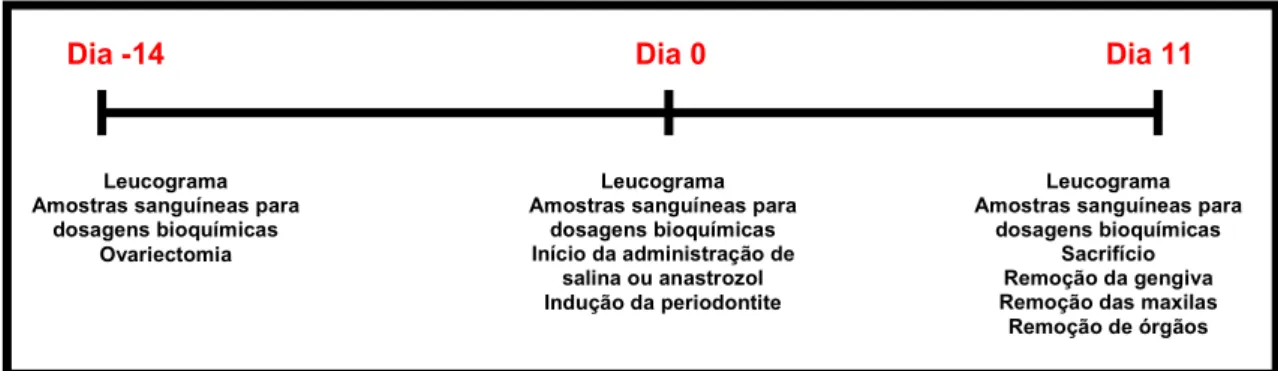
Initially, on the day -14, the animals had their ovaries removed or only exposed for the simulation of surgical stress. The ovariectomy surgery was performed as Anbinder et al. (2007) [34] with some modification, since a portion of the uterus was not removed. After anesthesia with chloral hydrated (300 mg/kg, i.p.), and trichotomy of the abdominal region, the skin and musculature were incised longitudinally below the last rib, and the ovary was identified and exposed. In the ovariectomized animals, after hemostasis through ligation of uterine tubes, ovaries were excised together surrounding fatty tissue. In the sham ovariectomized animals, the ovaries were replaced. After 14 days of ovariectomy [35], on day 0, the periodontitis was induced as described by Lima
et al. (2000) [36]. A nylon (000) thread ligature was placed around the cervix of the second left and right upper molar of rats anesthetized with chloral hydrated (300 mg/kg, i.p.). The ligature was then knotted on the vestibular side of the tooth, resulting in a subgingival position on the palatal side and supragingival on the buccal side. The animals were sacrificed after 11 days of periodontitis induction or 25 days of ovariectomy by offset cervical, on day 11.
Experimental groups
Initially, animals were divided into three groups, Normal, S-OVX and OVX. The Normal (n = 7) group did not undergo ovariectomy; S-OVX (n = 7) and OVX (n = 28) were submitted to sham surgery and to bilateral ovarietomy, respectively. Briefly the periodontitis induction, the OVX group was divided into four groups with seven animals each: saline (SAL), that received by gavage sterile saline solution 0.9% (2 ml/kg) 30 minutes before the ligature, and daily for ten days following; anastrazole 0.02 mg/kg (ANA 0.02), anastrazole 0.1 mg/kg (ANA 0.1) and anastrozole 0.5 mg/kg (ANA 0.5), which received by gavage anastrozole (ArimidexTM, Astrazeneca, Newark, DE, USA) dissolved in 0.9% saline solution at doses of 0.02, 0.1 and 0.5 mg/kg [37, 38], respectively, 30 minutes before the ligature, and daily for 10 days following, always at the same time. The animals of S-OVX group also received by gavage sterile saline solution 0.9% (2 ml/kg) 30 minutes before the ligature, and daily for 10 days following. The Normal group did not receive any treatment or saline.
Serum dosage of estradiol
Macroscopic study of bone tissue
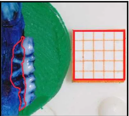
The animals were sacrificed after 11 days of periodontitis induction by offset cervical, and maxillae removed and fixed in 10% neutral formalin for 24 h. The left maxillary half of all groups was then dissected, and stained with aqueous methylene blue (1%) in order to differentiate bone from teeth. In order to quantify alveolar bone loss (ABL), hemimaxillae were suitably placed in microscope slides to be photographed with a digital camera Nikon™ (Nikon D40, Melville, NY, USA). The acquired image was sent to the computer program Image J (ImageJ 1.44p, National Institute of Health; EUA) for alveolar bone loss analysis, which was measured using a modified method described by Goes et al. (2010) [39]. For this, measurements were made along the region between the cemento-enamel junction and the alveolar bone crest by an experienced and calibrated examiner in a single-blind fashion. All obtained images were compared to millimeter well-known area (0.5x0.5 mm2).
Histological procedures
The animals were sacrificed after 11 days of periodontitis induction by offset cervical, and maxillae removed and fixed in 10% neutral formalin for 24 h. The right maxillary half of Normal, S-OVX, SAL and ANA 0.5 were demineralized in 7% formic acid (Merck®, Jacarepaguá, RJ, Brazil) for 10 days. Following this, the specimens were dehydrated, embedded in paraffin, and sectioned (sections of 4 m thickness) along the molars in a mesio-distal plane for Hematoxylin and Eosin stain.
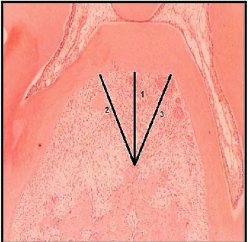
Histometric analysis
The furcation area of the second molars was evalueted by light microsocopy (x40) and an image was taken of this region. The acquired image was sent to the computer program Image J (ImageJ 1.44p, National Institute of Health; EUA) for alveolar bone loss analysis. For this, three linear measurements were made between the bone crest and the tooth surface. All obtained images were compared to millimeter well-known measurement (1 mm).
Histological analysis
The histological analysis was made as previously described [36, 40]. The area between the first and second molars, where a ligature had been placed, was evaluated by light microscopy (x40 and x400). Parameters such as inflammatory cell influx, osteoclast number, and alveolar bone and cementum integrity were analyzed by an experienced and calibrated histologist in a single-blind fashion and graded using a well described score system as follows: Score 0: absence of or only discrete cellular infiltration (inflammatory cellular infiltration is
sparse and restricted to the region of the marginal gingiva), few or absence of osteoclasts, preserved alveolar process and cementum; Score 1: moderate cellular infiltration (inflammatory cellular infiltration present all over the insert gingiva), presence of some osteoclasts, minor alveolar process resorption and intact cementum; Score 2: accentuated cellular infiltration (inflammatory cellular infiltration present in both gingival and periodontal
ligament), presence of osteoclasts, accentuated degradation of the alveolar process, and partial destruction of cementum; Score 3: accentuated cellular infiltration and total destruction of alveolar process and cementum. The
Myeloperoxidade (MPO) activity
MPO activity, a marker for neutrophil presence in inflamed tissue, was also evaluated in sample of gingival tissue, using methodology described by Lima et al. (2005) [41]. At sacrifice, Normal, S-OVX, SAL and ANA 0.5 groups of animals had a sample of their challenged gingival of left maxillary half removed for analysis of MPO activity. The specimens were stored at -80 oC until required for assay. For this, the gingival was weighed and triturated using a polytron Ultraturrax in ice-cold buffer solution [50 mM NaPO4, 0.5% (w/v) hexadecyltrimethylammonium bromide (H-TAB) solid] homogenized, frozen and thawed, and centrifuged at 4 ºC for 20 min (2,717 g). After that, the aliquot was centrifuged (2,717 g) for 10 minutes at 4 °C and held the determination of the MPO activity following the kinetics of the reaction front to hydrogen peroxide. In a 96-channels 7 µl of supernatant from each sample and 200 µl of coloring reagent were added and prepared just before use and composed of ortho-dianisidine hydrochloride (0.167 mg/ml) and hydrogen peroxide (H2O2) 0.0005% (w/v) in phosphate buffer (50 mM, pH 6.0). Then, the MPO activity was determined by reading absorbance at 450 nm and values were expressed as amount of MPO activity per gram of tissue.
Systemic parameters
Serum dosage of Transaminases (AST and ALT)
On day 11 of the assay, blood samples were collected from orbital plexus of Normal, S-OVX, SAL and ANA 0.5 groups of animals by heparinized microcapillary tubes, centrifuged (1,800 g x 10 min), and the supernatants were stocked at -80 ºC until biochemical analysis. Liver function was evaluated through serum dosage of Aspartate aminotransferase (AST) and Alanine aminotransferase (ALT). Specific kits were used, and methodology followed manufacturer orientations (Labtest, Lagoa Santa, MG, Brazil).
Liver, renal and spleen indexes
At sacrifice, the liver, left kidney and spleen of Normal, S-OVX, SAL and ANA 0.5 groups of animals were removed and weighed. Values were expressed as the index of the respective organ (wet weight of each organ divided by the weight of the animal on the day of sacrifice) [42].
Leukogram
On days -14, 0 and 11 the leukogram of Normal, S-OVX and OVX (SAL and ANA 0.5) groups of animals were made. The method used for the analysis of white blood cell counts, as well as its subpopulation (neutrophil and mononuclear cells) was as follows: 20 l of blood, taken from the rat tail, was added to 380 l Turk solution. Total white blood cell counts were performed using a Neubauer chamber and the differential counts were made using smears stained by rapid Instant Prov Stain Set® (Newprov Produtos para Laboratório; Pinhais, PR, Brazil).
Corporal mass variation
Statistical analysis
RESULTS
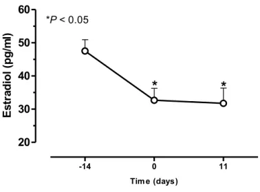
The removal of the ovaries significantly reduced (P < 0.05) the amount of circulating estrogen after 14 days of ovariectomy by 31.2%, when compared to baseline levels, and that lasted until the 11th day after the induction of periodontitis by 33.2% (Fig. 1). Macroscopically, the experimental periodontitis induced by ligature around the cervix of second upper molars of rats during 11 days caused a significant (P < 0.05) alveolar bone loss in Sham ovariectomized (S-OVX) animals when compared to Normal group (Table 1) inducing root exposition and furcation lesion (Fig. 2a and d). The reduction of seric estrogen promoted by ovariectomy did not increase alveolar bone loss in ovariectomized animals that received saline (SAL), when compared to S-OVX group (Table 1), with macroscopic aspect similar to that observed in S-OVX group (Fig 2d and g). To corroborate these data, the second molar furcation area was evaluated by histometry, and it was observed that the ligature induced a significant (P < 0.05) alveolar bone loss in S-OVX animals by 287.5%, when compared to normal group (Table 1). Besides, ovariectomy did not increase bone loss in the furcation area compared to S-OVX (Table 1). Histological analysis (Table 1) also showed significant (P < 0.05) differences in the area between the first and second molars of rats submitted to periodontitis and Normal animals. The ligature caused an important cell infiltration and intense destruction of alveolar bone, cementum and periodontal ligament after 11 days of periodontitis in S-OVX (Fig. 2e and f) and SAL (Fig. 2h and i) animals , receiving median scores of 3 (1-3) and 2 (1-3), respectively, compared to Normal maxillae (Table 1), which presented minimal cell infiltration and alveolar bone, cementum and periodontal ligament preservation (Fig. 2 b and c), receiving a median score of 0 (0-1). There were no significant histological differences between groups S-OVX and SAL (Table 1). In addition, the 11 days of experimental periodontitis showed an increase (P < 0.05) of myeloperoxidase (MPO) activity in gingival tissues in both S-OVX and SAL groups, when compared to normal rats (Fig. 3), by 409.2% and 411.8%, respectively. The reduction in seric estrogen induced by ovariectomy seemed in SAL group did not increase MPO activity when compared to S-OVX group, even under periodontitis (Fig. 3).
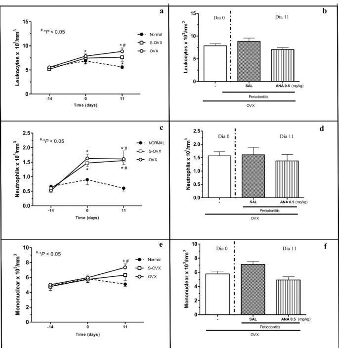
Systemically,the analysis of serum levels of transaminase aspartate aminotransferase (AST) and alanine aminotransferase (ALT) showed no change after 25 days of experiment when compared to Normal group, which was not submitted to ovariectomy nor to periodontitis (Table 2). In accordance to these findings S-OVX and SAL groups showed no significant changes in liver index (Table 2). The renal and spleen indexes of the S-OVX and SAL animals also showed no change in these organs (Table 2).The sham ovariectomy and ovariectomy surgeries caused an important (P < 0.05) leukocytosis observed from day 0, day of periodontitis induction, until day 11 compared to day -14 and to normal group on the 11th day (Fig. 4a). The leukocytosis in S-OVX group was due to neutrophilia on days 0 and 11 (Fig. 4c), whereas in ovariectomized animals (SAL) on day 11 lyphomonocytosis was also observed (Fig. 4e). The experimental peridontitis did not increase the number of total leukocytes (Fig. 4a and b), neutrophils (Fig. 4c and d) or mononuclear cells (Fig. 4e and f) on day 11 when compared to the time before the ligature (day 0). After sham ovariectomy and ovariectomy surgeries, the animals showed reduction of corporal mass, following the normal curve of weight in the 5th and 6th days after this procedure for SAL and S-OVX animals, respectively (Fig. 5a).Besides, SAL group gained more weight (P
after placement of the ligature, but while the S-OVX animals only followed the normal curve on the 3rd day after ligature, SAL animals remained with the weight similar to the normal group (Fig. 5a).
Effect of anastrozole (ANA)
Table 1 shows the macroscopic aspect of the effect of anastrozole (ANA 0.02, 0.1 or 0.5 mg/kg-v.o.) in ovariectomized animals submitted to 11 days of experimental periodontitis. It was observed that anastrozole did not increase alveolar bone loss compared to ovariectomized animals that received saline (SAL) only, showing root exposition and furcation lesion similar to that observed in SAL group (Fig. 2g and j). Complementarily to these analyses, the histometric study confirmed that the reduction of seric estrogen promoted by ovariectomy combined with anastrozole did not increase bone loss in the furcation area compared to SAL (Table 1). This table also shows that the highest dose of anastrazole (ANA 0.5 mg/kg-v.o.) in the periodontium did not increase the cell infiltration, destruction of the alveolar bone, the cementum or the periodontal ligament in interproximal region on the 11th day, when compared to SAL group. However, the daily administration of ANA 0.5 mg/kg-v.o. by the same time demonstrated an important (P < 0.05) increase in myeloperoxidase (MPO) activity in gingival tissues, when compared to SAL animals (Fig. 3), by 269.3%.
DISCUSSION
In this study, we have shown that 14 days after ovariectomy, the serum levels of estradiol decreased significantly and remained low for the next 11 days. These data are in accordance with a previous study demonstrating that one week after removal of the ovaries, the serum levels of estradiol dropped rapidly and remaind stable for 3 weeks following [35]. Biosynthesis of this hormone is catalyzed by aromatase enzyme [10], which is expressed in a variety of tissues, not only in the ovaries [18]. Thus, the use of anastrozole (Arimidex™), a potent orally active, highly selective, non-steroidal and third-generation aromatase inhibitor promotes a further reduction in the concentration of estrogen in postmenopausaul women [20]. Considering that the estrogen deficiency affects the bone metabolism, improving the process of bone resorption by increased osteoclast formation and life-span of this cell [4, 5], we decided to evaluate the effects of anastrozole in a short-term ligature-induced periodontitis in ovariectomized rats. The periodontitis was induced by ligature two weeks after ovariectomy as previously reported by Liu et al. (2010) [35] and coinciding with low levels of estrogen, but the reduction of the seric estrogen promoted only by ovariectomy did not increase the alveolar bone loss, nor cementum and periodontal ligament destruction or inflammatory infiltration in rats submitted to 11 days of periodontitis. Although studies show that estrogen deficiency in ovariectomized rats is associated with increased alveolar bone loss [35, 43], other studies did not find significant differences between ovariectomized and sham ovariectomized rats under periodontitis with respect to the alveolar bone loss, even with 30 days of ligature [44, 45]. Besides, it was found that the reduction in the synthesis of estrogen promoted by ovariectomy associated to anastrozole did not increase bone loss, cell infiltration, destruction of cementum and periodontal ligament. The effect of anastrozole had not yet been evaluated in a periodontitis model, then these data are important because although clinical studies show that this drug is associated to a greater number of fractures [21-25] and lower bone density [26], its use did not affect a short-term of experimental periodontits. Additionally, in contradiction with the clinical results this drug and letrozole applied in intact female rats for 16 weeks did not change femoral and vertebral bone mineral density [38]. Besides, in ovariectomized rats, the anastrozole did not abrogate androstenedione-induced bone protective effect [46]. Thus, the clinical and experimental findings to anastrozole are apparently different.Since there was no difference between doses of anastrozole in the macroscopic study of alveolar bone loss, the highest dose tested of this drug was chosen for remaining analyzes, including for systemic evaluation.
anastrozole administration, this drug increased MPO activity in this period, suggesting that anastrozole increased neutrophil presence in gingival tissue. Besides, this cell participates in the process of bone destruction that is hallmark of periodontitis through release of oxygen radicals, which are considered responsible for osteoclst activation [49]. Therefore, the short-time administration of anastrozole in spite of not having caused greater alveolar bone loss, increased the inflammatory response underlying to resorptive bone process of periodontitis.
Systemically, although the literature reports three cases of hepatoxicty due to anastrozole [50-52] the low levels of estrogen induced by ovariectomy alone or combined with anastrozole did not cause any alteration in liver function, demonstrated by the respective index and by liver transaminases levels. The ovariectomy alone or combined with anstrozole also did not induce any changes in renal and splenic functions, showed by their indexes. The surgical procedures of sham ovariectomy and ovariectomy promoted an important leukocytosis due to an increase in neutrophils, suggesting that surgical manipulation of the animals caused these changes. However, on day 11, the amount of mononuclear cells in ovariectomized animals was significantly higher when compared with normal animals or with their baseline values. This increase in the number of mononuclear cells can be explained by the estrogen deficiency effect in the production of lymphocytes. Although our results did not show changes in spleen index, studies have shown that ovariectomy increases the frequency of T and B cells in the bone marrow and in the secondary lymphoid organ as spleen [2, 53]. Besides, the periodontitis induction and combination of anastrozole with ovariectomy did not promote any alteration in the number of leukocytes, thus the data indicate that the administration of anastrozole for 11 days did not increase the effect of estrogen reduction on leukogram of animals. The sham ovariectomy and ovariectomy surgeries even as periodontitis induction induced an initial corporal mass loss. In fact surgical procedures can induce corporal mass loss in animals, in accordance to this Lima et al. (2004) [40] observed that periodontitis induction caused weight loss. However, ovariectomized rats showed a tendency to gain more corporal mass than normal animals and gained more weight than S-OVX rats. Moreover, anastrozole group presented similar body weight curve to ovariectomized animals that received saline. Other experimental studies show that low levels of estrogen are associated with more gain corporal mass and obesity in female and male animals [54, 55]. Stubbins et al. (2011) [56] also show that ovariectomized mice had a higher propensity of gaining weight and adipocyte diameter even as estrogen supplementation caused minimal change in body weight, similar to non ovaricetomized mice, and decreased adipocyte diameter.
REFERENCES
1. Ames MS, Hong S, Lee HR, Fields HW, Johnston WM, Kim DG (2010) Estrogen deficiency increases variability of tissue mineral density of alveolar bone surrounding teeth. Arch Oral Biol 55(8):599-605 2. Tyagi AM, Srivastava K, Sharan K, Yadav D, Maurya R, Singh D (2011) Daidzein prevents the
increase in CD4+CD28null T cells and B lymphopoesis in ovariectomized mice: a key mechanism for anti-osteoclastogenic effect. PLoS One 6(6):e21216
3. Koh KK, Soh JW, Ahn JY, Lee SK, Hwang HY, Kim DS, Jin DK, Ahn TH, Shin EK (2001) Effect of hormone replacement therapy on nitric oxide bioactivity and monocyte chemoattractant protein-1 levels. Int J Cardiol 81(1):43-50
4. Martin-Millan M, Almeida M, Ambrogini E, Han L, Zhao H, Weinstein RS, Jilka RL, O'Brien CA, Manolagas SC (2010) The estrogen receptor-alpha in osteoclasts mediates the protective effects of estrogens on cancellous but not cortical bone. Mol Endocrinol 24(2): 323-334
5. Nakamura T, Imai Y, Matsumoto T, Sato S, Takeuchi K, Igarashi K, Harada Y, Azuma Y, Krust A, Yamamoto Y, Nishina H, Takeda S, Takayanagi H, Metzger D, Kanno J, Takaoka K, Martin TJ, Chambon P, Kato S (2007) Estrogen prevents bone loss via estrogen receptor alpha and induction of Fas ligand in osteoclasts. Cell 130(5):811-823
6. Roggia C, Gao H, Cenci S, Weitzmann MN, Toraldo G, Isaia G, Pacifici R (2001) Up-regulation of TNF-producing T cells in the bone marrow: a key mechanism by which estrogen deficiency induces bone loss in vivo. Proc Natl Acad Sci U S A 98(24):13960-13965
7. Hofbauer LC, Khosla S, Dunstan CR, Lacey DL, Spelsberg TC, Riggs BL (1999) Estrogen stimulates gene expression and protein production of osteoprotegerin in human osteoblastic cells. Endocrinology 140(9): p. 4367-4370
8. Shevde NK, Bendixen AC, Dienger KM, Pike JW (2000) Estrogens suppress RANK ligand-induced osteoclast differentiation via a stromal cell independent mechanism involving c-Jun repression. Proc Natl Acad Sci U S A 97(14): 7829-7834
9. D'Amelio P, Grimaldi A, Di Bella S, Brianza SZ, Cristofaro MA, Tamone C, Gribaldi G, Ulliers D, Pescarmona GP, Isaia G (2008) Estrogen deficiency increases osteoclastogenesis up-regulating T cells activity: a key mechanism in osteoporosis. Bone 43(1):92-100
10. Simpson ER, Mahendroo MS, Means GD, Kilgore MW, Hinshelwood MM, Graham-Lorence S, Amarneh B, Ito Y, Fisher CR, Michael MD, Mendelson CM, Bulun SE (1994) Aromatase cytochrome P450, the enzyme responsible for estrogen biosynthesis. Endocr Rev 15(3):342-355
11. Antonio-Rubio NR, Guerrero-Estévez SM, Lira-Romero E, Moreno-Mendoza N (2011) Expression of 3beta-HSD1 and P450 Aromatase enzymes during mouse gonad differentiation. J Mol Histol 42(6): 535-543
12. Kellis JTJ, Vickery LE (1987) Purification and characterization of human placental aromatase cytochrome P-450. J Biol Chem 262(9): 4413-4420
14. Nawata H, Tanaka S, Tanaka S, Takayanagi R, Sakai Y, Yanase T, Ikuyama S, Haji M (1995) Aromatase in bone cell: association with osteoporosis in postmenopausal women. J Steroid Biochem Mol Biol 53(1-6):165-174
15. O'Neill JS, Elton RA, Miller WR (1988) Aromatase activity in adipose tissue from breast quadrants: a link with tumour site. Br Med J (Clin Res Ed) 296(6624): p. 741-743
16. Balthazart J, Baillien M, Charlier TD, Cornil CA, Ball GF(2003) Multiple mechanisms control brain aromatase activity at the genomic and non-genomic level. J Steroid Biochem Mol Biol 86(3-5):367-379 17. Castagnetta LA, Agostara B, Montalto G, Polito L, Campisi I, Saetta A, Itoh T, Yu B, Chen S, Carruba
G (2003) Local estrogen formation by nontumoral, cirrhotic, and malignant human liver tissues and cells. Cancer Res 63(16):5041-5045
18. Czajka-Oraniec I, Simpson ER (2010) Aromatase research and its clinical significance. Endokrynol Pol 61(1):126-134
19. Chetrite GS, Cortes-Pietro J, Philippe JC, Wright F, Pasqualini JR (2000) Comparison of estrogen concentrations, estrone sulfatase and aromatase activities in normal, and in cancerous, human breast tissues. J Steroid Biochem Mol Biol 72(1-2):23-27
20. Geisler J, King N, Dowsett M, Ottestad L, Lundgren S, Walton P, Kormeset PO, Lonning PE (1996) Influence of anastrozole (Arimidex), a selective, non-steroidal aromatase inhibitor, on in vivo aromatisation and plasma oestrogen levels in postmenopausal women with breast cancer. Br J Cancer 74(8):1286-1291
21. Baum M, ATAC Trialist' Group (2002) Anastrozole alone or in combination with tamoxifen versus tamoxifen alone for adjuvant treatment of postmenopausal women with early breast cancer: first results of the ATAC randomised trial. Lancet 359(9324):2131-2139
22. Baum M, ATAC Trialist' Group (2003) Anastrozole alone or in combination with tamoxifen versus tamoxifen alone for adjuvant treatment of postmenopausal women with early-stage breast cancer: results of the ATAC (Arimidex, Tamoxifen Alone or in Combination) trial efficacy and safety update analyses. Cancer 98(9):1802-1810
23. Howell A, ATAC Trialist' Group (2005) Results of the ATAC (Arimidex, Tamoxifen, Alone or in Combination) trial after completion of 5 years' adjuvant treatment for breast cancer. Lancet 365(9453):60-62
24. Forbes JF, ATAC Trialist' Group (2008) Effect of anastrozole and tamoxifen as adjuvant treatment for early-stage breast cancer: 100-month analysis of the ATAC trial. Lancet Oncol 9(1):45-53
25. Cuzick J, ATAC Trialist' Group (2010) Effect of anastrozole and tamoxifen as adjuvant treatment for early-stage breast cancer: 10-year analysis of the ATAC trial. Lancet Oncol 11(12):1135-1141
26. Rastelli AL, Taylor ME, Gao F, Armamento-Vilarreal R, Jamalabadi-Majidi S, Napoli N, Ellis MJ (2011) Vitamin D and aromatase inhibitor-induced musculoskeletal symptoms (AIMSS): a phase II, double-blind, placebo-controlled, randomized trial. Breast Cancer Res Treat 129(1):107-116
27. Burt B (2005) Position paper: epidemiology of periodontal diseases. J Periodontol 76(8):1406-1419 28. Page RC, Kornman KS (1997) The pathogenesis of human periodontitis: an introduction. Periodontol
29. Kinane DF, Preshaw PM, Loos BG (2011) Host-response: understanding the cellular and molecular mechanisms of host-microbial interactions - consensus of the Seventh European Workshop on Periodontology. J Clin Periodontol 38(Suppl 11):44-48
30. Chen L, Wei B, Li J, Liu F, Xuan D, Xie B, Zhang J (2010) Association of Periodontal Parameters With Metabolic Level and Systemic Inflammatory Markers in Patients With Type 2 Diabetes. J Periodontol 81(3):364-371
31. Mirrielees J, Crofford LJ, Lin Y, Kryscio RJ, Dawson DR, Ebersole JL, Miller CS (2010) Rheumatoid arthritis and salivary biomarkers of periodontal disease. J Clin Periodontol 37(12):1068-1074
32. Haas AN, Rosing CK, Oppermann RV, Albandar JM, Susin C (2009) Association among menopause, hormone replacement therapy, and periodontal attachment loss in southern Brazilian women. J Periodontol 80(9):1380-1387
33. Liang L, Yu JF, Wang Y, Ding Y (2008) Estrogen Regulates Expression of Osteoprotegerin and RANKL in Human Periodontal Ligament Cells Through Estrogen Receptor Beta. J Periodontol 79(9):1745-1751
34. Anbinder AL, Prado F de A, Prado M de A, Balducci I, Rocha F (2007) The influence of ovariectomy, simvastatin and sodium alendronate on alveolar bone in rats. Braz Oral Res 21(3):247-252
35. Liu S, Cheng Y, Fan M, Chen D, Bian Z (2010) FSH aggravates periodontitis-related bone loss in ovariectomized rats. J Dent Res 89(4):366-371
36. Lima V, Bezerra MM, Alencar VBM, Vidal FDP, Rocha FAC, Brito GAC, Ribeiro RA (2000) Effects of chlorpromazine on alveolar bone loss in experimental periodontal disease in rats. Eur J Oral Sci 108(2):123-129
37. Hozumi Y, Hakamata Y, Sasanuma H, Ogura S, Nagai H (2002) Effects of anastrozole on lipid metabolism compared with tamoxifen in rats. Breast Cancer Res Treat 76(2):131-136
38. Kumru S, Yildiz AA, Yilmaz B, Sandal S, Gurates B (2007) Effects of aromatase inhibitors letrozole and anastrazole on bone metabolism and steroid hormone levels in intact female rats. Gynecol Endocrinol 23(10):556-561
39. Goes P, Lima APS, Melo IM, Rêgo ROCC, Lima V (2010) Effect of Atorvastatin in radiographic density on alveolar bone loss in wistar rats. Braz Dent J 21(3):193-198
40. Lima V, Vidal FD, Rocha FA, Brito GA, Ribeiro RA (2004) Effects of tumor necrosis factor-alpha inhibitors pentoxifylline and thalidomide on alveolar bone loss in short-term experimental periodontal disease in rats. J Periodontol 75(1):162-168
41. Lima V, Brito GAC, Cunha FQ, Rebouças CG, Falcão BAA, Augusto RF, Souza MLP, Leitão BT, Ribeiro RA (2005) Effects of the tumour necrosis factor-alpha inhibitors pentoxifylline and thalidomide in short-term experimental oral mucositis in hamsters. Eur J Oral Sci 113(3):210-217
42. Silva LMCM, Lima V, Holanda ML, Pinheiro PG, Rodrigues JAG, Lima MEP, Benevides NMB (2010) Antinociceptive and anti-inflammatory activities of lectin from marine red alga Pterocladiella capillacea. Biol Pharm Bull 33(5):830-835
44. Marques MR, da Silva MA, Manzi FR, Cesar-Neto JB, Nociti FH Jr, Barros SP (2005) Effect of intermittent PTH administration in the periodontitis-associated bone loss in ovariectomized rats. Arch Oral Biol 50(4):421-429
45. Vaziri H, Naserhojjati-Roodsari R, Tasili-Fahadan N, Khojasteh A, Mashhadi-Abbas F, Eslami B, Dehpour AR (2007) Effect of simvastatin administration on periodontitis-associated bone loss in ovariectomized rats. J Periodontol 78(8):1561-1567
46. Lea CK, Flanagan AM (1998) Physiological plasma levels of androgens reduce bone loss in the ovariectomized rat. Am J Physiol 274(2 Pt 1):E328-335
47. Verdu EF, Deng Y, Bercik P, Collins SM (2002) Modulatory effects of estrogen in two murine models of experimental colitis. Am J Physiol Gastrointest Liver Physiol 283(1):G27-36
48. Albayrak A, Uyanik MH, Odabasoglu F, Halici Z, Uyanik Z, Bayir Y, Albayrak F, Albayrak Y, Polat B, Suleyman H (2011) The effects of diabetes and/or polymicrobial sepsis on the status of antioxidant enzymes and pro-inflammatory cytokines on heart, liver, and lung of ovariectomized rats. J Surg Res 169(1): 67-75
49. Nussbaum G, Shapira L (2011) How has neutrophil research improved our understanding of periodontal pathogenesis? J Clin Periodontol 38(Suppl 11):49-59
50. Inno A, Basso M, Vechio FM, Marsico VA, Cerchiaro E, D'Argento E, Bagalà C, Barone C (2011) Anastrozole-related acute hepatitis with autoimmune features: a case report. BMC Gastroenterol 11:32 51. Zapata E, Zubiaurre L, Bujanda L, Piérola A (2006) Anastrozole-induced hepatotoxicity. Eur J
Gastroenterol Hepatol 18(11):1233-1234
52. de La Cruz L, Romero-Vazquez J, Jiménez-Sáenz M, Padron JR, Herrerias-Gutierrez JM (2007) Severe acute hepatitis in a patient treated with anastrozole. Lancet 369(9555):23-24
53. Garcia-Perez MA, Noguera I, Hermenegildo C, Martínez-Romero A, Tarín JJ, Cano A (2006) Alterations in the phenotype and function of immune cells in ovariectomy-induced osteopenic mice.
Hum Reprod 21(4):880-887
54. Chen Y, Heiman ML (2001) Increased weight gain after ovariectomy is not a consequence of leptin resistance. Am J Physiol Endocrinol Metab 280(2): E315-22
55. Christoffersen BO, Gade LP, Golozoubova V, Svendsen O, Raun K (2010) Influence of castration-induced testosterone and estradiol deficiency on obesity and glucose metabolism in male Gottingen minipigs. Steroids 75(10):676-684
Figure 1
-14 0 11
20 30 40 50 60
*
*
*P< 0.05
Tim e (days)
E
s
tr
a
d
io
l
(p
g
/m
l)
Figure 2
Fig. 2 Macroscopic and Histological aspects of Normal rat maxilla (a, b and c) or periodontium of Sham
ovariectomized (d, e and f) animals submitted to ligature-induced periodontitis or Ovariectomized animals also submitted to ligature-induced periodonititis, receiving by gavage (v.o.) Saline (g, h and i), or Anastrozole 0.5 mg/kg (ANA) (j, k and l), respectively. Animals received Saline or ANA, 30 min before periodontitis induction, and daily for 11 d. After the animals were killed, the left maxillary half were then processed for macroscopic analysis comparing the area between the cemento-enamel junction and the alveolar bone crest to a well known area (0.5 x 0.5 mm2). For histological analyses, the right maxillary half maxillae were processed for Hematoxylin and Eosin staining (40 x and 400 x magnification). g, pl, c, d and ab mean gengivae, periodontal ligament, cementum and alveolar bone, respectively. Asterisks indicate leukocyte infiltration.
a
b
c
d
e
f
g
h
i
j
k
l ab
pl g d
c
*
*
*
*
*