Lentiviral Mediating Genetic Engineered Mesenchymal
Stem Cells for Releasing IL-27 as a Gene Therapy
Approach for Autoimmune Diseases
Shohreh Hajizadeh-Sikaroodi, Ph.D.1, Ahmad Hosseini, Ph.D.2, 3, 4*, Ali Fallah, M.Sc.5, Hajar Estiri, M.Sc.6, Zahra Noormohammadi, Ph.D.7, Mohammad Salehi, Ph.D.2, 3,
Sayyed Mohammad Hossein Ghaderian, Ph.D.8, Haleh Akhavan Niaki, Ph.D.9, Masoud Soleimani, Ph.D.10, Bahram Kazemi, Ph.D.2, 11
1. Science and Research Branch, Islamic Azad University, Tehran, Iran
2. Cellular and Molecular Biology Research Center, Shahid Beheshti University of Medical Sciences, Tehran, Iran 3. Mehr Infertility Research Center, Rasht, Iran
4. Department of Cell Biology and Anatomical Science, Faculty of Medicine, Shahid Beheshti University of Medical Sciences, Tehran, Iran
5. Systems and Synthetic Biology Group, Mede Bioeconomy Company, Tehran, Iran
6. Department of Molecular Biology and Genetic Engineering, Stem Cell Technology Research Center, Tehran, Iran 7. Department of Biology, Faculty of Basic Sciences, Science and Research Branch, Islamic Azad University,
Tehran, Iran
8. Department of Medical Genetics, Faculty of Medicine, Shahid Beheshti University of Medical Sciences and Health Services, Tehran, Iran
9. Cellular and Molecular Biology Research Center, Babol University of Medical Sciences, Babol, Iran 10. Department of Hematology, Faculty of Medical Sciences, Tarbiat Modares University, Tehran, Iran 11. Department of Biotechnology, Faculty of Medicine, Shahid Beheshti University of Medical Sciences,
Tehran, Iran
*Corresponding Address: P.O.Box: 193954719, Cellular and Molecular Biology Research Center, Shahid Beheshti University of Medical Sciences, Tehran, Iran
Email: [email protected]
Received: 6/Jan/2013, Accepted: 12/Aug/2013
Abstract
Objective: Autoimmune diseases precede a complex dysregulation of the immune sys-tem. T helper17 (Th17) and interleukin (IL)-17 have central roles in initiation of inlam-mation and subsequent autoimmune diseases. IL-27 signiicantly controls autoimmune diseases by Th17 and IL-17 suppression. In the present study we have created genetic engineered mesenchymal stem cells (MSCs) that mediate with lentiviral vectors to release IL-27 as an adequate vehicle for ex vivo gene therapy in the reduction of inlammation and autoimmune diseases.
Materials and Methods: In this experimental study, we isolated adipose-derived MSCs (AD-MSCs) from lipoaspirate and subsequently characterized them by differentiation. Two subunits of IL-27 (p28 and EBI3) were cloned in a pCDH-513B-1 lentiviral vector. Expres-sions of p28 and EBI3 (Epstein-Barr virus induced gene 3) were determined by real time polymerase chain reaction (PCR). MSCs were transduced by a pCDH-CMV-p28-IRES-EBI3-EF-copGFP-Pur lentiviral vector and the bioassay of IL-27 was evaluated by IL-10 expression.
Results: Cell differentiation confirmed true isolation of MSCs from lipoaspirate. Re-striction enzyme digestion and sequencing verified successful cloning of both p28 and EBI3 in the pCDH-513B-1 lentiviral vector. Real time PCR showed high expres-sions level of IL-27 and IL-10 as well as accurate activity of IL-27.
Conclusion: The results showed transduction of functional IL-27 to AD-MSCs by means of a lentiviral vector. The lentiviral vector did not impact MSC characteristics.
Keywords: Autoimmune Disease, Gene Therapy, IL-27, Mesenchymal Stem Cells
Cell Journal(Yakht eh), Vol 16, No 3, Aut um n 2014, Pages: 255- 262
Introduction
In recent years stem cell therapy has become a pri-mary aspect of numerous research and clinical pro-jects (1). Stem cell types such as pluripotent (ES, iPS), fetal and adult stem cells are most commonly used as treatments, however despite the advantages, in numerous diseases it is necessary to make genetic alterations to these cells by over expressing or knock-ing down genes (2). Mesenchymal stem cells (MSCs) with their basic properties can be a good source for cell therapy. In addition these cells have unique fea-tures for moderating cell attack and immune system reactions (3). Adipose-derived MSCs (AD-MSCs) are the best source for MSCs that can be used for cell therapy (4). AD-MSCs can be easily isolated from lipoaspirate and possess a stable karyotype as well as high capability for self-renewal in comparison to other sources of MSCs (5). Stem cells in adipose tis-sue usually comprise up to 3% of the entire cell popu-lation, which is 2500 fold more than the frequency of stem cells in bone marrow (6).
Autoimmune diseases are multi-factorial dis-orders with complicated immune system dys-regulation mediated by immune cytokines and immune cells (7). In many autoimmune diseases transforming growth factor beta (TGF-B) and interleukin (IL)-6 induce T helper17 (Th-17) causing IL-23 and IL-17 secretion (8). IL-23 and IL-17 can persuade special CD4+ with CCR2+ and
CCR5-effector T cells which have been identiied
as major agents for inducing autoimmune disease in a mouse model (9). As mentioned, down-regu-lation of Th17 or IL-17 can be an effective therapy for treatment of many autoimmune diseases (10).
Previous studies have conirmed that IL-27 is a
strong suppressor of Th17 and IL-17. Therefore overexpression of IL-27 may be a good optional therapy against autoimmune diseases (11).
There are many autoimmune diseases which all have the same mechanism of pathogenicity, thus one approach can be used as a general treatment for these diseases (12). In the present study we re-port a construct that can be used for a gene therapy approach based on the suppression of 17 by IL-27 producer cells.
Materials and Methods
IL-27 construct in the lentiviral vector
In this experimental study, we purchased two
subunits of mouse IL-27 (p28 and EBI3) cDNA from Open Biosystems (Huntsville, AL, Unit-ed States). Both genes were clonUnit-ed in pCDH-513B-1 (System Bioscience, Mountain View, CA, United States ) combined with an internal ribosome entry site (IRES) sequence arranged as p28-IRES-EBI3 under a cytomegalovirus (CMV) promoter. pCDH-513B-1 have copGFP (copepod green fluorescent protein) for fluo-rescent tagging and puromycin for selecting stably transduced cells. All cloning proce-dures were performed according to the com-mon digestion-ligation protocol. Polymerase chain reaction (PCR) was carried out for three fragments with Xba1-p28-Nhe1, Nhe1-IRES-BamH1 and Nhe1-IRES-BamH1-EBI3-Not1 overhanging. Then, all fragments were separately cloned in pCDH-513B-1. We verified the pCDH-CMV-p28-IRES-EBI3-EF1-copGFP-Pur construct by digestion and subsequent sequencing.
Recombinant lentiviral production
Recombinant lentivirus was produced accord-ing to the TRONOLAB protocol with some modifications (13). Briefly, 1×10⁶ HEK-293T
cells (Invitrogen, Carlsbad, CA, United States) were cultured in a 10 cm plate in Dulbecco’s Modified Eagle’s Medium (DMEM) (GIBCO-BRL, Tokyo, Japan) with 10% Fetal bovine serum (FBS) (GIBCO-BRL, Tokyo, Japan) one day prior to transfection. We replaced the medium 2 hours before transfection with fresh medium. Ca3(PO4)2 buffer that contained 21 µg of pCDH-CMV-p28-IRES-EBI3-EF1-copGFP-Pur, 21 µg of pCMV-dR8.2, 10.5 µg of pMD2, 33 µl of TE 1X, 105 µl of 2.5 M CaCl2, and 1050 µl of 2x Hank’s buffered salt solution (HBSS) was used for one 10 cm plate. We added the 2X HBSS during the time the solution was vor-texed. Transfection medium was replaced with fresh medium within 14 hours after transfec-tion. Medium with viruses was collected after 24, 48 and 72 hours and centrifuged at 15000 rpm, then filtered through a 0.45 µm filter be-fore transduction.
Mesenchymal stem cell (MSCs) isolation from human adipose tissue, culture and differentiation
lipoaspi-rate plastic surgery performed at clinics ac-cording to a Bioethics Agreement of the Shahid Beheshti University of Medical Science and Stem Cell Research Center Committee. Adi-pose tissue was washed three times with phos-phate buffered saline (PBS) that contained 3X penicillin/streptomycin and amphotericin until a clear tissue was attained. DMEM medium that contained dispase (50 U/ml)-Collagenase I (250 U/ml; Sigma-Aldrich, St. Louis, MO) were added to the adipose tissue, after which the solution was shaken for 30 minutes at 37˚C. The solution was centrifuged at 1500 rpm and the supernatant was discarded. The plated cells were kept. RBC was lysed by erythrocyte ly-sis buffer for 5 minutes at 37˚C, and then cen -trifuged at 1200 rpm for 5 minutes. The plated cells were suspended in DMEM and distributed in flasks with DMEM that contained 10% FBS for 3 days. For adipogenic differentiation, cells were cultured in DMEM that contained 10% FBS, 0.5 mM isobutylmethylxanthine (IBMX), dexamethasone (10-7 M), insulin (66 nM), and
indomethacin (0.2 mM). For osteocyte differ-entiation the cells were cultured in DMEM that contained 10% FBS, dexamethasone (10-7M),
β-glycerol-phosphate (10 mM), and ascorbic
acid 2-phosphate (50 µg/ml).
Adipose-derived mesenchymal stem cells (AD-MSCs) transduction by lentivirus
Second passage AD-MSCs were cultured in six-well cell culture plates, and then washed with PBS before adding fresh recombinant virus. In order to remove all FBS proteins to enable better trans-duction we used the spinfection method at 2000 rpm for 60 minutes at a temperature of 25˚C. After centrifuging, the plate was placed in a 37˚C incui-bator; the medium was changed 14-20 hours after spinfection.
Expression of IL-27 and self-renewing assay with Oct-4
Total RNA extraction and cDNA synthesis from 2×10⁶ MSCs and lentiviral engineered
AD-MSCs were carried out by Qiagen (Alameda, CA, United States), RNA extraction and cDNA kits (Waltham, MA, United States), respectively, ac-cording to the manufacturers’ protocols. cDNA was used for quantitative real time PCR. Expres-sions of octamer-binding transcription factor 4 (Oct-4), IL-27 and EBI3 were evaluated in AD-MSCs and lentiviral engineered AD-AD-MSCs. TA-TA-binding protein (TBP) expression was consid-ered to be the endogenous reference gene. Primer sequences used this studied are provided in table 1.
Table 1: Primer sequences used for quantitative real time PCR
Reverse Forward
Primer
5´ CCTCCTCCTTTGAACATTT 3´ 5´ AGACTCTGCTTCCTCGCTA 3´
IL-27
5´ GTTTCCCATAATCTGTGAGG 3´ 5´ TGAGCGAATCATCAAGCC 3´
EBI3
5´ GGTGATCCTCTTCTGCTTC 3´ 5´ CGGCGTATGAGTTGTGTG 3´
Oct-4
5´ ACGACCAAATCCGTTGACTC 3´ 5´ CTCTCTGCTCCTGTTCG 3´
TBP
Bioassay of IL-27
Secretion and function of IL-27 were examined by bioassay using naive T cells from a C57BL/6 mouse that was co-cultured with COS-7 cells transduced with recombinant virus derived from pCDH-CMV-p28-IRES-EBI3-EF1-copGFP-Pur and se-lected for puromycin at a concentration of 2 µg/cc. The C57BL/6 mouse was killed according to the laboratory animal protocol. The spleen was removed and cut in small pieces then digested with dispase-collagenase (100U/ml) for 10 minutes and passed
through 0.45 μM ilter. Cells were collected by cen -trifugation at 1200 rpm and RBCs were lysed by a RBC lysis buffer. After three days, naive T cells were cultured in fresh RPMI 1640 medium and co-cul-tured with COS-7 that was engineered by a recom-binant virus (COS7/IL-27). Total RNA was extracted from T cells co-cultured with COS-7 and COS-7/ IL-27. Expression of IL-10 was appraised as a down-stream gene in the IL-27 signal transduction pathway by real time PCR. Glyceraldehy3-phosphate de-hydrogenase (GAPDH) expression was used as an endogenous reference gene. The following primers were used for IL-10 and GAPDH: IL-10 forward: 5´AATAAGAGCAAGCCAGTG3´ and reverse: 5´CCAGCAGACTCAATACAC3'; and GAPDH forward: 5'CCACAACTCTTCCATTCTC3' and re-verse: 5'CCAAGATTCACGGTAGATAC 3'.
Ethical considerations
Human adipose tissue was obtained following in-formed consent in accordance with the Declarations of the Shahid Beheshti University of Medical Sci-ence and Stem Cell Research Center Committee.
Results
IL-27 lentiviral construct and recombinant viral particle production
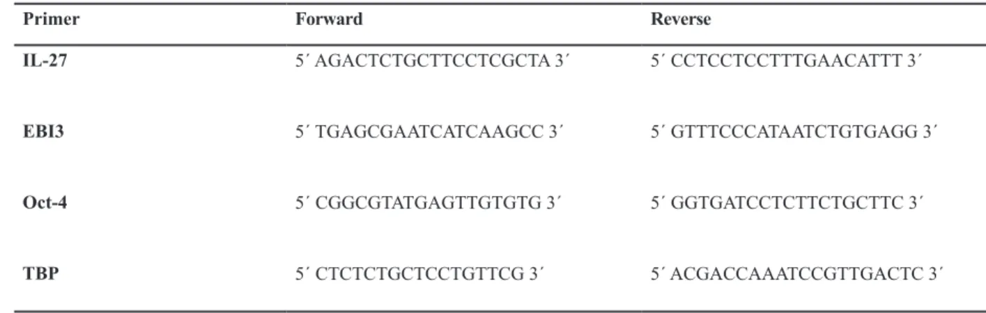
We cloned mouse p28 and EBI3 cDNA in a pCDH-513B-1 lentiviral vector. Digestion with XbaI-NotI showed that cloning was successful (Fig l). The construct was co-transfected with the helper packaging vector mediated Ca3(PO4)2 protocol. The transfect efficiency was more than 90% (Fig 2). The viral particle titer was approximately 1.5-2×10⁶.
Fig 1: p28 and EBI3 genes inserted into the pCDH-513B-1 lentiviral vector. Digestion with XbaI-NotI showed an 8189 bp length of the pCDH-513B-1 backbone. The presence of a 1611 bp segment related to p28-IRES-EBI3and a segment
of an 8189 bp related to pCDH-513B-1 conirmed that the cloning was established. A genetic map conirmed these data.
Fig 2: Transfection of 293T for achieving viral particles. Panel A shows the 293T culture. Panel B represents HEK-293T at 18 hours after transfection by pCDH-CMV-p28-IRES-EBI3-EF1-copGFP-Pur. High expression of GFP in HEK-HEK-293T shows the high rate of transfection.
Adipocyte stem cell isolation, transduction of adipose-derived mesenchymal stem cells (AD-MSCs) with recombinant lentiviral particles
Adipocyte cells were isolated from liposuction tissue with mechanical and enzyme digestion.
Multipotency of the cells was conirmed by their
differentiation into adipocyte and osteocyte cells.
Alizarin Red staining conirmed the presence of
osteocytes (Fig 3A) and oil red staining showed
the adipocyte properties after differentiation (Fig
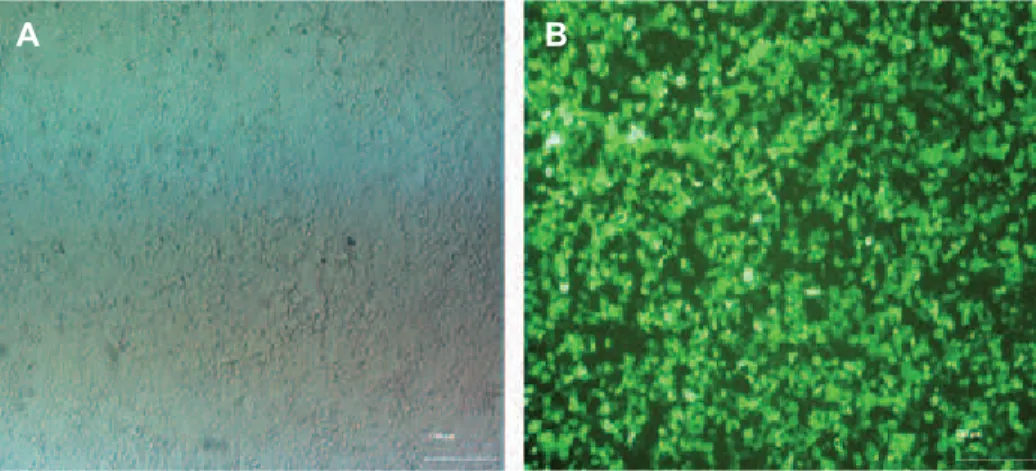
3B). AD-MSCs showed over 70% eficiency when
transduced by lentiviral particles (Fig 4). Trans-duced AD-MSCs were selected via puromycin. The selection curve determined that 2 µg of
puro-mycin was suficient to generate approximately
95% pure transduced cells after 3 days. The GFP marker provided a good index for the transfection,
transduction and puriication processes.
Fig 3: Isolation of mesenchymal stem cells (MSCs) from adipose tissue and characterized by differentiation. Panel A shows passage 2 adipose-derived MSCs (AD-MSCs), Panel B represents oil red staining of passage 2 AD-MSCs that differentiated into adipocytes. The vesicle that contained oil is visible in cells which showed adipogenic differentiation and Panel C shows the passage 2 AD-MSCs that were cultured in osteogenic differentiation medium and stained with alizarin red. Alizarin red stained
the calcium deposits which conirmed osteogenic differentiation.
Fig 4: Transduction of adipocyte-derived mesenchymal stem cells (AD-MSCs) by lentiviral particles. Panel A shows AD-MSCs prior to transduction and Panel B shows transduced AD-MSCs by pCDH-CMV-p28-IRES-EBI3-EF1-copGFP-Pur lentiviral vector. The numerous green cells and GFP expression indicate a high level of transduction.
B A
B
Gene expression proiles
Real time PCR showed expression of p28 in-creased 2000-fold and EBI3 inin-creased 650-fold in transduced AD-MSCs compared with the control AD-MSCs (Fig 5). IRES sequencing between p28 and EBI3 had a 3-fold decrease in EBI3 expression in reference to p28. Expression of Oct-4 in transduced
AD-MSCs conirmed that AD-MSCs preserved their
self-renewing potency. Lentiviral transduction did not affect the mesenchymal properties.
IL-27 functional assay
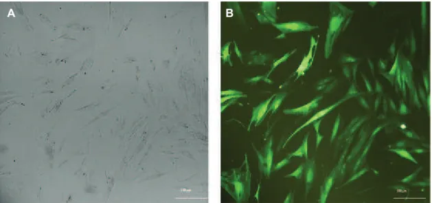
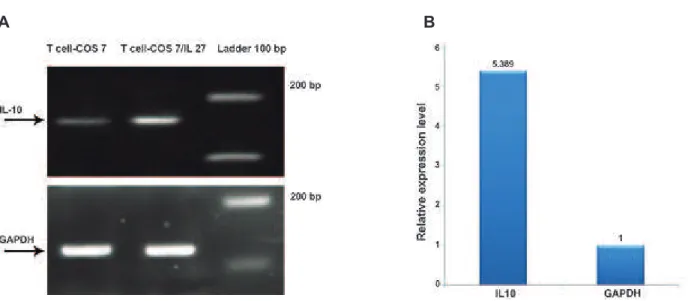
We examined the biological activity of IL-27 that was secreted from COS-7 cells. Naive T cells produced larger amounts of IL-10 when co-cul-tured with COS-7/IL-27 cells compared to naive T cells co-cultured with COS-7 (Fig 6). Real time PCR showed a 5-fold higher expression of IL-10 in T cells co-cultured with COS-7/IL-27 compared with T cells co-cultured COS-7 (Fig 7).
Fig 5: Expressions of IL-27, EBI3, and Oct-4 in adipose-derived mesenchymal stem cells (MSCs) and transduced
AD-MSCs/IL-27. Panel A shows the result of real time-PCR that conirmed the presence of a deinite transcript by gel resolution.
Expressions of p28 and EBI3 increased in transduced AD-MSCs/IL-27 compared with control AD-MSCs. Oct-4 expression did
not show any signiicant difference. TBP was used as the RNA integrity control. Panel B represents the level of p28, EBI3 and
Oct-4 expressions in AD-MSCs and AD-MSCs/IL-27 compared by real time PCR. The level of p28 expression increased 2000-fold, whereas EBI3 expression increased 650-fold.
Fig 6: T cell isolation and co-culture with COS-7. Panel A shows COS-7 cells prior to transduction. Panel B shows the trans-duced COS-7 by pCDH-CMV-p28-IRES-EBI3-EF1-copGFP-Pur lentiviral vector. COS-7 and COS-7/IL-27 were inactivated by mitomycin C. Panel C shows T cells isolated from the spleen of a C57BL/6 mouse.
B
A C
Fig 7: Expression of IL10 as an IL-27 biological activity assay. Panel A shows the results of real time-PCR. IL-10 is overex-pressed in T cell-COS7/IL-27 compared with T cell-COS7. GAPDH served as the endogenous reference gene. Panel B shows the level of IL10 expression in T cells cultured with COS-7/IL-27 increased 5-fold compared with COS-7 by real time PCR.
B A
Discussion
Autoimmune diseases are complex disorders with an immunological basis that are dependent on cytokines that are good targets for gene therapy (14). A great deal of research has shown that the use of MSCs as a therapy can be possible (15). In this study, we have shown that human MSCs de-rived from adipose tissue can be considered as a cellular vehicle for ex-vivo gene therapy. We in-serted two subunits of IL-27 (p28 and EBI3) into a lentiviral vector that has a bright form of green
luorescent protein (GFP). Numerous studies have
shown that copGFP as a new version of GFP with
boosted luorescent is more useful for enhanced in vivo and in vitro visualization (16). The indings of this study have shown that high transduction effectiveness was approximately 95% based on
the puromycin selection which is beneicial for an
ideal therapeutic application.
Real time PCR veriied overexpressed IL-27
because of the leniviral CMV promoter. P28 and EBI3 combined with IRES, thus the expression of the EBI3 subunit decreased by one third compared to p28. This result was similar to other published studies related to IRES effectiveness on gene ex-pression (17). p28 is a core subunit of IL-27 and
suficient for anti-inlammation function whereas
EBI3 is a transmembrane protein to have eficient
secret and purpose of IL-27 (18), therefore low ex-pression level of EBI3 does not make any effect on IL-27 functional activity.
Our results and those of other similar studies (19, 20) did not show any negative effects of len-tiviral transduction and transgene expression on AD-MSCs pluripotency properties. Transcription factor Oct-4 expression was similar in both AD-MSCs and AD-AD-MSCs/IL-27.
Murugaiyan et al. have shown that human IL-27 induces generation of T cells which secrete large amounts of IL-10 (21). Real time PCR can show
expression level of genes however bioassay dei
-nitely veriies gene expression effectiveness. There
are a small number of reports that have used
bioas-says for functional activity conirmation (22). The
present study has demonstrated overexpression of IL-10 in T cells cultured with COS-7/IL-27 which represented the correct functionality of IL-27.
Conclusion
Acknowledgments
This study was generously supported by a grant from the Cellular and Molecular Biology Research Center (CMBRC), Babol University of Medical Sciences, Babol, Iran which the authors express
their appreciation. There is no conlict of interest
in this article.
References
1. Lin HT, Otsu M, Nakauchi H. Stem cell therapy: an exer-cise in patience and prudence. Philos Trans R Soc Lond B Biol Sci. 2013; 368(1609): 20110334.
2. Varga G, Bori E, Kállo K, Nagy K, Tarján I, Rácz GZ. Novel possible pharmaceutical research tools: stem cells, gene delivery and their combination. Curr Pharm Des. 2013; 19(1): 133-141.
3. Yi T, Song SU. Immunomodulatory properties of mesen-chymal stem cells and their therapeutic applications. Arch Pharm Res. 2012; 35(2): 213-221.
4. Orbay H, Tobita M, Mizuno H. Mesenchymal stem cells isolated from adipose and other tissues: basic biological properties and clinical applications. Stem Cells Int. 2012; 2012: 461718.
5. Aust L, Devlin B, Foster SJ, Halvorsen YD, Hicok K, du Laney T, et al. Yield of human adipose-derived adult stem cells from liposuction aspirates. Cytotherapy. 2004; 6(1): 7-14.
6. Strioga M, Viswanathan S, Darinskas A, Slaby O, Michalek J. Same or not the same? comparison of adipose tissue-derived versus bone marrow-tissue-derived mesenchymal stem and stromal cells. Stem Cells Dev. 2012; 21(14): 2724-2752.
7. Cho JH, Gregersen PK. Genomics and the multifactorial nature of human autoimmune disease. N Engl J Med. 2011; 365(17): 1612-1623.
8. Zhou L, Ivanov II, Spolski R, Min R, Shenderov K, Ega-wa T, et al. IL-6 programs T(H)-17 cell differentiation by promoting sequential engagement of the IL-21 and IL-23 pathways. Nat Immunol. 2007; 8(9): 967-974.
9. Chen Z, Laurence A, O’shea JJ. Signal transduction path-ways and transcriptional regulation in the control of Th17 differentiation. Semin Immunol. 2007; 19(6): 400-408.
10. Costa VS, Mattana TC, da Silva ME. Unregulated IL-23/ IL-17 immune response in autoimmune diseases. Diabe-tes Res Clin Pract. 2010; 88(3): 222-226.
11. Batten M, Li J, Yi S, Kljavin NM, Danilenko DM, Lucas S, et al. Interleukin 27 limits autoimmune encephalomyelitis by suppressing the development of interleukin 17-produc-ing T cells. Nat Immunol. 2006; 7(9): 929-936.
12. Kuchroo VK, Ohashi PS, Sartor RB, Vinuesa CG. Dysreg-ulation of immune homeostasis in autoimmune diseases. Nat Med. 2012; 18(1): 42-47.
13. Klages N, Zufferey R, Trono D. A stable system for the high-titer production of multiply attenuated lentiviral vec-tors. Mol Ther. 2000; 2(2): 170-176.
14. O’shea JJ, Ma A, Lipsky P. Cytokines and autoimmunity. Nat Rev Immunol. 2002; 2(1): 37-45.
15. Makar TK, Trisler D, Bever CT, Goolsby JE, Sura KT, Bal-asubramanian S, et al. Stem cell based delivery of IFN-beta reduces relapses in experimental autoimmune en-cephalomyelitis. J Neuroimmunol. 2008; 196(1-2): 67-81. 16. Day RN, Davidson MW. The luorescent protein palette:
tools for cellular imagining. Chem Soc Rev. 2009; 38(10): 2887-2921.
17. Chan HY, V S, Xing X, Kraus P, Yap SP, Ng P, et al.
Com-prasion of IRES and F2A-based locus-speciic multicis -tronic expression in stable mouse lines. PLoS One. 2011; 6(12): e28885.
18. Jankowski M, Kopiński P, Goc A. Interleukin-27: biological properties and clinical application. Arch Immunol Ther Exp (Warsz). 2010; 58(6): 417-425.
19. Kucerova L, Altanerova V, Matuskova M, Tyciakova S, Altaner C. Adipose tissue-derived human mesenchymal stem cells mediated prodrug cancer gene therapy. Cancer Res. 2007; 67(13): 6304-6313.
20. Morizono K, De Ugarte DA, Zhu M, Zuk P, Elbarbary A, Ashjian P, et al. Multilineage cells from adipose tis-sue as gene delivery vehicles. Hum Gene Ther. 2003; 14(1): 59-66.
21. Murugaiyan G, Mittal A, Lopez-Diego R, Maier LM, An-derson DE, Weiner HL J. IL-27 is a key regulator of IL-10 and IL-17 production by human CD4+ T cells. J Immunol. 2009; 183(4): 2435-2443.