Drosophila
Oogenesis
Alexandre Costa1., Cecilia Pazman2., Kristina S. Sinsimer1
, Li Chin Wong1, Ian McLeod3, John Yates, 3rd3, Susan Haynes2, Paul Schedl1,4*
1Department of Molecular Biology, Princeton University, Princeton, New Jersey, United States of America, 2Department of Biochemistry and Molecular Biology, Uniformed Services University of the Health Sciences, Bethesda, Maryland, United States of America,3Department of Cell Biology, The Scripps Research Institute, La Jolla, California, United States of America,4Institute of Gene Biology, RAS, Moscow, Russia
Abstract
The determination of cell fate and the establishment of polarity axes duringDrosophilaoogenesis depend upon pathways that localize mRNAs within the egg chamber and control their on-site translation. One factor that plays a central role in regulating on-site translation of mRNAs is Orb. Orb is a founding member of the conserved CPEB family of RNA-binding proteins. These proteins bind to target sequences in 39UTRs and regulate mRNA translation by modulating poly(A) tail length. In addition to controlling the translation of axis-determining mRNAs like grk, fs(1)K10, and osk, Orb protein autoregulates its own synthesis by binding toorbmRNA and activating its translation. We have previously shown that Rasputin (Rin), theDrosophila homologue of Ras-GAP SH3 Binding Protein (G3BP), associates with Orb in a messenger ribonucleoprotein (mRNP) complex. Rin is an evolutionarily conserved RNA-binding protein believed to function as a link between Ras signaling and RNA metabolism. Here we show that Orb and Rin form a complex in the female germline. Characterization of a newrinallele shows thatrinis essential for oogenesis. Co-localization studies suggest that Orb and Rin form a complex in the oocyte at different stages of oogenesis. This is supported by genetic and biochemical analyses showing thatrinfunctions as a positive regulator in theorbautoregulatory pathway by increasing Orb protein expression. Tandem Mass Spectrometry analysis shows that several canonical stress granule proteins are associated with the Orb-Rin complex suggesting that a conserved mRNP complex regulates localized translation during oogenesis inDrosophila.
Citation:Costa A, Pazman C, Sinsimer KS, Wong LC, McLeod I, et al. (2013) Rasputin Functions as a Positive Regulator of Orb inDrosophilaOogenesis. PLoS ONE 8(9): e72864. doi:10.1371/journal.pone.0072864
Editor:Andre´ Paul Gerber, University of Surrey, United Kingdom
ReceivedOctober 14, 2011;AcceptedJuly 22, 2013;PublishedSeptember 12, 2013
This is an open-access article, free of all copyright, and may be freely reproduced, distributed, transmitted, modified, built upon, or otherwise used by anyone for any lawful purpose. The work is made available under the Creative Commons CC0 public domain dedication.
Funding:AC was supported by a fellowship from CNPq, Brazil. This work was supported by Uniformed Services University of the Health Sciences Intramural grants C071FB and C071GP; initial studies were supported by the Intramural Research Program of the NIH/NICHD. JY would like to acknowledge support from P41 RRO11823. PS would like to acknowledge support from GM043432. PS would also like to acknowledge support from a grant to the Gene Biology Institute by the Russian Federation Ministry of Education and Science (14.B25.31.0022) and from grant#RFBR 13-0400761. The funders had no role in study design, data collection and analysis, decision to publish, or preparation of the manuscript.
Competing Interests:The authors have declared that no competing interests exist. * E-mail: [email protected]
.These authors contributed equally to this work.
Introduction
Translational regulation of maternal mRNAs localized within eggs and embryos directs many key decisions during animal development. One of the mechanisms these cells use to activate the translation of localized mRNAs is cytoplasmic polyadenylation. It is mediated by Cytoplasmic Polyadenylation Element Binding (CPEB) proteins, a family of sequence-specific RNA-binding proteins which bind to the cytoplasmic polyadenylation element (CPE) located in the 39 untranslated region (39 UTR) of target mRNAs. CPEBs were initially identified because of their role in RNA localization and translational regulation during gametogen-esis in Drosophila [1], [2] andXenopus[3] and later shown to be involved in many other biological processes ranging from synaptic plasticity and learning and memory to cell division and cellular senescence [4].
The Drosophila genome encodes two CPEB homologues, orb (oo18 RNA-binding) andorb2. Whileorb2 has just recently been described [5–7], the function of the orb gene has been well characterized in the female germline.orb is required at multiple
steps during oogenesis, including formation of the 16-cell cyst, oocyte differentiation and the establishment of both dorsoventral (DV) and anteroposterior (AP) axes in the egg and embryo [2], [8], [9]. The establishment of the DV axis in developing eggs is directed by translational activation of localizedgurken(grk) mRNA, which encodes a transforming growth factor a (TGF-a) homo-logue [10]. Mutations that disrupt the DV axis pathway produce ventralized eggs in which the dorsal respiratory appendages are either missing or fused.orbmutations disrupt both localization and translational regulation of two mRNAs in this pathway,fs(1)K10 andgrk, and result in ventralized eggs [8–11].orbis also required for the proper localization of mRNAs encoding the posterior determinant Oskar (Osk) to the posterior pole of the oocyte and for activating the on-site translation of these mRNAs [8], [12].
Orb targets by promoting the accumulation of high levels of Orb protein in subcellular compartments where its activity is required. We have previously identified four proteins that associate with Orb in an RNase-resistant messenger ribonucleoprotein (mRNP) complex [14]: theDrosophila homologue of the Fragile-X Mental Retardation protein (dFMR1/FXR1) [15], [16], Lingerer [17], Caprin (CG18811), and Rasputin (Rin), which is the homologue of the Ras-GAP SH3 domain Binding Protein (G3BP). G3BP was originally identified as a putative effector of Ras signaling through interactions with the SH3 domain of Ras-GTPase activating protein (Ras-GAP) [18], [19]. The G3BPs are evolutionarily conserved RNA binding proteins involved in an array of biological activities ranging from cell-cycle regulation to mRNA metabolism and stress granule assembly [20]. G3BP proteins are overexpressed in human cancers [21], [22] and interact with pathways implicated in cancer, including Ras, NFkB, and the ubiquitin proteasome system [23–25]. G3BPs have been shown to function as sequence-specific endoribonucleases [20], [26], [27] and to harbor non-processive DNA and RNA helicase activities in vitro[28]. These biochemical activities of G3BPs are thought to be important for repressing the translation of specific mRNAs [29]. Its known targets includeb-F1 ATPase, Tau, and c-Myc mRNAs [26], [29], [30]. In addition, G3BP both self-associates into aggregates and interacts with specific protein partners like Caprin-1 and the ubiquitin protease USP10 [25], [31]. These protein-protein interactions are thought to be important in the formation of stress granules. Stress granules (SGs) are storage sites for abortive translation initiation complexes formed in the cytoplasm of cells subjected to environmental stress (reviewed in [32]). mRNAs stored in SGs are directed to either degradation or translation re-initiation depending on the composition of the mRNP complex in which they are packaged. Similar granules are found in mammalian neurons (neuronal granules) and early embryos (polar and germinal granules) where they regulate the localized translation of associated mRNAs. Knockdown of G3BP or its ubiquitin protease partner USP10 blocks the assembly of stress granules, whereas overexpression of G3BP or Caprin promotes it [31], [33].
While mammals express three G3BPs encoded by two genes, Drosophila encodes a single G3BP homologue, Rasputin (Rin), which shares 40% identity and 60% similarity with mammalian G3BPs. Genetic studies support a role for Rin in Ras signaling [34]. Males and females homozygous for null mutations inrinare viable but sterile, and display defects in photoreceptor recruitment and ommatidial polarity in the eye [34]. Genetic interactions betweenrinand Ras signaling pathway components suggest that Rin functions downstream of Ras, but independently of the MAPK pathway. Moreover, a specific genetic interaction between rinandRhoAsuggests that Rin may provide a link between Ras and Rho signaling [34].
Here we report that Orb and Rin physically associate in an RNase-resistant complex in the female germline. Consistent with the physical association, confocal imaging studies confirm that Orb and Rin partially co-localize in developing egg chambers. To determine the functional significance of this association, we first isolated and characterized a new null allele ofrinthat affects only the ringene. Genetic and biochemical analysis revealed that rin functions as a positive regulator in theorbautoregulatory pathway and is required for Orb protein expression. Tandem Mass Spectrometry analysis identified eighteen proteins that are associated with both Orb and Rin in Drosophila ovaries. Most proteins are components of the translational apparatus or are known translational regulators. Several are also found in cytoplasmic granules involved in RNA localization, stability, and
translation in diverse species. The identities of these proteins suggest that they, like Rin, may impact orb activity during oogenesis.
Results
Orb and Rin associate as part of an RNase-resistant mRNP complex
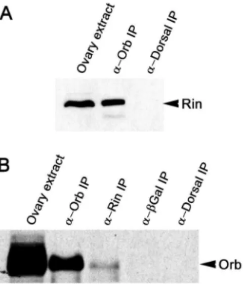
To identify factors involved in Orb protein activity and/ororb autoregulation, we searched for proteins that physically associate with Orb in vivo. Ovary extracts were immunoprecipitated with anti-Orb or control antibodies and isolated proteins were fractionated by SDS polyacrylamide gel electrophoresis. Bands specific to the Orb immunoprecipitates were then analyzed by mass spectrometry. One of the proteins specifically associated with Orb was Rin [14]. To further confirm this association, ovarian proteins were immunoprecipitated in the presence of RNase-A with Orb antibodies and probed with antibodies against Rin on Western blots (Figure 1A). As expected, Rin co-immunoprecipi-tates with Orb but not with control antibodies against another fly protein, Dorsal. We further verified the association between Orb and Rin by reverse immunoprecipitation of Orb with anti-Rin antibodies. As shown in Figure 1B and Figure S1A, Orb is detected in the Rin immunoprecipitates but fails to co-immuno-precipitate with antibodies against Dorsal or a heterologous protein (b-Galactosidase).
Expression ofrinmRNA
To learn more about rin function during oogenesis, we first examined the expression and localization ofrinmRNA. We found that the pattern of rin mRNA accumulation (data not shown) differs in a number of respects from that seen fororb[1]. First,rin mRNA is detected in both the germline and somatic follicle cells. orb, by contrast, is expressed exclusively in the germline. Second, during the previtellogenic stages, rin mRNA appears to be distributed more or less uniformly in the nurse cells and oocyte. In contrast, during this same period orb mRNA preferentially accumulates at the posterior pole of the oocyte, while there is only
Figure 1. Orb and Rin are components of an RNase-resistant complex.Western blot of proteins immunoprecipitated (IP) with the indicated antibodies and probed on Western blots for Rin (A) and Orb (B). Immunoprecipitations were performed using equal amounts of wild-type ovarian extracts and in the presence of RNase A.
little mRNA elsewhere in the egg chamber. Third, after the onset of vitellogenesis,rinmRNA expression appears to be upregulated and it accumulates to high levels in the nurse cells but not in the oocyte. At this point in oogenesis,orbmRNA is localized along the anterior margin of the oocyte. Although rin mRNA is largely absent from the oocyte in stage 9–10 egg chambers, much of it moves into the oocyte during nurse cell transport (‘‘dumping’’). This finding would be consistent with our previous results [34], which showed that high levels of presumably maternalrinmRNA are present in early embryos.
Accumulation of Rin protein
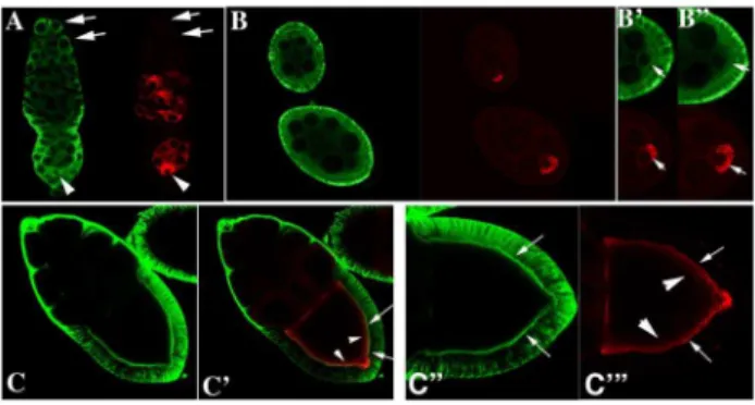
We next examined the pattern of Rin protein accumulation in wild type ovaries using confocal microscopy. Whereas Orb is germline-specific and concentrates in the oocyte (Figure 2, red), Rin is expressed both in the germline and somatic follicle cells (Figure 2, green). In the germline, Rin is present in stem cells and cystoblasts (arrows in Figure 2A, green) at the tip of the germarium as well as in the cysts in region 1 where there is little or no Orb protein (arrows in Figure 2A, red). Detectable levels of Orb are first evident in newly formed 16-cell cysts in region 2 where it accumulates in a subset of cells (Figure 2A, red). In contrast, Rin expression seems diffuse in the germline cysts in region 2 and is more heavily concentrated in the somatic follicle cells (Figure 2A). In region 3, both Orb and Rin are found in all germline cells but appear to be enriched in the oocyte located at posterior pole of the chamber (Figure 2A arrowhead). Stage 1 egg chambers pinch off from the germarium and continue development through 14 morphologically distinct stages which are divided into previtello-genesis (Stages 1–7) and vitelloprevitello-genesis (Stages 8–14). During previtellogenesis, Orb concentrates in the oocyte with the highest levels at the posterior end (Figure 2B-red; see arrows in enlargements in Figure 2B9 and 2B0-red) while Rin is enriched in the follicle cells, and in the cortical cytoplasm of the nurse cells (Figure 2B) and oocyte (arrows in Figure 2B9 and 2B0-green). During vitellogenesis, there is very little Orb or Rin in the nurse cells, while both proteins are found in the oocyte (Figure 2C and C9red and green) and Rin is enriched in the follicle cells. Within the oocyte Orb and Rin accumulate preferentially along the oocyte cortex (see arrows in Figure 2C0and C-), with Rin being more tightly associated with the cortex than Orb. Although the two proteins appear to overlap along the edge of the oocyte cortex (see arrows in Figure 2C), the Orb protein that is more loosely associated with the cortex (see arrowheads in Figure 2C) does not appear to be in close proximity to Rin. The overlapping yet distinct patterns of Rin and Orb accumulation within the oocyte would be consistent with the co-immunoprecipitation data. Moreover, it suggests the existence of protein complexes that include both Rin and Orb proteins as well as complexes in which only one of the two proteins is present (see below).
Generation and characterization of rin3
To learn more about the function of the Rin protein in Drosophila we isolated and characterized a null allele of rinthat affects only the ringene. Existing rinmutations included a weak hypomorphic allele (rin1) caused by a P-element insertion (P4957) in the 59UTR ofrin(Figure 3A) and an imprecise excision allele (rin2
) that deleted multiple genes [34]. To isolate a null allele affecting onlyrin, we generated and screened additional excisions and selected putative excision lines on the basis of female sterility as homozygotes and hemizygotes, as described previously [34]. PCR and Southern analysis of the newrin3allele revealed that it has a 3.3 kb deletion of most of the coding region (Figure 3A). Homozygous and hemizygous rin3 flies are viable and do not
produce anyrintranscripts (data not shown) or protein as shown by Western analysis of homozygousrin3flies (Figure 3B and Figure S1B). We also looked at the tissue distribution of Rin by probing Western blots of extracts derived from dissected ovaries and ovarectomized adult females. As shown in Figure 3B, high levels of Rin protein are found in ovaries. This would be consistent with our previous studies, which revealed the presence of appreciable amounts of Rin in early embryos [34] and suggested that rin transcripts and protein are maternal contributions to the embryo. Althoughrinmutants are homozygous and hemizygous viable, rin3 homozygous flies are underrepresented. The reduction in adult viability depended on the strength of the allele (Table S1). Hemizygousrin1/Df(3R)urdflies emerged at a lower than expected frequency compared to their heterozygous siblings. Homozygous and hemizygous nullrin3flies showed a 40% reduction in viability compared to their heterozygous siblings. The reduction in viability was largely rescued by the Tub-rin transgene. Additionally, homozygousrin3flies had a noticeably shorter lifespan than their heterozygous siblings. Analysis of longevity of adult flies revealed a severe reduction in lifespan that correlated with the lack of Rin protein (Figure S2). Homozygousrin3flies began to die starting 3– 4 days after hatching from pupae; after the second and third weeks approximately 35% and 70% of the adult flies were dead, respectively. In contrast, homozygous rin1, heterozygous rin3/ TM3, andTub-rin;rin3/rin3flies had normal life spans.
rinmutants have reduced fertility
To investigate the effects ofrinmutations on female fertility we measured the hatching rate of larvae from eggs laid byrinmutant females. The severity of the allelic combination correlated with the level of reduction in fertility (Table 1). The hatching rate of embryos laid by homozygous and hemizygous females was greatly reduced as compared to wild type females. Furthermore, both homozygous rin3
and hemizygous rin3
/Df(3R)urd and rin3 / Df(3R)l26cfemales were completely sterile, but the fertility ofrin3 females carrying the Tub-rintransgene was close to that of wild type.
Figure 2. Rin protein expression in ovaries.Confocal analyses of Rin (green) and Orb (red) in the germarium (A), stages 4 and 6 egg chambers (B, B9and B0), and a stage 10 egg chamber (C, C9, C0and C-). Note that Orb is expressed only in the germline and concentrates in the oocyte whereas Rin is expressed both in the germline and surrounding somatic follicle cells. Arrows in A show that Rin is present in stem cells, cystoblasts and 2, 4 and 8 cell cysts while Orb is not. Arrows in B9and B0
show accumulation of Rin and Orb in the oocyte of stage 4 and 6 cell chambers. Arrows and arrowheads in C9, C0 and C- mark overlap between Rin and Orb at the edge of the oocyte cortex in stage 10 egg chambers.
rinmutants have a spectrum of oogenesis defects We found that rin mutants have a range of incompletely penetrant phenotypes that likely together contribute to the female sterility. One of the earliest is a rather unusual defect that affects the proper encapsulation of the germline-derived cells by a single follicular epithelium. In wild type, the egg chamber consists of 15 nurse cells and a single oocyte (Figure 4A). As shown in Figure 4B and C,rin3ovarioles often have adjacent egg chambers with too few nurse cells. In panel B, the smaller chamber (arrow) has a single nurse cell nucleus, while the larger chamber has 14 nurse cell nuclei. In panel C, the normal complement of 15 nurse cell nuclei and the oocyte appears to be distributed between 3 adjacent incomplete chambers.
A second defect that becomes evident at or before the beginning of vitellogenesis is the abnormal packaging of chromatin in nurse cell nuclei. In wild type, the endoreplicated nurse cell chromo-somes have a polytene-like chromosome structure and are organized into 5 domains or blobs until stage 5. At this point, homolog pairing is disrupted and the newly replicated
chromo-somes disperse throughout the nurse cell nuclei (Figure 4D). Inrin mutants, the nurse cell chromosomes do not disperse appropri-ately, and instead of diffuse DNA staining, the chromosomes have a readily discernable structure. In the examples shown in Figure 4E and F, the polytene-like organization of the nurse cell chromo-somes persists and discrete chromosomal blobs are still evident within the nurse cell nuclei (arrows). In some cases, the chromosomes appear to line up around the periphery of the nucleus, giving a doughnut-like staining pattern (arrowheads in panel E).
As might be expected from previous studies which showed genetic interactions withRhoA[34],rinmutations appear to disrupt the actin cytoskeleton in developing egg chambers. Staining with rhodamine-conjugated phalloidin of rin3 egg chambers reveals Figure 3. Molecular characterization of therin3allele.(A) The
3.3 kb deletion in therin3allele was generated by imprecise excision of
the P-element P4957 originally isolated from the EMBL lethal collection. The deletion removes DNA encoding the translation start codon, the entire NTF2-like N-terminus, as well as the proline-rich (P-rich) and glutamine-rich (Q-rich) central portions of Rin. The deletion partially affects the RNA Recognition Motif (RRM) at the C-terminus, but leaves the coding region of the arginine/glycine-rich domain (RG-rich) intact. (B)rin3is a null allele. Western blots of protein extracts prepared from
dissected ovaries and ovarectomized wild type (wt) andrin3females were probed with antibodies against the RRM domain of Rin. Similar results were obtained with antibodies raised against the N-terminal part of Rin (data not shown). Equal amounts of protein were loaded per lane. doi:10.1371/journal.pone.0072864.g003
Table 1.Fertility Assays.
Genotype % hatched Eggs hatched/total
Wild type 89.8% 1762/1963
rin1 51.8% 2243/4329
rin1/Df(3R)urd 24.0% 1125/4693
rin1/rin3 38.7% 1988/5137
rin3 0% 0/2223
rin3/Df(3R)urd 0% 0/1504
rinTub-rinrin3/rin3 86.0% 418/486
rin3is a homozygous recessive, fully penetrant female sterile mutation. Females of the indicated genotypes were mated with wild-type males. Eggs were collected and allowed to develop for 25 hours at 25uC, and scored for embryo hatching.
doi:10.1371/journal.pone.0072864.t001
Figure 4. Defects in egg chamber packaging and chromosome morphology.Ovarioles from wild type (A, D) andrin3(B,C,E,F) females
were stained with DAPI. A–C) Wild type chambers always contain 15 nurse cells and 1 oocyte, butrin3chambers may have fewer nurse cells
defects in the organization of the nurse cell actin cytoskeleton (Figure 5). Actin distribution in nurse cells is abnormal and actin filaments fail to form in stage 10–11 rin3
nurse cells. There are often large actin-free gaps in rin3 egg chambers, and subcortical actin is discontinuous. Perhaps the most striking defects are in the ring canals. Ring canals provide channels for transport through the nurse cell complex to the oocyte and are composed of actin plus a number of specialized cytoskeleton proteins including Kelch and Hu li tai shao (Hts). When they are first formed during the mitotic cycles the ring canals are between 0.5 and 1.0 bm in diameter. As oogenesis proceeds, the ring canals grow in size and reach a diameter of about 10bm by stage 11. As illustrated for actin (red) and phosphotyrosine (green) in Figure 5B, wild type ring canals have a regular circular structure with the different components organized in a sterotypic layering pattern. There are a variety of ring canal defects in rin3 mutant egg chambers that become more prevalent in later stages of oogenesis. Figure 5D shows a ring canal that has fragmented and only a part of the actin-phosphotyrosine ring remains. Actin strings can be seen extending away from this incomplete ring structure. In addition to fragmented or incomplete (arrowhead Figure 5F) ring canals, ring canals that are much larger than normal (arrow Figure 5F) are also observed. As multinucleated nurse cells are often seen in rin mutant egg chambers (Figure 5C), it is possible that the nurse cells may fuse following ring canal failure. Perhaps not surprisingly, given the abnormalities in ring canal morphology and the presence of fused nurse cells, one of the terminal phenotypes is dumpless (see Figure S3A, B). As expected from its ability to rescue viability and female fertility, theTub-rintransgene also rescues the defects in partitioning, chromosome structure and ring canals seen inrin mutant ovaries (Figure S4A–C).
rininteracts genetically with orb
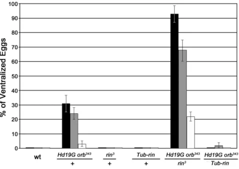
To determine whether the complexes containing Orb and Rin detected in ovaries have functional significance we tested for genetic interactions. For this purpose, we took advantage of the fact thatorbis weakly haploinsufficient in thegurkendorsal-ventral signaling pathway and 5–10% of the eggs laid by females heterozygous for a strong allele like orb343 have ventralized chorions due to defects in the localization and translation of grk mRNA [8]. These defects in DV polarity can be further exacerbated by an hsp83 transgene (HD19G) which expresses a hybrid mRNA consisting of the E. coli b-galactosidase protein coding sequence fused to the orb 39 UTR [13]. The transgene behaves like a dominant negative because the Orb binding sites in the 39 UTR of the chimeric mRNA compete with the target sequences in endogenousorb mRNAs. This compromises theorb positive autoregulatory loop and reduces Orb protein expression. When a single copy of the transgene is introduced into orb343/+ females it increases the frequency of ventralized eggs. Hd19G orb343/
+females lay between 3–30% ventralized eggs depending on the temperature. Loss-of-function mutations in genes that function to downregulateorbexpression or activity are expected to suppress the DV polarity defects in this assay, while mutations in genes that upregulate orb expression or activity are expected to exacerbate the polarity defects.
Because rin homologues in mammals repress translation of target mRNAs [29], we anticipated that rin mutations would reduce the frequency of ventralized eggs. Instead, precisely the opposite result was observed: the frequency of ventralized eggs is increased 3-fold or more depending on the temperature when Hd19G orb343/+mothers are also heterozygous forrin3(Figure 6). For example, at 18uC about 30% of the eggs produced byHd19G orb343/+mothers are ventralized, while over 90% are ventralized
whenrinis heterozygous. An even stronger interaction is observed at 29uC. About 3% of the eggs produced by Hd19G orb343/+ mothers are ventralized, while over 20% are ventralized when Hd19G orb343/+mothers are also heterozygous forrin.
Unlike orb, rinis not haploinsufficient in the grkDV signaling pathway (Figure 6). However, the strong genetic interactions between orb and rin suggested that rin activity is likely to be required in the DV signaling pathway. To test this we examined the eggs produced byrin3/rin2 mothers. As shown in Figure 7, about 10% have a collapsed eggshell phenotype. Many of these are likely to be derived from the dumpless chambers that are seen in rinmutant ovaries. Approximately 30% of the eggs are ventralized, indicating that like orb, rin activity is required for normal DV patterning. While the remainder of the eggs has seemingly normal eggshells, cuticle preparations of the embryos produced byrin3/ rin2 mothers indicate severe defects in embryonic development. For example, many of the embryos are undeveloped or have only scraps of cuticle. Similar embryonic phenotypes have been observed for theorbhypomorphic allele,orbmel.
rinfunctions as a positive regulator oforb
One plausible mechanistic connection between the ventralized eggs produced by rin mutant mothers and the orb-rin genetic interactions is thatrinfunctions inorbautoregulation, promoting Orb activity and/or expression. Several different approaches were used to explore this possibility. We first investigated Orb protein expression in the absence of rin using confocal microscopy. As Figure 5. Defects in ring canals.A–D) Wild type (A, B) andrin3(C, D) ovarioles were probed for actin (red) and phosphotyrosine (green) and stained with DAPI (blue). In wild type (A) each nurse cell has a defined number of ring canals (depending on the number of cell divisions) and the ring canals have a regular circular organization of components. In therin3chambers the ring canals often fail to maintain a regular circular
structure and may disintegrate (D), possibly resulting in nurse cell fusion (C). Ring canal defects, varying in severity, were observed in most vitellogenic stagerinchambers. E, F) Wild type (E) andrin3chambers probed with phosphotyrosine (PY) antibodies. Note the enlarged (arrow) and fragmented (arrowhead) ring canals in therin3chamber.
shown in Figure 8A, Orb protein levels were appreciably reduced in rin3 ovaries throughout all stages of oogenesis. This finding together with the strongorb-ringenetic interactions suggests thatrin might, at least under special circumstances, be a limiting factor in theorbautoregulatory loop. One such special circumstance would be when the autoregulatory loop is compromised, as it is inHd19G orb343/+ovaries. Ifrinis limiting in this genetic background, then it should be possible to suppress theHd19G orb343/+DV defects by increasingrinactivity using theTub-rinrescue transgene. As shown in Figure 6, the frequency of ventralized eggs is reduced to near background levels at all temperatures when Hd19G orb343/+ mothers have a single copy of theTub-rintransgene.
To further test the relationship between rin and orb, we examined the effects of different levels of rin on Orb protein
accumulation in wild type and sensitized Hd19G orb343/+ egg chambers. We first compared the effects of reducingrinactivity in an otherwise wild type background or in Hd19G orb343/+. As shown in Figure 8B and C, Orb protein levels are not changed very much if at all by reducingrinactivity (rin3/+) in a wild typeorb background (Figure 8B and C, lane 3). On the other hand, there is a noticeable drop in Orb protein when the rin mutation is introduced intoHd19G orb343/+females (compare Figure 8B and C, lane 5 with lane 2). Consistent with a role forrinin upregulating orb activity/expression, increasing rin activity augments Orb protein levels. Whereas Orb protein levels in the ovaries of Hd19G orb343/+ females are only about 1/4th that of wild type, Orb protein levels inHd19G orb343/+ females that also carry the Tub-rintransgene are even greater than wild type (Figure 8B and C, lane 4). Moreover Orb protein levels can be increased in wild type females by introducing theTub-rin transgene. These results support the conclusion thatrinfunctions as a positive regulator in theorbautoregulatory pathway and argue thatrinimpacts DV axis specification at least in part by regulating Orb expression/activity. Note that given the range of other phenotypes evident in rin mutants, it is quite possible thatrinmay have functions in DV axis specification beyond helping to ensure that there is sufficient Orb protein.
Identification of additional components of the Rin-Orb mRNP complex
To identify other potential components of the Orb-Rin mRNP complex, we performed Tandem Mass Spectrometry on ovarian proteins co-immunoprecipitated with antibodies against either Orb, Rin or a negative control, Dorsal, in the presence of RNase-A. We identified 22 proteins that associate with Rin only, 132 proteins that immunoprecipitate exclusively with Orb, and 18 proteins that associate with both Orb and Rin (see Table 2 and Table S2). Two of the proteins that are common to both Rin and Figure 6.orbandrininteract genetically.The percentage of dorsal-ventral polarity defects in eggs laid by females with different doses of Orb and Rin is shown. Females of the indicated genotypes were crossed to wild-type males at 18uC (black bars), 25uC (gray bars) and 29uC (white bars). Each of the crosses at the indicated temperature was repeated three or more times and a total of between 1,000 to 2000 eggs were scored. Fused dorsal appendage phenotypes range from fusion at the base to fusion along the entire length of the two appendages.Hd19Gis a dominant negative transgene carrying sequences of the orb 39UTR bound by endogenous Orb and sufficient to recapitulate the pattern of localization of the endogenousorbtranscript [13];orb343,orbnull allele [1];Tub-rin, transgene carrying a wild type copy ofrinunder control of thetubulinpromoter [34]. doi:10.1371/journal.pone.0072864.g006
Figure 7.rinmothers produce ventralized or collapsed eggs.
Egg collections (n = 752) from rin3/rin2 mothers were scored for
ventralized eggshells and collapsed eggs. No eggs hatched. The frequency of defective eggs of either type in wild type collections is typically less than a percent.
Orb immunoprecipitates, dFMR1 and Lingerer, were identified previously in Orb immunoprecipitates [14] (Table 2). dFMR1 is a KH-domain RNA binding protein which associates with poly(A) containing mRNAs and functions to negatively regulate transla-tion [35]. It also appears to be a component of the RNAi machinery. In fly ovaries, we found that it negatively regulatesorb activity [14]. Lig is expressed at high levels in the nervous system and gonads and was initially identified because of defects in mating behavior evident in hypomorphic lig mutant males [17]. More recent studies indicate thatligfunctions in the ovary as a negative regulator of gurken mRNA translation [36]. The remaining 16 proteins (Table 2) include many known translational factors, namely five 60S and three 40S ribosomal proteins, one elongation factor, and two RNA-binding proteins (Ataxin-2 (Atx-2) and Poly(A) Binding Protein (PABP)). In addition, four known proteins (Poly (ADP-ribose) polymerase (PARP), Heat Shock 70 kDa Protein Cognate 5 (HSC70-5), Rudimentary, and Twenty-four (Twf)) and one novel protein (CG5726) co-immunoprecipitated with both Orb and Rin. Several other potentially relevant proteins are present in Orb, Rin but also Dorsal immunoprecipitates. These include Caprin which is known to be associated with G3BP in other species (and was found previously in Orb but not control immunoprecipitates [14]), multiple ribosomal proteins, and translational regulators like Cup and Lost that have been implicated in DV or AP polarity (Table S3).
With the exception of Rudimentary all of the proteins found associated with both Rin and Orb function in translation,
translational regulation and/or RNA metabolism. Moreover, several of the factors besides Rin (Atx-2, dFMR1, PABP, and Caprin) have been previously implicated in translational repres-sion and stress granule formation [32]. Our current and previous studies show that some proteins associated with Orb can either promote or repress its expression or activity. dFMR1 negatively regulatesorb[14], whereas bothrinand PABP function as positive regulators oforb expression and/or activity (this paper; [37]). To explore the functional significance of other proteins associated with both Orb and Rin, we tested several for genetic interactions withorbin the sensitizedHd19G orb343/+background. As shown in Figure S5, reducingligactivity greatly increases the frequency of ventralized eggs produced by females transheterozygous forligand Hd19G orb343, similar to what was observed betweenHd19G orb343 andrin. In contrast, reducingparpactivity suppresses the polarity defects induced byHd19G orb343, as was observed fordfmr1[14]. As foratx-2, the allele we tested shows at most only a very modest interaction withorb(data not shown).
Discussion
Previous studies have shown that orb autoregulation promotes localized accumulation of Orb protein in subcellular compart-ments where its activity is required [13]. To investigate the mechanisms underlyingorb autoregulation we sought to identify proteins that physically associate with Orb and thus potentially help regulate its expression and/or activity. We have previously Figure 8. Rin positively regulates Orb expression.(A) Orb expression is downregulated inrin3ovaries. Confocal analysis of wild type (wt) and
homozygousrin3egg chambers at different stages of oogenesis. Samples were processed in parallel and microscopy was carried out under identical settings. (B) Western blot analysis of ovarian protein extracts derived from the indicated genotypes and probed with antibodies against Orb, Rin, and
shown that theDrosophilaFragile-X protein (dFMR1) is found in complexes with Orb in Drosophila ovaries and functions to negatively regulate orb accumulation and activity [14]. Here we show that Rin, theDrosophilaG3BP homologue, is also associated with Orb in ovaries. However, in contrast to dFMR1, Rin functions as a positive regulatory factor, helping to promote Orb accumulation and activity.
Our results show that Rin associates with Orb as part of an RNase-resistant complex. The RNase-resistance aspect of this interaction suggests that their association is mediated by protein-protein interactions rather than, or in addition to, binding to the same mRNA species. While it is possible that Orb and Rin interact directly with each other, an equally plausible scenario is that their association is mediated by one or more proteins found in both Orb and Rin immunoprecipitates. For example, mammalian G3BP has been shown to interact directly with Caprin-1, and the fly Caprin protein is found in both Rin and Orb immunoprecipitates. Two other findings would also seem to argue in favor of an indirect, rather than a direct interaction. First the overlap between Rin and Orb, especially in vitellogenic chambers is quite limited. Second, only a small subset of the proteins associated with Rin or Orb are common to both. It is also possible that the initial association between Orb and Rin could depend upon binding to the same target mRNAs and their subsequent interaction could depend upon a short stretch of RNA that is hidden in the complex and protected from RNase activity. In this case, the limited co-localization observed in egg chambers would imply that only a subset of their mRNA targets are in common.
GB3Ps in mammals are thought to have two functions. The first is repressing the translation of target mRNAs by mechanisms that depend upon their helicase and RNase activities, while the second is in the assembly of stress granules under conditions of
environmental stress such as heat shock or drug treatment. For this reason, we anticipated thatrin, likedfmr1, would function to negatively regulateorb activity and/or expression. However, we observed exactly the opposite result. Instead of suppressing the DV polarity defects in eggs fromHd19G orb343/+females, reducingrin activity increases the frequency of DV defects. Conversely, DV polarity defects are suppressed by providing excess rin activity. While it is clear from the phenotypic effects seen in mutants that rin has multiple functions in oogenesis (and these will be independent oforb), the most plausible explanation for the effects of decreasing and increasingrinactivity on DV polarity inHd19G orb343/+ females is that they arise because of changes in the expression/accumulation of Orb protein. Consistent with this explanation, we find that we can change the level of Orb protein by manipulatingrinactivity. If orb is wild type, reducing therin dose by half has little if any effect on Orb protein accumulation. However, whenorbactivity is compromised by theHd19G orb343 combination, heterozygosity forrin results in Orb protein levels that are less than 5% that of wild type. Conversely, increasingrin activity in a wild type background elevates Orb protein levels almost two-fold over wild type, while in background compromised by theHd19G orb343 combination adding extra rinrestores Orb protein levels to that of wild type. Taken together, these findings argue thatrinfunctions as a positive regulator oforb. At this point it is not clear howrinmight control the accumulation of Orb protein. Since Rin and Orb are associated with each other, the simplest model is that Rin helps activate the translation oforbmRNA and thus functions as a co-factor inorbautoregulation. The substantial reduction in Orb protein levels evident inrin3ovaries is consistent with this idea. This view would also be supported by the DV polarity defects evident in eggs laid by rin mutant females. However, other less direct models (e.g.,rinrepresses the translation Table 2.Proteins Present in Orb and Rin but not Dorsal Immunoprecipitates.
UniProtKB Orb Rin
Protein Accession Seq ct Spec ct % Cov Seq ct Spec ct % Cov
Lingerer (CNS; behavior; Grk signaling) (CG8715) Q86S05 74 329 41.3% 25 55 23.7%
Drosophila Fragile X Mental Retardation (dFMR1) (CNS: translation factor; Orb regultor) (CG6203)
Q9NFU0 31 65 41.2% 5 9 11.1%
CG5726 (RNAi) Q7JRH5 30 62 56.1% 17 38 36.8%
Poly (ADP-ribose) polymerase (PARP) (CG40411) P35875 16 20 22.1% 3 5 5.8%
Elongation factor 1a48D (CG8280) P08736 12 17 36.1% 2 2 11.4%
Poly(A)-binding protein (PABP) (CG5119) P21187 8 8 18.8% 10 13 24.1%
twenty-four (circadian translation factor) (CG485) Q9W4M7 3 3 1.7% 33 72 23.0%
Ataxin-2 (microRNAs,) (CG5166) Q8SWR8 2 3 3.8% 9 31 20.4%
Rudimentary (pyrimidine biosynthesis (CG18572) P05990 3 3 3.6% 2 2 2.9%
Heat shock 70 kDa protein cognate 5 (CG8542) P29845 2 2 4.1% 5 8 14.8%
40S ribosomal protein S2 (String of pearls) P31009 7 17 30.0% 4 10 19.1%
40S ribosomal protein S14 (CG1524) P14130 6 7 37.1% 3 7 28.5%
40S ribosomal protein S24 (CG3751) Q9W229 2 3 20.6% 2 3 20.6%
CG13096 (Ribosome component) Q9VLK2 5 9 6.9% 2 2 6.0%
60S ribosomal protein L10 (CG17521) O61231 3 6 19.3% 2 5 13.8%
60S ribosomal protein L11 (CG7726) P46222 3 3 12.5% 2 2 16.8%
60S ribosomal protein L13A (CG6459) Q9VNE9 4 8 17.1% 2 3 12.7%
of some factor that inhibitsorbmRNA translation orrinis required to stabilize Orb protein) can’t be excluded at this time. Likewise, rinmay have other targets besidesorbin the establishment of DV polarity.
Potentially arguing in favor of a role for Rin in the translation of orb mRNA and/or in the activity of Orb protein is the fact that mutations in genes encoding several of the other proteins found in both Orb and Rin immunoprecipitates also show genetic interactions with orb. Thus, dfmr1 and parp suppress the DV polarity defects in eggs laid byHd19G orb343/+females, whilepabp and lig enhance the polarity defects. We have shown previously that dfmr1 also exerts its effects, at least in part, by altering the expression of Orb protein [14]. Further connecting the effects of rinanddfmr1to translation, we also found that Orb, dFMR1 and Rin fractionate with polysomes in sucrose gradients (data not shown). If the proteins detected in Orb and Rin immunoprecip-itates are part of the same complex rather than different complexes, then the gene dose effects we have observed would suggest that they act coordinately to regulate Orb expression and that the relative balance between positive (e.g., Rin) and negative (e.g., dFMR1) factors in the egg chamber helps set the level of Orb accumulation. Further studies will be required to understand precisely how rininfluences Orb protein accumulation and how some of the other factors associated with Rin and Orb like dFMR1, Lig and PABP function in this process.
Although our studies implicaterinas a positive regulator oforb, this is clearly not the only role for rinin the ovary. Instead, the phenotypic effects ofrinmutations point to a potentially diverse array of functions, not only in gem cells but also in the surrounding somatic follicle cells. For example, the fragmentation of ring canals, the failure to properly disperse the endoreplicated nurse cell chromosomes, and the dumpless phenotype are not observed in orb mutants. Moreover, ring canal and chromatin dispersal defects suggest that rin has functions in nurse cells, which is a compartment that has only little Orb protein. Likewise, the encapsulation defects could be of somatic origin whererin, but not orbis expressed. Further studies will be required to identify therin regulatory targets in these and potentially other processes.
Materials and Methods
Fly stocks and genetic analysis
Wild typew1118andorb343have been described previously [1] as have therin1and rin2mutant stocks andTub-rinrescue construct [34]. TheHD19Gtransgene containing DNA sequences from the orb39UTR has been described in [38].
3–5 day old females of various genotypes were mated tow1118 males in cages at 18uC, 25uC or 29uC. Beginning on the second day, eggs were collected for seven days and at least 200 eggs/day were scored for a ventralized phenotype as described in [13].
In situ hybridization
Whole-mount in-situ hybridization was performed as described [14] on wild type ovaries. DIG-labeledrincDNA was used as a probe and was labeled according to the manufacturer’s instruc-tions (Roche).
Immunostaining
Ovaries were dissected in ice-cold 16 PBS and ovarioles microdissected with needles. Ovarioles were fixed with 4% paraformaldehyde in 16PBS for 20 minutes at room temperature followed by three washes in 16PBS. Ovarioles were rinsed once in PBST (0.1% Triton X-100 in 16PBS) and blocked in PBSTTB (0.1% Triton X-100, 0.05% Tween 20, 10% BSA in 16PBS) for
1–2 hours at room temperature. Labeling was performed with anti-Orb antibodies (6H4 obtained from the Developmental Studies Hybridoma Bank) at 1:30 and anti-Rin antibodies [34] at 1:1000 in 16PBSTTB overnight at 4uC. After several washes in 16PBSTT, ovarioles were incubated for 2 hours in 16PBSTTB with Alexa Fluor 568 goat anti-mouse IgG2a (Molecular Probes, Inc.) against Orb 6H4 antibodies, and Alexa Fluor 647 goat anti-rabbit (Molecular Probes, Inc.) against Rin antibodies. After several washes in 16PBSTT, ovarioles were mounted in 15–30% glycerol in 16 PBS and visualized by confocal microscopy. Microscopy was performed with an inverted Zeiss LSM510 confocal microscope.
Immunoprecipitation and western analysis
Ovaries of well-fed 1- to 4-day old wild-type females were hand-dissected in ice-cold 16PBS, frozen in dry ice and stored at
280uC. Ovary extracts were prepared by homogenizing 100 ovaries in 200ml of ice-cold IP buffer (20 mM Hepes, pH 7.5, 150 mM NaCl, 2.5 mM MgCl2, 250 mM sucrose, 0.05% Tergitol, 0.5% Triton X-100, 1mg/ml pepstatin A, 10mg/ml aprotinin, 1mg/ml leupeptin, 1 mM dithiothreitol, 1 mM PMSF, 1 mM NaF, 40mM NaVO3, 40mM Na3VO4) supplemented with 500mg of RNase A. Homogenates were cleared twice by centrifugation at 750g for 5 minutes at 4uC and supernatants were transferred to a fresh microfuge tube. Supernatants were diluted to 500ml with ice-cold IP buffer and mixed with 60ml of a 50% slurry of Protein G Plus/Protein A-agarose beads (Calbio-chem) to which antibodies have been crosslinked at 2 mg/ml. After overnight rotation at 4uC, beads were precipitated by centrifugation at 100gfor 30 seconds and washed 4 times with 100 volumes of ice-cold IP buffer without MgCl2.
For western blotting, 5ml of washed beads (Figure 1) or 20mg of total protein (Figure 8B) were electrophoresed through an 11% SDS-polyacrylamide gel, transferred onto Immobilon-P PVDF membranes (Millipore), and probed with anti-Orb antibodies (6H4 and 4H8 obtained from the Developmental Studies Hybridoma Bank) at 1:30 or anti-a-tubulin antibodies (Sigma) at 1:2500 followed by secondary peroxidase-conjugated goat anti-mouse antibodies (Jackson ImmunoResearch Laboratories) at 1:2000. Membranes were also probed with anti-Rin antibodies [34] at 1:3000 followed by secondary peroxidase-conjugated goat anti-rabbit antibodies (Jackson ImmunoResearch Laboratories) at 1:2000. Proteins were detected by chemiluminescence according to manufacturer’s instructions (ECL, Amersham Pharmacia Biotech).
For the western blots shown in Figure 3B, ovaries or female carcasses were homogenized in RIPA buffer and the extract was quantitated using the Bio-Rad protein assay (Bio-Rad Laborato-ries) to ensure equal amounts of sample per lane. After separation on 4–12% polyacrylamide gels, proteins were transferred to ECL-nitrocellulose (Amersham Pharmacia Biotech), and probed with anti-Rin antibodies as described above.
Supporting Information
Figure S1 A) Rin Immunoprecipates.Ovary extracts were immunoprecipitated with b-Gal or Rin antibodies. In the top blot, the immunoprecipitate (IP) was probed with Orb antibody. In the bottom blot the immunoprecipitate was probed with Rin antibody. B) Westerns ofrin3mutant ovaries.Western blots of ovary extracts from wild type (WT) orrin3mutant ovaries were probed as indicated.
(TIF)
Figure S2 Effects of rin mutations on lifespan. Newly emerged adult flies of each genotype were collected, placed in fresh vials with normal yeast cornmeal media (10 flies per vial and 10 vials of each genotype), and monitored for survival every 3 days for a period of 40 days at 25uC. The graph shows the average of two experiments. Heterozygotes were indistinguishable from wild type.
(TIF)
Figure S3 A subset of the rin3 chambers have the dumpless phenotype.Wild type (A) andrin3(B) egg chambers. As illustrated in B) a subset (5–10%) of the late stagerin3chambers are dumpless. Arrow marks the dorsal appendages.
(TIF)
Figure S4 Rescue ofrin3oogenesis defects by the Tub-rintransgene.TheTub-rintransgene not only rescues fertility,
but also fully rescues the partitioning, nuclear structure and ring canal defects ofrin3(Tub-rin;rin3/rin3females). A) DAPI staining showing rescue of the nuclear chromosomal phenotype. B) Actin staining shows rescue of the partitioning defects. C) Phosphotyr-osine antibody shows rescue of the ring canal defects.
(TIF)
Figure S5 Genetic interactions between orb and genes encoding proteins common to Orb and Rin immunopre-cipitations.The percentage of dorsal-ventral polarity defects in eggs laid at 25pC by femalestrans-heterozygous forHd19G orb343 (orb) and genes (lig,parp, andatx-2) encoding proteins detected in both Orb and Rin immunoprecipitates is shown. Fused dorsal appendage phenotypes range from fusion at the base to fusion
along the entire length of the two appendages. Hd19G is a dominant negative transgene carrying sequences of theorb39UTR bound by endogenous Orb and sufficient to recapitulate the pattern of localization of the endogenousorbtranscript [13];orb343, orbnull allele [2].Hd19G orb343/atx-2results are from a different experiment and the frequency of DV polarity defects in the control Hd19G orb343/+ females is less that seen in other control experiments. Even so the effects ofatx-2are modest.
(TIF)
Table S1 Viability of flies carrying rin mutations. Percent of viability is calculated as the number of observed/ number of expectedrinmutant flies (n). The observed number of rin/+ siblings was used to calculate the number of expectedrin mutant flies.Df(3R)urddeletesrinand adjacent loci.
(DOC)
Table S2 Proteins detected in Dorsal, Orb and Rin immunoprecipitates from ovary extracts. At least two peptides were detected for all proteins on the list.
(DOC)
Table S3 Potentially relevant proteins present in Orb, Rin and also Dorsal Immunoprecipitates. In addition to ribosomal proteins, a number of these proteins have been found associated with Rin homologs in other species or have been implicated in translation regulation.
(DOC)
Acknowledgments
We thank J. Goodhouse for technical assistance with confocal microscopy. We would also like to thank our many colleagues for their helpful suggestions and comments.
Author Contributions
Conceived and designed the experiments: AC CP LCW KSS SH PS. Performed the experiments: AC CP LCW KSS SH IM. Analyzed the data: AC CP LCW KSS SH IM JY. Contributed reagents/materials/analysis tools: IM JY. Wrote the paper: AC CP LCW KSS SH PS.
References
1. Lantz V, Ambrosio L, Schedl P (1992) TheDrosophilaorb gene is predicted to encode sex-specific germline RNA-binding proteins and has localized transcripts in ovaries and early embryos. Development 115, 75–88.
2. Lantz V, Chang JS, Horabin JI, Bopp D, Schedl P (1994) TheDrosophilaorb RNA-binding protein is required for the formation of the egg chamber and establishment of polarity. Genes Dev 8: 598–613.
3. Hake LE, Richter JD (1994) CPEB is a specificity factor that mediates cytoplasmic polyadenylation during Xenopus oocyte maturation. Cell 79, 617– 627.
4. Richter JD (2007) CPEB: a life in translation. Trends Biochem Sci 32, 279–285. 5. Keleman K, Krutner S, Alenius M, Dickson BJ (2007) Function of theDrosophila CPEB protein Orb2 in long-term courtship memory. Nat Neurosci 10: 1587– 1593.
6. Mastushita-Saki T, White-Grndley E, Samuelson J, Seidel C, Si K (2010) DrosophilaOrb2 targets genes involved in neuronal growth, synapse formation and protein turnover. PNAS, 107, 11987–11992.
7. Hafer N, Xu S, Bhat KM, Schedl P (2011) TheDrosophilaCPEB protein Orb2 has a novel expression pattern and is important for asymmetric cell division and nervous system function. Genetics, 189, 901–921.
8. Christerson LB, McKearin DM (1994) orb is required for anteroposterior and dorsoventral patterning duringDrosophilaoogenesis. Genes Dev 8: 614–628. 9. Huynh JR, St Johnston D (2000) The role of BicD, Egl, Orb and the
microtubules in the restriction of meiosis to theDrosophilaoocyte. Develop-ment.127:2785–94.
10. Roth S, Schu¨pbach T (1994) The relationship between ovarian and embryonic dorsoventral patterning inDrosophila. Development. 120:2245–57.
11. Chang JS Tan L Wolf MR, Schedl P (2001) Functioning of theDrosophilaorb gene ingurkenmRNA localization and translation. Development 128:1159–69.
12. Chang JS, Tan L, Schedl P (1999) TheDrosophila CPEB homolog, orb, is required for oskar protein expression in oocytes. Dev Biol 215: 91–106. 13. Tan L, Chang JS, Costa A, Schedl P (2001) An autoregulatory feedback loop
directs the localized expression of the DrosophilaCPEB protein Orb in the developing oocyte. Development 128: 1159–69.
14. Costa A, Wand Y, Dockendorf TC, Erdjument-Bromage H, Tempst P, et al. (2005) TheDrosophilafragile X protein functions as a negative regulator in the orb autoregulatory pathway. Dev Cell. 8: 331–42.
15. Wan L, Dockedorff TC, Jongens TA, Dreyfuss G (2000) Characterization of dFMR1, aDrosophila melanogaster homolog of the fragile X mental retardation protein. Mol Cell Biol. 20:8536–47
16. Morales J, Hiesinger PR, Schroder AJ, Lume K, Verstreken P, et al. (2002) Drosophilafragile X protein, DFXR, regulates neuronal morphology and function in the brain. Neuron 34:961–72.
17. Kuniyoshi J, Baba K, Ueda R, Kondo S, Awano W, et al (2003) Expression analysis of the lingerer gene in the larval central nervous system ofDrosophila melanogaster. J Neurogenet. 17:117–37.
18. Parker F, Maurier F, Delumeau I, Duchesne M, Faucher D, et al (1996) A Ras-GTPase-activating protein SH3-domain-binding protein. Mol Cell Biol. 6: 2561–9.
19. Erickson SL, Lykke-Anderson J (2011) Cytoplasmic mRNP granules at a glance. J Cell Sci. 124: 293–7.
20. Irvine K, Stirling R, Hume D, Kennedy D (2004) Rasputin, more promiscuous than ever: a review of G3BP. Int J Dev Biol. 48:1065–77.
22. Barnes CJ, Li F, Mandal M, Yang Z, Sahin AA, et al (2002) Heregulin induces expression, ATPase activity, and nuclear localization of G3BP, a Ras signaling component, in human breast tumors. Cancer Res. 62:1251–5.
23. Mallumbres M, Pellicer A (1998) RAS pathways to cell cycle control and cell transformation. Front Biosci. 6: 887–912.
24. Chen G, Goeddel DV (2002) TNF-R1 signaling: a beautiful pathway. Science. 296:1634–5.
25. Soncini C, Berdo I, Draetta G (2001) Ras-GAP SH3 domain binding protein (G3BP) is a modulator of USP10, a novel human ubiquitin specific protease. Oncogene 20, 3869–3879.
26. Gallouzi IE, Parker F, Chebli K, Maurier F, Labourier E, et al. (1998) A novel phosphorylation-dependent RNase activity of GAP-SH3 binding protein: a potential link between signal transduction and RNA stability. Mol Cell Biol. 18:3956–65.
27. Tourrie`re H, Chebli K, Zekri L, Courselaud B, Blanchard JM, et al. (2003) The RasGAP-associated endoribonuclease G3BP assembles stress granules. J Cell Biol. 160:823–31.
28. Costa M, Ochem A, Staub A, Falaschi A (1999) Human DNA helicase VIII: a DNA and RNA helicase corresponding to the G3BP protein, an element of the ras transduction pathway. Nucleic Acids Res. 27:817–21.
29. Oretega AD, Willaers IM, Sala S, Cuezva JM (2010) Human G3BP1 interacts with beta-F1-ATPase mRNA and inhibits its translation. J Cell Sci. 123:2685– 96.
30. Atlas R, Behar L, Elliott E, Ginzburg I (2004) The insulin-like growth factor mRNA binding-protein IMP-1 and the Ras-regulatory protein G3BP associate with tau mRNA and HuD protein in differentiated P19 neuronal cells. J Neurochem. 89:613–26.
31. SolomonS, Xu Y, Wang B, David MD, Schubert P, et al. (2007) Distinct structural features of caprin-1 mediate its interaction with G3BP-1 and its
induction of phosphorylation of eukaryotic translation initiation factor 2a, entry to cytoplasmic stress granules, and selective interaction with a subset of mRNAs. Mol Cell Biol. 27, 2324–2342.
32. Anderson P, Kedersa N (2009) RNA granules: post-transcriptional and epigenetic modulators of gene expression. Nat Rev Mol Cell Biol. 10:430–6. 33. Ohn T, Kedersha N, Hickman T, Tisdale S, Anderson P (2008) A functional
RNAi screen links O-GlcNAc modification of ribosomal proteins to stress granule and processing body assembly. Nature Cell Biol. 10:1224–1231. 34. Pazman C, Mayes C, Fanto M, Haynes SR, Mlodzik M (2000) Rasputin, the
Drosophilahomologue of the RasGAP SH3 binding protein, functions in ras- and Rho-mediated signaling. Development 127:1715–25.
35. Bassell GJ, Warren ST (2008) Fragile X syndrome: loss of local mRNA regulation alters synaptic development and function. Neuron 60: 201–14. 36. Geng C, MacDonald PM (2007) Identification of genes that influence gurken
expression. Fly 1: 259–267.
37. Wong LC, Costa A, McLeod I, Sarkeshik A, Yates J 3rd, et al. (2011) The Functioning of theDrosophilaCPEB Protein Orb Is Regulated by Phosphory-lation and Requires Casein Kinase 2 Activity. PLoS One 6:e24355. 38. Lantz V Schedl P (1994) Multiple cis-acting targeting sequences are required for
orb mRNA localization during oogenesis. Mol Cell Biol. 14: 2235.
39. Bern M, Goldberg D, McDonald WH, Yates JR 3rd (2004) Automatic quality assessment of peptide tandem mass spectra. Bioinformatics. 20 Suppl 1:i49–54. 40. Link AJ, Eng J, Schieltz DM, Carmack E, Mize GJ, et al. (1999) Direct analysis
of protein complexes using mass spectrometry. Nat Biotechnol. 17:676–82. 41. Tabb DL, McDonald WH, Yates JR 3rd (2002) DTASelect and Contrast: tools