JSCS–4789 Original scientific paper
Poly(methyl methacrylate) denture base materials modified with
ditetrahydrofurfuryl itaconate: Significant applicative
properties
PAVLE SPASOJEVIĆ1*#, VESNA PANIĆ1, SANJA ŠEŠLIJA2#, VLADIMIR NIKOLIĆ3#,
IVANKA G. POPOVIĆ4# and SAVA VELIČKOVIĆ4
1Innovation Centre, Faculty of Technology and Metallurgy, University of Belgrade, Karnegijeva 4, 11000 Belgrade, Serbia, 2Institute of Chemistry, Technology and Metallurgy,
University of Belgrade, Njegoševa 12, 11000 Belgrade, Serbia, 3Innovation Centre, Faculty of Chemistry, University of Belgrade, Studentski trg 12–16, 11000 Belgrade, Serbia
and 4Faculty of Technology and Metallurgy, University of Belgrade, Karnegijeva 4, 11000 Belgrade, Serbia
(Received 23 January, revised 9 April, accepted 22 April 2015) Abstract: The aim of this work was to examine the possibility of modification of commercial denture base materials with itaconic acid esters, in order to obtain materials with lower toxicity and higher biocompatibility. Despite their relatively higher price compared to methacrylates, itaconic acid and itaconates are materials of choice for environmentally friendly applications, because they are not produced from petrochemical sources, but from plant products. A com-mercial system based on poly(methyl methacrylate) was modified using ditetra-hydrofurfuryl itaconate (DTHFI), whereby the ratio of DTHFI was varied from 2.5 to 10 % by weight. Copolymerization was confirmed using FTIR spectro-scopy, while SEM analysis showed the absence of micro defects and pores in the structure. The effects of the itaconate content on the absorption of fluids, the residual monomer content, thermal, dynamic-mechanical and mechanical properties (hardness, toughness, stress and elongation at break) were inves-tigated. It was found that the addition of DTHFI significantly reduced the amount of residual methyl methacrylate, which made these materials less toxic. It was shown that increasing the DTHFI content resulted in materials with decreased glass transition temperatures, as well as with decreased storage mod-ulus, ultimate tensile strength and impact fracture resistance; however the mech-anical properties were in the rang prescribed by ADA standards, and the mat-erials could be used in practice. The deterioration in mechanical properties was therefore worthwhile in order to gain lower toxicity of the leached monomer. Keywords: dental; itaconic; methyl methacrylate; absorption, tensile.
* Corresponding author. E-mail: pspasojevic@tmf.bg.ac.rs
# Serbian Chemical Society member.
INTRODUCTION
Poly(methyl methacrylate) (PMMA) is one of the most significant acrylic polymers. Although it was discovered and commercialized many years ago, PMMA is still the subject of intense scientific research. Poly(methyl metha-crylate) is used as a substitute for transparent glass and dielectric films,1 acrylic
paints,2 micro-cell foam,3etc. However, biomedicine represents the most
attract-ive application area where this material is used to create denture bases,4 contact
lenses, bone cement,5 inhalers,6etc. Materials based on PMMA are often used as
biomaterials due to their good biocompatibility, non-toxicity, stability of colour and shape,7 the absence of taste, smell and irritation of the surrounding tissue,8
good adhesion to teeth, insolubility in body fluids, the ease of handling and design, as well as good aesthetic properties.9 In spite of these advantages of the
material, dental prosthesis based on PMMA have several drawbacks. The most important of these drawbacks are toxicity of residual monomer,10–13
suscept-ibility to distortions as well as limitations in terms of mechanics.14 As a result of
these deficiencies, residual monomer may leak out by diffusion from prosthesis and irritate the surrounding tissue. This leakage creates cracks and other struc-tural damages to the dental prosthesis that could lead to mechanical fracture of the prosthesis and create an environment suitable for the development of differ-ent types of bacteria, moulds and fungi.
In order to overcome the drawbacks and the limitations of PMMA denture base materials, the possibility of modifying a commercial PMMA-based formul-ation using itaconic acid derivatives was investigated. Itaconic acid is structurally very similar to methacrylic acid, except that at the α-carbon atom a carboxyl group is attached instead of the H atom. Despite the slightly higher market price compared to methacrylic acid, itaconic acid and itaconates are more acceptable in terms of ecology and sustainable development.15,16 The reason for this lies in the
fact that the itaconic acid is obtained from plants (mostly by enzymatic trans-formations of molasses17), while methacrylic acid is derived from petrochemical
sources. As a dibasic acid, itaconic acid provides more options when making its esters compared to methacrylic acid. Due to the many similarities of itaconates with the corresponding methacrylates and the mentioned advantages, itaconates represent an interesting alternative to methacrylates in the synthesis of a variety of materials.
In dentistry, itaconic acid and its esters are known. They have been widely used in the production of glass ionomer cements.18–20 Itaconic acid and its
deri-vatives are used as components in many systems for controlled drug release.21–26
It is important to note that itaconates have been extensively used in varies medi-cal applications because of their very low toxicity.19 Furthermore, itaconic acid and
its derivatives are increasingly used in the preparation of paints and coatings,27–29
Due to the structural similarities of the methacrylates and the itaconates, numerous studies concerning copolymers of methyl methacrylate and dialkyl itaconates have been published.11,12 Fernandez-Garcia and Madruga13 examined
the effect of copolymer composition on the glass transition temperature and came to the conclusion that the glass transition temperature of the copolymers dec-reased with increasing amount of itaconate, as well as with the increasing alkyl chain length of the ester group. Investigation of the thermal stability of the copolymers of the methyl methacrylate and dialkyl itaconates showed that the relative thermal stability increased with increasing proportion of methyl metha-crylate in the copolymer, following a similar trend as the change of glass tran-sition temperature.14
In a previous study, the residual monomer content and water sorption for PMMA denture base materials modified with dimethyl itaconate and dibutyl ita-conate were investigated.35 It was shown that the addition of itaconate led to a
reduction in the water uptake and greatly reduced the residual methyl metha-crylate content. In this way, the applicative properties and biocompatibility of PMMA denture base materials for the production of dental prostheses were greatly improved.
In order to further investigate the effect of the substitution of methyl metha-crylate in denture base materials with the esters of itaconic acid, in this study, a commercial denture base material was modified by ditetrahydrofurfuryl itaconate (DTHFI). The applicative properties of significance of the novel materials were investigated.
EXPERIMENTAL Materials
A commercial system for denture base materials Biokril® (Galenika AD, Serbia) was
used as received. The system was delivered as two-components; one part was solid and the other was liquid. Liquid component included monomer (MMA) and ethylene glycol dimetha-crylate (EGDMA) as crosslinker, while solid component included PMMA powder and ben-zoyl peroxide (BPO) as initiator. Itaconic acid (2-methylidenebutanedioic acid) was a com-mercial product (Fluka), while ditetrahydrofurfuryl itaconate (DTHFI) was synthesized as previously described in the literature.16
Synthesis of PMMA denture base materials modified with itaconates
The liquid component was mixed with a precisely defined amount of ditetrahydrofurfuryl itaconate. The amount of DTHFI that was added in liquid component was determined in a manner that mixture obtained by mixing modified liquid component with solid commercial component had satisfying characteristics (“working hours” and curing time). The mass percent of DTHFI in the new procedures were 0, 2.5, 5.0, 7.5 and 10 (Table I).
Polymerization under heating in a water bath
moulds for the measurement of elongation, viscoelastic properties and toughness had a rectangular shape with the dimensions of 60 mm× 10 mm× 4 mm, 60 mm× 12 mm× 2 mm and 80 mm× 10 mm× 4 mm, respectively.
TABLE I. Feed composition
Sample Biokril® PMMA Component
powder, % Biokril
® liquid
component, % DTHFI, %
PMMA 66.0 34.0 –
PMMA/2.5PDTHFI 64.5 33.0 2.5
PMMA/5PDTHFI 63.0 32.0 5.0
PMMA/7.5PDTHFI 61.5 31.0 7.5
PMMA/10PDTHFI 59.5 30.5 10
Determination of the amount of residual monomer in the obtained materials by high pressure liquid chromatography with a UV detector (HPLC-UV)
The amount of the residual monomer after polymerization was determined by high pressure liquid chromatography with a UV detector (HPLC-UV). The residual monomers were extracted from samples with an average mass of 0.5 g. The samples were placed in cups and then immersed in 20 mL of methanol. To complete the extraction, the cups were closed, covered with Parafilm® and stored in the dark for 10 days. A Surveyor HPLC system (Thermo
Fisher Scientific, Waltham, MA, USA) was used for the determination of the amount of the residual monomers (MMA and DTHFI). The chromatographic separation of monomers was realised on the Zorbax Eclipse® XDB-C18 column that was preceded by a pre-column of the
same producer. The mobile phase consisted of methanol (A) and deionised water (B). Chro-matographic separation of MMA and DTHFI was performed isocratically for a duration of 10 min and mobile phase consisted of 70 % A and 30 % B at a constant flow of 1.1 mL min-1. A
10-μl sample was injected into the HPLC system. The UV spectra of monomers were obtained by a Surveyor PDA detector. Only one absorption maximum for both monomers (λ = 210 nm) was observed in the obtained UV spectra. This absorption maximum was used for quantitative determination of the monomers.
Fourier transform infrared spectroscopy (FTIR)
FTIR spectra were recorded by Bomen MB 100 spectrophotometer (Hartmann and Braun, Canada). The KBr pellet technique was used whereby the KBr:sample ratio was 10:1. The Spectra were recorded with 10 repetitions at a resolution of 4 cm-1. Win Bomen software
was used for the processing of the spectra. Scanning electron microscopy (SEM)
Prior to SEM analysis, the samples were covered with platinum vapour in a Leica SCD005 nebulizer. The SEM observations were performed using a Jeol JSM 5800 scanning electron microscope at an acceleration voltage of 20 kV.
Water absorption
reached. Before measuring, the excess water was removed by wiping the samples with filter paper. All of the calculations were realised using data from the second absorption cycle. The second absorption cycle lasted 28 days. Water-soluble oligomers and other impurities were leached from the sample during the first adsorption cycle and hence, the results obtained from the second cycle were accepted as relevant for the water absorption–desorption process.
Equilibrium absorption (M∞ / %) was calculated using the equation:
(
0)
0
100 m m M m ∞ ∞ − = (1)
where m0 is mass of the dry sample and m∞ is the mass of the sample at equilibrium. Water diffusion coefficient
The diffusion of water into the synthesized materials was modelled by the Higuchi Equation: t n M Kt M∞ = (2)
where Mtis the % adsorption by the sample at time t, K is a kinetic constant and n is the diffusion exponent. In case of Fickian diffusion, n is 0.5, but in literature values between 0.43 and 0.5 could be found.36 A value of n higher than 0.5 is indicative of an anomalous diffusion
mechanism, while in case when n = 1, the diffusion kinetics is zero order, i.e., time-inde-pendent.
According to the Fickian second law, at the initial period of diffusion (Mt/M∞), diffusion through a solid material could be calculated using the equation:37
1/2 2 2 π t M Dt
M∞ L
=
(3)
where D is the effective diffusion coefficient and 2L is the sample thickness.
The dependence between Mt/M∞ and t1/2 is linear and the diffusion coefficient can be calculated from the linear part using Eq. (3).
Differential scanning calorimetry (DSC)
The glass transition temperatures (Tg) of the samples were determined by differential scanning calorimetry on a Perkin Elmer DSC-2 instrument under an inert nitrogen atmo-sphere. The heating and the cooling rates were 20 °C min-1 in the temperature range of 50 to
200 °C. The measurements were performed in two cycles in order to remove unreacted monomers in the first cycle. All calculations were performed using the data from the second cycle.
Dynamic mechanical analysis (DMA)
The dynamic mechanical properties of the samples were tested on a Rheometrics mech-anical spectrometer 605. The samples were exposed to a constant shear stress of 0.3 % at a frequency of 1 Hz. The heating rate was 3 °C min-1 in the temperature range from 25 to 180
Tensile properties
The analysis of the mechanical tensile properties was performed on an Instron-1332 (FastTrack 8800 control system, High Wycombe, UK) testing machine. The specimens (Fig. S-1 of the Supplementary material to this paper) were clamped by mechanical jaws. The testing speed was 0.5 mm min-1. For each sample five measurements were performed. The
average values of the ultimate stress and deformation at break, as well as the standard deviations, were calculated. During the test, the deformations were continuously registered as a function of the stress.
Hardness
The hardness of derived materials was measured on an Instron D-XD durometer (Instron, Norwood, USA). Five repetitions were performed for each sample.
Impact strength
Impact strength was measured on Charpy pendulum using samples with a V notch (Fig. S-2 of the Supplementary material). Length of pendulum was 60 mm with impact energy of 8.73 J. The impact edge of the cone-shaped pendulum with an angle of 30±1°, rounded with a radius R1= 2±0.5 mm, was made of hard steel. All tests were performed at 23 °C and repeated 5 times.
For the calculation of the Charpy impact strength for notched specimens, acN / KJ m-2, the following equation was used:
3 cN
N
10 w
a
hb
=
(4)
where w / J is the corrected energy absorbed by the fracture of the specimen, h / mm is the thickness and bN / mm is the remaining width of the specimen at the notch.
RESULTS AND DISCUSSION
A commercial formulation of denture base material based on PMMA was modified with DTHFI. The modification was performed in order to obtain a mat-erial with better biocompatibility, minor risks for an immune response and pre-served significant physicochemical properties. Polymerization in moulds was used for the syntheses of the modified denture base materials in which a part of the MMA in liquid phase was replaced with specified amounts of itaconate (Table I).
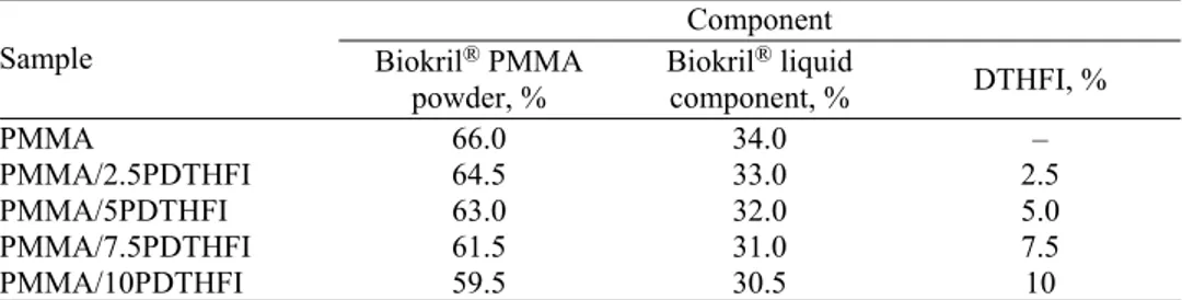
The samples prepared this way, including PMMA and PDTHFI homopoly-mers, were characterized by FTIR spectroscopy. The FTIR spectra of PMMA and PDTHFI homopolymers are shown in Fig. 1, while the FTIR spectra of PMMA/PDTHFI containing 2.5 and 10 wt. % itaconate are presented in Fig. 2. From Fig. 1, it could be seen that the FTIR spectra of PMMA and PDTHFI were similar and the characteristic peaks were at the same wavenumbers (3000 and 2950 cm–1 for the C–H stretching vibrations of the methyl group, 1730 cm–1 for
the C=O stretching vibrations of the ester group, 1450 and 1300 cm–1 for the
asymmetric and symmetric C–H deformation vibrations, 1165 cm–1 for the
of ring C–H vibrations). The FTIR spectrum of PDTHFI contained an absorption peak at 1266 cm–1 (C–O–C asymmetric stretching vibrations) but in the case of
PMMA, this peak was moved toward slightly a lower wavenumber. From Fig. 2, it could be seen that if the amount of DTHFI residues in monomer feed was increased, the absorption peak at 1266 cm–1 had a higher intensity; this led to the
conclusion that polymerization between MMA and DTHFI had occurred.
Fig. 1. FTIR spectra of the synthesized PMMA and PDTHFI homopolymers.
Fig. 2. FTIR spectra of the synthesized copolymers PMMA/PDTHFI containing 2.5 and 10 wt. % of itaconate.
justified the continuation of the testing and detailed characterization of the PMMA denture base materials modified with DTHFI.
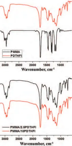
Fig. 3. SEM micrographs of the denture base materials a) PMMA/2.5DTHFI and b) PMMA/10DTHFI; bar: 200 µm.
The residual monomer contents of the synthesized PMMA/PDTHFI samples, as well as of the referent PMMA sample, were analyzed using high-performance liquid chromatography with ultraviolet detection (Table II).
TABLE II. Residual MMA and DTHFI contents and total residual monomer (TRM) content of the PMMA and PMMA/PDTHFI denture base materials
Sample Residual monomer, % TRM / %
MMA DTHFI
PMMA 1.27 – 1.27
PMMA/2.5DTHFI 0.79 0.95 1.74
PMMA/5DTHFI 0.62 1.66 2.28
PMMA/7.5DTHFI 0.27 2.37 2.64
PMMA/10DTHFI 0.33 2.97 3.30
The results presented in Table II showed that the highest amount of residual MMA was found in case of the referent sample and its value was in accordance with literature data.27,38,39 It is clear from Table II that the addition of DTHFI
decreased the residual MMA content in the synthesized samples. Considering the lower toxicity of itaconates compared to methacrylates,40 the replacement of
reaction proceeded, the ratio of MMA to DTHFI decreased. From Table II it could easily seen be that even the minimum modification of denture base mat-erial with DTHFI (2.5 wt. %) led to a decrease in the residual MMA content by 37.8 %. Modification with higher amounts of itaconate (> 7.5wt. %) led to the minimal residual MMA content. The values of the residual MMA and DTHFI content were similar as in the case of the polymerization reaction between DMI and MMA, as well as DBI and MMA.35
The use of denture base materials implies absorption of water and others oral fluids. If the absorption is very pronounced, the dimensions of dental prosthesis could be significantly increased so they would not fit properly. The absorbed molecules act as plasticizers and hence they affect the mechanical properties of the material and the rate of aging of the material. Furthermore, the fluids abs-orbed in micropores make the perfect environment for the propagation of many microorganisms. For these reasons, it is very important to determine the mech-anism and rate of absorption.
The adsorption–desorption characteristics of the denture base material modi-fied with DTHFI were investigated (Fig. S-3 of the Supplementary material). The water diffused from both the top and the bottom surfaces of the samples until equilibrium. Many authors have used the weight gain as a parameter defining the capability of a material to absorb water, but this phenomenon needs a more detailed insight. Namely, the weight of the samples after desorption were smaller than the initial weight, which indicates that a certain amount of impurities had leached from the sample during the first adsorption cycle. As already mentioned, the denture base materials produced by free radical polymerization have a small percentage of residual monomer as an undesirable part of the product. After the immersion of the samples, two processes occurred – entry of water into the sample, and desorption of residual monomer, water-soluble oligomers and other impurities.27 Since the variation in weight is the cumulative result of both the
increase in weight due to water penetration, and the decrease in weight due to elution of low-molecular weight components, it is impossible to conclude the exact amount of absorbed water only by measuring the increase in weight. Seve-ral studies showed that the greatest amount of residual monomers leached during the first seven days of immersion, while the rest leached out over a longer period of time.28 According to this, in the present study, all results concerning the
maxi-mum water uptake and the diffusion coefficients were calculated using the data obtained from the second absorption cycle, as it was reasonable to assume that most of the impurities had diffused out from the samples during the first 28 days of absorption.
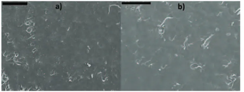
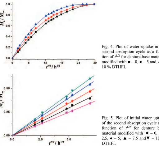
The plots of water uptake in the second absorption cycle for the denture base materials modified with DTHFI as a function of t1/2 / h1/2 are shown in Figs. 4
better clarity, curves are shown for only three samples. The plots were initially linear with the respect to t1/2 (Fig. 5), and thus, it could be concluded that the uptake was diffusion controlled.
Fig, 4. Plot of water uptake in the second absorption cycle as a func-tion of t1/2 for denture base material modified with ■ – 0, ● – 5 and ▲ – 10 % DTHFI.
Fig. 5. Plot of initial water uptake of the second absorption cycle as a function of t1/2 for denture base material modified with ◄ – 0, ■ – 2.5, ● – 5, ▲ – 7.5 and▼ – 10 % DTHFI.
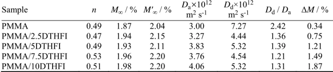
The results for the diffusion exponent (n), the weight loss (ΔM / %), the
maximum degree of absorption (M∞ / %) and desorption (M∞/ %), as well as the
calculated values of the diffusion coefficient for absorption (Da / m2 s–1) and
desorption (Dd / m2 s–1) are summarised in Table III for the denture base
mater-ials modified with DTHFI. The ratio Dd/Da is also presented, which is an index
of the degree of the concentration dependence of the diffusion coefficient.
Dd/Da> 1 indicates that the diffusion coefficient decreases with concentration.
Weight loss for the denture base materials (ΔM) involved a single
correlations of the total residual monomer content and weight loss as a function of DTHFI content were found (Fig, S-4 of the Supplementary material). Further-more, the slopes of these linear correlations were almost the same indicating that the weight loss in this kind of materials depended mostly on the total residual monomer content.
TABLE III. Kinetic parameter (n), weight loss (ΔM), the maximum degree of absorption (M∞) and desorption (M∞), and diffusion coefficients for absorption (Da) and desorption (Dd) for the studied denture base materials
Sample n M∞ / % M′∞ / % Dma×2 s10-112 Dmd×102 s-112 Dd / Da ΔM / %
PMMA 0.49 1.87 2.04 3.00 7.27 2.42 0.34
PMMA/2.5DTHFI 0.47 1.94 2.15 3.27 4.44 1.36 0.75 PMMA/5DTHFI 0.49 1.93 2.11 3.83 5.32 1.39 1.21 PMMA/7.5DTHFI 0.53 1.96 2.20 3.76 4.54 1.21 1.49 PMMA/10DTHFI 0.51 1.98 2.20 4.06 5.32 1.31 1.87
The thermal properties of PMMA denture base materials modified with DTHFI were investigated via differential scanning calorimetry (Fig. S-5 of the
Supplementary material). The commercial PMMA denture base material had a single glass transition temperature (Tg) at around 123 °C. The PMMA materials
modified with DTHFI also showed a single Tg indicating that copolymers were
homogeneous in the range of 10–30 nm.41 Substitution of a part of MMA with
DTHFI lowered the values of the glass transition temperature of the polymer. The greater the amount of DTHFI, the lower was the Tg. However, the observed
decrease in the Tg was not as pronounced as in the case of modification of PMMA
denture base materials with monomers such as dimethyl itaconate (DMI) and dibutyl itaconate (DBI).42 This could be attributed to the presence of the
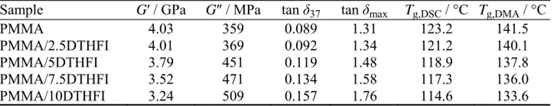
tetra-hydrofuran ring in the structure of DTHFI, which is a not so movable side group. During the use of a denture, it undergoes diverse tensions that appear at different places and have enormously wide magnitude of intensities. The most common reason for denture deterioration is due to its breakage, and therefore, it is of a great importance to investigate the dynamic-mechanical properties of new materials. The dependences of the storage modulus (G / GPa) on temperature for
The values of the storage modulus (G / GPa), loss modulus (G / MPa) and
damping factor (tan 37) at 37 °C, the damping factor (tan max) and glass
tran-sition temperatures calculated from DMA (Tg,DMA / °C) and DSC (Tg,DSC / °C)
measurements for the PMMA denture base materials modified with DTHFI are listed in Table IV.
TABLE IV. Dynamic-mechanical parameters at 37 °C and glass transition temperatures cal-culated from DMA (Tg,DMA) and DSC (Tg,DSC) measurements for the PMMA denture base materials modified with DTHFI
Sample G / GPa G / MPa tan 37 tan max Tg,DSC / °C Tg,DMA / °C
PMMA 4.03 359 0.089 1.31 123.2 141.5
PMMA/2.5DTHFI 4.01 369 0.092 1.34 121.2 140.1
PMMA/5DTHFI 3.79 451 0.119 1.48 118.9 137.8
PMMA/7.5DTHFI 3.52 471 0.134 1.58 117.3 136.0 PMMA/10DTHFI 3.24 509 0.157 1.76 114.6 133.6
The “ideal” material for a denture base should exhibit great rigidity and strength (large G), moderate deformation under the stress (G ) and the ability to
return to the previous shape shortly after removal of a load (low tan ). It could
be noticed in Table IV that increasing the DTHFI content in the PMMA denture base materials led to decreases in the values of the storage modulus and increases in the loss modulus and tan . These results indicated reduced stiffness and inc-reased deformation under the load in the case of the materials modified with DTHFI, which was due to the plasticizing effect of the side group of the emp-loyed itaconate. Despite the noticed effect, the addition of small amounts of DTHFI (see sample with 2.5 wt. % of DTHFI) caused only negligible decreases in G′ (< 1 %) and ensured the same dynamic-mechanical properties as those of
the commercial PMMA denture base material.
It could be noticed that the Tg values determined from DMA measurements,
as temperature corresponding to the maximal value of damping factor (tan max),
were somewhat higher than those obtained by DSC measurement, but the trend in the change in Tg was the same for both methods. A similar observation was
reported earlier.42 The differences in the Tg values arise from the fact that the
Tg,DSCrepresents the temperature at which the material undergoes the maximum
change in polymer chain mobility, which corresponds to the chemical definition of the Tg, while Tg,DMA describes the damping characteristics of the material and
has historical significance.
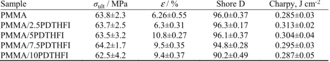
elon-gation at break ( / %), the Shore D hardness and the Charpy impact strength were determined and are listed in Table V.
TABLE V. Values of the ultimate tensile strength (σult), elongation at break ( ), Shore D
hardness and Charpy impact strength of the investigated denture base materials
Sample σult / MPa ε / % Shore D Charpy, J cm-2
PMMA 63.8±2.3 6.26±0.55 96.0±0.37 0.285±0.03
PMMA/2.5PDTHFI 63.7±2.5 6.3±0.31 96.3±0.17 0.313±0.02 PMMA/5PDTHFI 63.5±3.2 10.8±0.27 96.1±0.37 0.304±0.04 PMMA/7.5PDTHFI 64.2±1.7 9.5±0.35 94.8±0.28 0.295±0.03 PMMA/10PDTHFI 62.5±4.2 9.4±0.37 90.2±0.49 0.287±0.05
The tensile properties of modified denture base materials were investigated on an Instron testing machine. Modification of commercial PMMA denture base material with DTHFI gave materials with slightly lower values of the ultimate tensile strength. On the other hand, the modified materials elongated more at break. This behaviour could be explained by the fact that the DTHFI molecule possesses an oxygen atom in its structure that might establish hydrogen bonds with hydrogen atoms. Formed H-bonds were very weak and had no effect on the values of stress at break but affected the values of elongation at break. It should be noted that for all samples, the values of tensile strength were within the framework of the prescribed standards.43
The mean values of the Shore D hardness for PMMA denture base materials modified with DTHFI ranged between 90.2 and 96.3 and thus fulfilled the required hardness values for denture base materials, as prescribed by the Amer-ican Dental Standards Institute.43 Bearing in mind that many material properties,
such as composition, surface porosity, residual monomer concentration, etc.,
affect its hardness, it could be concluded that investigated modifications of PMMA denture base material did not have an effect on the Shore D hardness.
Inclusion of DTHFI in commercial PMMA denture base formulation led to increase in the impact resistance of prepared samples (Table V) except in the case of the sample with the highest DTHFI content (10 wt. %). It was observed in all impact tests experiments that the specimens broke with a sharp fracture, exhibiting typical brittle fracture behaviour characterized by a lack of distortion of the broken parts.
CONCLUSIONS
and, therefore, made the base material significantly more biocompatible and drastically reduced the risk of a variety of immune responses. The glass transition temperatures of the synthesized samples were shifted to lower values, indicating that the side groups of DTHFI acted as plasticizers. By DMA, it was found that increasing the DTHFI content led to a lowering the values of the storage modulus and stress at break. However, analysis of the mechanical properties showed that all the modified materials possessed characteristics prescribed by ADA stan-dards, and could be used in practice. The magnitude of the measured values indi-cated that the PMMA denture base materials modified with DTHFI could be developed into a less toxic, more environmentally and patient-friendly product than commercial pure PMMA denture base material. The optimal mechanical properties were exhibited by the sample with the minimal DTHFI modification (2.5 wt. %).
SUPPLEMENTARY MATERIAL
The geometry of the specimens and additional analysis are available electronically from http://www.shd.org.rs/JSCS/, or from the corresponding author on request.
Acknowledgements. The authors acknowledge funding from the Ministry of Education, Science and Technological Development of the Republic of Serbia, Project No. 172062 “Syn-thesis and characterization of novel functional polymers and polymeric nanomaterials”.
И З В О Д
ПОЛИ(МЕТИЛМЕТАКРИЛАТНИ) МАТЕРИЈАЛИЗАБАЗУПРОТЕЗАМОДИФИКОВАНИ ДИТЕТРАХИДРОФУРФУРИЛ-ИТАКОНАТОМ: СВОЈСТВАВАЖНАЗАПРИМЕНУ
ПАВЛЕСПАСОЈЕВИЋ1, ВЕСНАПАНИЋ1, САЊАШЕШЛИЈА2, ВЛАДИМИРНИКОЛИЋ3, ИВАНКАГ. ПОПОВИЋ4
иСАВАВЕЛИЧКОВИЋ4
1Иновационицентар, Технолошко–металуршкифакултет, УниверзитетуБеограду, Карнегијева 4,
11000 Београд, 2Институтзахемију, технологијуиметалургију, УниверзитетуБеограду, Његошева
12, 11000 Београд, 3Иновационицентар, Хемијскифакултет, УниверзитетуБеограду, Студентски
трг 12–16, 11000 Београди4Технолошко–металуршкифакултет, УниверзитетуБеограду,
Карнегијева 4, 11000 Београд
имали задовољавајуће механичке карактеристике. Нађено је да са повећањем удела DTHFI долазидосмањењавредноститемпературеостакљивања, модуласачуванеенер -гије, напонакидањаиударнежилавости, међутиммеханичкекарактеристикесуидаље уграницамапрописанимАDАстандардиматакодасеновиматеријалимогукористити упракси.
(Примљено 23. јануара, ревидирано 9. априла, прихваћено 22. априла 2015)
REFERENCES 1. J. F. McCabe, R. M. Basker, Br. Dent. J. 140 (1976) 347 2. C. Y. K. Lung, B. W. Darvell, Dent. Mater. 21 (2005) 1119 3. D. C. Smith, Brit. Dent. J. 111 (1961) 9
4. A. Moshaverinia, N. Roohpour, S. Ansari, M. Moshaverinia, S. Schricker, J. A. Darr, I. U. Rehman, Dent. Mater. 25 (2009) 1240
5. A. Moshaverinia, S. Ansari, Z. Movasaghi, R. W. Billington, J. A. Darrhtesham, U. Rehman, Dent. Mater. 24 (2008) 1381
6. K. Kinashita, J. Chem. Soc. 50 (1929) 583
7. B. Axelsson, G. Nyquist, Odontol. Revy 27 (1962) 370
8. J. K. Anusavice, Phillips’ Science of Dental Materials, W. B. Saunders Co., Philadelphia, PA, 2003, p. 176
9. D. C. Smith, M. E. Bains, Br. Dent. J. 98 (1955) 55
10. B. E. Tate, Vinyl and Diene Monomers, Wiley-Interscience, New York, 1979, p. 205 11. M. Fernandez-Garcia, E. L. Madruga, Polymer 37 (1996) 263
12. E. L. Madruga, M. Fernandez-Garcia, Polymer 35 (1994) 4437 13. M. Fernandez-Garcia, E. L. Madruga, Polymer 38 (1997) 1367
14. M. Fernandez-Garcia, J. L. Fuente, E. L. Madruga, Polym. Eng. Sci. 41 (2001) 1616 15. J. Velickovic, S. Vasovic, Makromol. Chem. 153 (1972) 207
16. ISO 20795-1:Dentistry -- Base polymers -- Part 1: Denture base polymers, 2013 17. G. Giavaresi, E. B. Minelli, M. Sartori, A. Benini, A. Parrilli, M. C. Maltarello, F.
Sala-manna, P. Torricelli, R. Giardino, M. Fini, J. Mater. Sci.: Mater. Med. 23 (2012) 1247 18. J. Zheng, Q. Su, C. Wang, G. Cheng, R. Zhu, J. Shi, K. Yao, J. Mater. Sci.: Mater. Med.
22 (2011) 1063
19. M. I. Shtilman, Polymeric Biomaterials, VSP BV, Utrecht, 2003, p. 47
20. S. Shen, W. Kiong, Z. Shi, L. Chia, K. G. Neoh, R. B. H. Tan, J. Mater. Sci.: Mater. Med.22 (2011) 2283
21. W. I. Higuchi, T. Higuchi, J. Am. Pharm. Assoc. Sci. Ed. 49 (1960) 598
22. R. W. Korsmeyer, R. Gurny, E. Doelker, P. Buri, N. A. Peppas, Int. J. Pharm. 15 (1983) 25 23. J. Crank, The Mathematics of Diffusion, Oxford University Press, London, 1999, p. 203 24. M. F. Burrow, S. Inokoshi, J. Tagami, Am. J. Dent. 12 (1999) 295
25. D. T. Turner, Polymer 28 (1987) 293
26. G. Bayraktar, B. Guvener, C. Bural, Y. Uresin, J. Biomed. Mater. Res., B 76 (2006) 340 27. P. K. Vallittu, V. Miettinen, P. Alakuijala, Dent. Mater. 11 (1995) 338
28. S. Baker, S. C. Brooks, D. M. Walker, J. Dent. Res. 67 (1988) 1295 29. C. Y. K. Lung, B. W. Darvell, Dent. Mater. 23 (2007) 88
30. K. Sato, K. Kodama, (Fuji Photo Film Co., Ltd) US 6,777,160 (2004)
31. K. Shirakawa, Y. Adegawa, S. Yasunami, (Fuji Photo Film Co., Ltd) US 6,773,862 (2004)
33. T. Watanabe, E. Ito, S. Tanikawa, S. Ichinohe, T. Yamazaki, M. Lamrani, (Menicon Co., Ltd.) US 6,770,728 (2004)
34. E. R. Lukenbach, C. Kaminski, S. Pascal-Suisse, M. Tahar, M. Ruggiero, (Johnson & Johnson Consumer Companies, Inc.) US 6,762,158 (2004)
35. P. Spasojevic, D. Stamenkovic, R. Pjanovic, N. Boskovic-Vragolovic, J. Dolic, S. Grujic, S. Velickovic, Polym. Int. 61 (2012) 1272
36. P. Neogi, Diffusion in polymers, Marcel Dekker, New York, 1996, p. 184
37. E. P. Lautenschlager, S. I. Stupp, J. C. Keller, Functional behavior of orthopedic bio-materials, CRC Press, Boca Raton, FL, 1984, p. 206
38. S. H. Mohamed, Alb. M. Al-Jadi, T. Ajaal, J. Phys. Sci. 19 (2008) 127 39. P. Pfeiffer, E. U. Rosenbauer, J. Prosthet. Dent. 92 (2004) 72
40. SIDS Initial Assessment Profile CAS No. 97-65-4, JETOC–Japan Chemical Industry Ecology-Toxicology and Information Center, Tokyo; Inchem, http://www.in-chem.org/documents/sids/sids/97654.html (29.09.2015)
41. L. A. Utracki, Polymer alloy and blends, Hanser Publishers, Munich, Germany, 1989, p. 189
42. R. J. Sayler, Assignment of the glass transition, American Society for Testing and Materials, Philadelphia, PA, 1994, p. 114