Original Research
Genipin cross-linked electrospun chitosan-based nanofibrous mat as tissue
engineering scaffold
Esmaeil Mirzaei1, Reza Faridi-Majidi1*, Mohammad Ali Shokrgozar2, Farnoush Asghari Paskiabi1
1
Department of Medical Nanotechnology, School of Advanced Medical Technologies, Tehran University of Medical Sciences, Tehran, Iran
2
National Cell Bank of Iran, Pasteur Institute of Iran, Tehran, Iran
Abstract
Objective(s):To improve water stability of electrospun chitosan/ Polyethylene oxide (PEO) nanofibers, genipin, a biocompatible and nontoxic agent, was used to crosslink chitosan based nanofibers.
Materials and Methods: Different amounts of genipin were added to the chitosan/PEO solutions, chitosan/PEO weight ratio 90/10 in 80 % acetic acid, and the solutions were then electrospun to form nanofibers. The spun nanofibers were exposed to water vapor to complete crosslinking. The nanofibrous membranes were subjected to detailed analysis by scanning electron microscopy (SEM), Fourier transform infrared-attenuated total reflection (FTIR-ATR) spectroscopy, swelling test, MTT cytotoxicity, and cell attachment.
Results: SEM images of electrospun mats showed that genipin-crosslinked nanofibers retained their fibrous structure after immerging in PBS (pH=7.4) for 24 hours, while the uncrosslinked samples lost their fibrous structure, indicating the water stability of genipin-crosslinked nanofibers. The genipin-genipin-crosslinked mats also showed no significant change in swelling ratio in comparison with uncrosslinked ones. FTIR-ATR spectrum of uncrosslinked and genipin-crosslinked chitosan nanofibers revealed the reaction between genipin and amino groups of chitosan. Cytotoxicity of genipin-crosslinked nanofibers was examined by MTT assay on human fibroblast cells in the presence of nanofibers extraction media. The genipin-crosslinked nanofibers did not show any toxic effects on fibroblast cells at the lowest and moderate amount of genipin. The fibroblast cells also showed a good adhesion on genipin-crosslinked nanofibers.
Conclusion: This electrospun matrix would be used for biomedical applications such as wound dressing and scaffold for tissue engineering without the concern of toxicity.
Keywords:Chitosan, Cytotoxicity, Electrospinning, Genipin, Structural stability
*Corresponding author: Reza Faridi-Majidi, Department of Medical Nanotechnology, School of Advanced Medical Technologies, Tehran University of Medical Sciences, Tehran, Iran.
Introduction
Electrospun nanofibers have attracted many interests for different applications in recent years because of their unique properties such as high surface to volume ratio and high porosity (1). These nanofibers have been studied for various applications including membranes and filtration (2), sensors (3) and biomedical applications such as drug delivery (4), wound dressing (5), tissue engineering, (6) and biosensing (7). Among many synthetic and natural polymers electrospun to nanofibers, chitosan has attracted many interest especially for biomedical applications owing to its unique properties. Chitosan is a biopolymer composed of repeated β-D-glucosamine and N-acetyl-β -D-glucosamine monomers and is obtained from deacetylation of chitin, the second most abundant natural polymer in the world. Chitosan has good biocompatibility and biodegradability as well as various biofunctionalities including antithrombo-genic, hemostatic, and wound healing properties (8). Chitosan-based electrospun nanofibers have shown potential for many biomedical applications owing to their
structural similarity to
glycosaminoglycans, a component of extra cellular matrix (ECM), and morphological proximity to fibrous collagen structures in the ECM at the scale of nanometers (50– 500 nm in diameter). The use of nanofibrous chitosan matrices is thus expected to mimic the natural ECM, in which cells can attach, proliferate, and differentiate (8, 9). Despite these desirable features, chitosan-based electrospun nanofibers are not stable in biological medium and they easily swell and lose their fibrous structure in contact with water. Therefore, chitosan-based nanofibers need to be crosslinked to maintain their structural integrity. Glutaraldehyde (GA) is a common reagent used for crosslinking electrospun chitosan nanofibers (10). However, GA is a toxic reagent and it is not suitable for
to crosslink pure chitosan nanofibers by genipin using TFA as solvent (21, 22). In one approach by Marjorie et al, genipin was added into chitosan/ TFA solution and mixed for 2 min immediately before electrospinning (21). In another approach by Frohbergh et al, chitosan nanofibers were first electrospun from a solution of chitosan dissolved in TFA and then the electrospun fibers were crosslinked with 0.1% (w/v) genipin dissolved in 1X PBS for 24 h (22).
In this study, a modification of Marjorie et al. approach was used to crosslink chitosan/PEO nanofibers with genipin. Crosslinking processes were done by adding various amounts of genipin into chitosan/PEO acetic acid solutions prior to electrospinning and then exposure of obtained nanofibers to water vapor at 30 °
C for 24 h. The morphology, structure, water stability, swelling behavior, and cytotoxicity of genipin-crosslinked nanofibers were investigated as well and compared with those of cross-linked by GA.
The attachment of human fibroblast cells were also investigated on genipin-crosslinked nanofibers.
Materials and Methods
Materials
Chitosan (CS) (low molecular weight, degree of deacetylation 91.2 %) was purchased from Easter Groups (DongChen Co., Ltd, China). Polyethylene oxide (PEO) (MW 900 kD) was purchased from Acros Organics Co. Glacial acetic acid was purchased from Merck Chemical. Genipin (GP), methyl-2-hydroxy-9-(hydroxymethyl)-3-oxabicyclonona-4, 8-diene-5-carboxylate, was obtained from Challenge Bioproducts Co. Ltd. (Touliu, Taiwan). Glutaraldehyde was purchased from Panreac (Spain). RPMI medium and zolium bromide) from Sigma-Aldrich, USA. Human fibroblast (AGO-1522) were
fetal bovine serum, FBS, were purchased from Gibco, USA and MTT (3-[4, 5-dimethylthiazol-2-yl]-2, 5-diphenyl-tetra
Preparation of chitosan/PEO nanofibers Chitosan solution (3.0 Wt. %) and PEO solution (3.0 Wt. %) were prepared separately by dissolving chitosan and PEO powders in aqueous acetic acid (80 V/V %) under magnetic stirring at 37 °C for 24 h.
The obtained solutions were then mixed together in weight ratio of CS/PEO, 90/10, as the required electrospinning solution with 3.0 Wt. % of total solid.
The electrospinning processes were carried out using Electroris (FNM, Tehran, Iran). For every run, the polymer solution was placed into a 5 mL plastic syringe with a blunt-ended 18 G stainless steel needle. An aluminum foil was wrapped on the Electroris rotating drum as collector and was located at the distance of 12 cm from the needle.
A syringe pump fed the solution to the needle tip at the injection rate of 3.0 ml/h. The wire of a DC positive high voltage was connected to the metallic needle and the collector to the ground.
The applied voltage, drum speed, and electrospinning time were fixed at 20 kV, 200 rpm, and 1 h, respectively.
Crosslinking of chitosan nanofibers by genipin and glutaraldehyde
To obtain genipin-crosslinked chitosan nanofibers, 10 w/v % genipin solutions were prepared by dissolving genipin powder in pure ethanol.
Four different volumes of the genipin solution were added to a constant amount of the 3 Wt. % polymer solutions (CS/PEO, 90/10) at room temperature with constant stirring for 5 minutes.
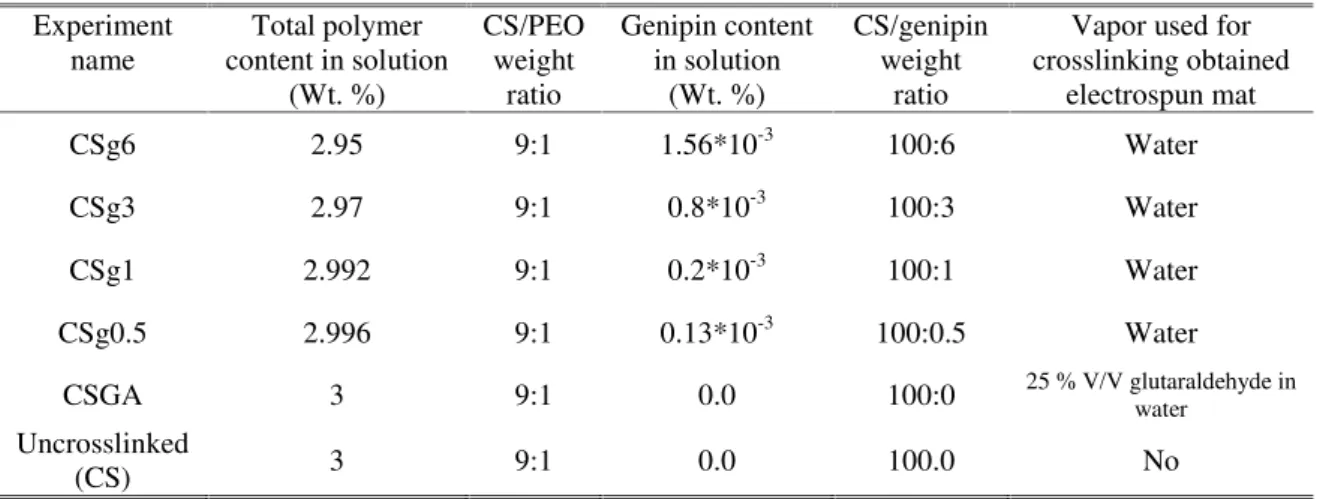
Table 1. Recipe used for preparing crosslinked and uncrosslinked chitosan nanofibers.
These solutions were immediately electrospun under conditions described in previous section. The obtained genipin-contained mats were removed from the collector and immediately exposed to water vapor in a desiccator at 30 °C. To produce water vapor in the desiccator, a glass Petri dish was filled with 5 ml of distilled water and placed on the bottom of the desiccator. After 24 hours, the mats were washed with ethanol and then were dried at 37°C for 24 h, and maintained for further analysis. The CS/PEO electrospun mat was also crosslinked with GA as control using a common method (25). The electrospun chitosan mat (without any added genipin) were exposed to vapor of 25 w/v % GA aqueous solution in desiccator at 30°C for 24 h. The resultant mat was then dried at 37°C for 24 h, and maintained for further analysis.The recipe used in this work for preparing chitosan crosslinked nanofibers is given in Table 1.
Characterization of chitosan nanofibers The size and morphology of produced nanofibrous mats were analyzed using scanning electron microscopy (SEM) (ZEISS DSM 960A Oberkochen, Germany). A small section of each nanofibrous mat was sputtered with a thin layer of gold and then analyzed by SEM. The Fourier transform infrared attenuated total reflection spectroscopy (FTIR-ATR)
instrument (Equinox 55, Bruker, Germany) in the range of 2500–500 cm-1. The water stability of nanofibers was investigated by observing SEM images of electrospun nanofibers before and after immerging in PBS (pH=7.4) at room temperature for 24 h.
The swelling degree of electrospun mats was determined by a gravimetric method. The electrospun mats were immerged in PBS (pH=7.4) at room temperature for 24 h. The samples were then taken out, the excess surface water was removed by filter paper, and the swelled mats were weighed. The swelling ratio was measured as following equation:
swelling ratio(%)=Ws-W0 W0
×100
Where Ws denotes the weight of the swelled mat and W0 denotes the weight of the mat in its dry state after 24 immerging in water.
MTT assay and cell viability
Cytotoxicity of the crosslinked mats was
examined by MTT
(3-(4,5-
Dimethylthiazol-2-yl)-2,5-diphenyltetrazolium bromide, a yellow tetrazole) assay of cell viability according to ISO 10993-5 standard test method. The electrospun mats were cut into 1 ͯ 1 cm square and sterilized by UV irradiation for
Experiment name
Total polymer content in solution
(Wt. %)
CS/PEO weight
ratio
Genipin content in solution
(Wt. %)
CS/genipin weight
ratio
Vapor used for crosslinking obtained
electrospun mat
CSg6 2.95 9:1 1.56*10-3 100:6 Water
CSg3 2.97 9:1 0.8*10-3 100:3 Water
CSg1 2.992 9:1 0.2*10-3 100:1 Water
CSg0.5 2.996 9:1 0.13*10-3 100:0.5 Water
CSGA 3 9:1 0.0 100:0 25 % V/V glutaraldehyde inwater
Uncrosslinked
and 2 ml of culture medium (RPMI) was added into each sample. The samples were then incubated at 37 °C for 3 incubation time intervals, i.e., 1, 3 and 6 days, and the extracts of each sample were collected for cell culture. Fresh culture medium was used as negative control. Human fibroblasts (AGO-1522) were seeded in 96-well tissue culture plate at a density of 1.2 ͯ 104 cells/well in RPMI medium supplemented with 10% fetal bovine serum, and incubated at 37 °C under 5% CO2/95% air condition for 24 h. After that, the culture medium was removed and replaced with the as-prepared extraction medium, supplemented with 10% fetal bovine serum and incubated for another 24 h, then the extraction medium was removed and 100 µL MTT solution (0.5 miligram/1 mililiter) was added to each well. After incubation at 37 °C and 5% CO2 for 4 h, the MTT solution was removed and 100 µL isopropanol was added to dissolve formazan crystals, formed by mitochondrial succinate dehydrogenase of alive cells from MTT. After 20 min incubation at 37 °C, the optical density of the formazan solution was detected by an ELISA reader (AWARENESS TECHNOLOGY, Stat fax-2100, USA) at 570 nm of wavelength. The mean absorption of each sample (10 well per each experiment) was divided to mean absorption of the control to calculate the percent of cell viability for each sample. The test was repeated 5 times for each sample.
Cell seeding and adhesion
The genipin-crosslinked mat (CSg1) were cut into 1 ͯ 1 cm square and sterilized by UV irradiation for 2 h. The samples were then washed three times with sterile PBS prior to transfer to individual 12-well tissue culture plates. The number of 15000 human fibroblasts (AGO-1522), suspended in RPMI, were seeded on the samples, cultured for 3 h and then 1 ml of RPMI supplemented with 10% FBS were added to each well. After 24 h of culture, cellular
constructs were harvested, rinsed twice with PBS to remove nonadherent cells, and subsequently fixed with 2.5 % glutaraldehyde at 4 °C for 4 h. After that, the samples were dehydrated through a series of graded ethanol solutions and air-dried overnight. Dry samples were sputtered with gold for observation of cell morphology on the surface of the scaffolds by SEM.
Statistical analysis
Analysis of variance (ANOVA) was used to compare the swelling ratio of chitosan nanofibers. The number of samples for each group was 5 and the data were represented as mean ± SD. The significance was considered as P.value < 0.05. The data of MTT assay were analyzed using one sample t test with bonferroni correction and differences from control were considered statistically significant atP.value < 0.01.
Results and Discussion
Appearance and morphology of mats In this work, genipin was used as a safe and biocompatible crosslinking agent instead of toxic but common crosslinking agent of chitosan nanofibers (i.e., glutaraldehyde).
The polymer solutions with different content of genipin (i.e., CSg.5, CSg1, CSg3, and CSg6) were electrospun and subsequently, the mats were immediately exposed to water vapor at 30°C for 24 h. Gross changes in the color of the samples after being exposed to water vapor were observed. The digital photographs of these mats compared to uncrosslinked (CS) and GA-crosslinked (CSGA) mats are shown in Figure 1.
The color of genipin-crosslinked chitosan mats changed from white to bluish green and dark blue depending on the genipin content (Figure 1. CSg.5, CSg1, CSg3 and CSg6), While, the color of GA-crosslinked mats (CSGA) turned from white to brown yellowish (Figure 1. CSGA).
(CSg6, CSg3) represented dark bluish color in comparison with mats with lower genipin content (CSg1, CSg.5) displaying bluish green color.
These color changes are associated with chitosan derivatives produced by the reaction of genipin with amino groups of chitosan (12). No color change were observed for the genipin contained nanofibers which not exposed to water vapor (data are not shown).
The morphology of the electrospun mats was investigated through the SEM micrographs.
The uncrosslinked chitosan mat had continuous and uniform nanofibrous structure (Figure 2a).
After crosslinking by various amount of genipin the total fibrous structure of the chitosan mats were kept constant, but there were a few structural deformations as shown in Fig. 3a, b, c, d, where a, b, c, and d are corresponded to Csg.5, CSg1, CSg3, and CSg6, respectively.
In comparison with uncrosslinked mats, the genipin-crosslinked mats showed fusion of adjacent nanofibers. On the other hand, crosslinking by glutaraldehyde caused no remarkable change in morphology of chitosan nanofibers (Figure 3e).
Figure1. digital photographs of uncrosslinked (CS), GA-crossliked (CSGA) and genipin-crossliked (CSg6,CSg3, CSg1, CSg.5) electrospun chitosan mats.
Figure 2.SEM images of uncrossliked nanofibers, (a) before and (b) after immerging in PBS (pH=7.4).
Figure 3. SEM images of genipin-corsslinked and GA-crosslinked chitosan nanofibers before (a, b, c, d, e) and after (a', b', c', d', e') immerging in PBS (pH=7.4).
Water stability and swelling behavior of electrospun mats
structure stable in aqueous binding amino groups of chit is necessary to mention that water vapor is essential contained nanofibers to retain structure after immerging in other words, the genipin nanofibers which were not water vapor lost their fibrous after immerging in water (da shown). To investigate absorption capability of nanofibers, the swelling b nanofibers was studied. The m ratio (%) related to the absorption capability.
The swelling ratio (%) electrospun mats is stated in Fi It can be seen that the swel chitosan mats did not significantly using genipin as agent, while crosslinking by the swelling ratio signi comparison with uncrossli (P.value < 0.05).
So, unlike crosslinking crosslinking by genipin does the water absorption capability nanofibers.
FTIR-ATR spectroscopy When CS systems are crossl genipin, conformational chang a result of structural rearra chains to form covalent bonds To investigate the chemical cha chitosan after crosslinking by g The FTIR-ATR analysis of unc and genipin-crosslinked chitosa nanofibers was carried out as show Figure 5.
Comparing with FTIR spe uncrosslinked chitosan nanof spectrum of genipin-crosslinke nanofibers revealed the crossl chitosan by genipin (Figure5). The C=O stretching band amide I at 1665 cm−1 (28) be after crosslinking due to over
ous media by hitosan (12). It hat exposure to l for genipin in their fibrous in water. In pin contained not exposed to brous structure (data are not the water of crosslinked behavior of more swelling he more water
of chitosan n Figure 4.
elling ratio of not decrease as crosslinking y GA reduced gnificantly in osslinked mats
ng by GA, does not decrease lity of chitosan
osslinked with hanges occur as rrangement of bonds (27).
changes of y genipin, uncrosslinked tosan
s shown in
spectrum of nofibers, FTIR sslinked chitosan rosslinking of 5).
nd of chitosan ) became broad overlapping with
C=C stretching in cy genipin at 1628 cm− 1 (2
Figure 4. Swelling ratio o crosslinked chitosan nanofibe in water. * represents signif uncrosslinked CS (P.value < as mean ± SD (n=5).
Figure 5. FTIR-ATR spectr and genipin-crosslinked (C CSg6) chitosan nanofibers.
The amino groups with genipin and form a heterocyclic amine in ge chitosan network (12). T absorption band of new 1650 (12) again overl absorption band of chito become broader. A new was appeared around crosslinking by genipi presence of ring-stretchi amine (12).The C‒ N st III at 1233 cm-1 (27) shi after crosslinking by geni The peak at 1740 cm−1
cyclic structure of 1 (20).
of uncrosslinked and ibers after 24 immerging nificant difference from < 0.05). Data are shown
ectrum of uncrosslinked (CSg.5, CSg1, CSg3, .
oups of chitosan react amide linkage and n genipin-crosslinked . The C=O stretching ew formed amide at overlaps with amid I hitosan and makes it new broad peak that ound 1415 cm-1 after nipin, indicated the ching of heterocyclic stretching of amide shifted to 1260 cm-1 enipin.
−1
the presence of carboxylic electrospun chitosan fi disappeared in the genipin chitosan fibers. This can be due of carboxylic groups from ge nanofibers when they expose vapor.
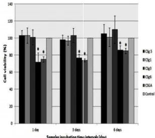
Cytotoxicity of crossliked nan The cell viability test using MT to evaluate cytotoxicity of the chitosan mats. Figure 6 re percent of cell viability after c cells with extraction media three different incubation time each sample (1, 3, and 6 days) were represented as mean ± S the chitosan mats crossl glutaraldehyde (CSGA), the c was significantly lower than control for all incubation tim (P.value < 0.01). The cell via sample was around 80 %. CSg1, and CSg3 the cell viabi similar to those of the cont culture medium), around 100 % However, The CSg6 showed lower cell viability (around 80 control (P.value < 0.01). Thi was seen for all incubation tim The lower cell viability of C attributed to the excess amount genipin that may not react w and released to the me incubation of samples in t From Figure 6, it could be a that for all crosslinked sampl viability was not decreased b incubation time of samples. This result implies that toxic released at first 24 h incubation after that no more toxic released by increasing incuba (from 1 day to 3 and 6 days) resulted from MTT assay that chitosan nanofibers with lowe genipin (3 Wt. % or lower b chitosan) cause no cytotoxicity
lic groups in fibers (29) pin-crosslinked due to the exit genipin-added pose to water
anofibers MTT was used
the crosslinked represents the er culturing the dia of mats for me intervals for ys). The values SD (n=5). For osslinked by he cell viability han that of the on time intervals viability of this . For CSg.5, ability (%) was control (fresh 100 %.
ed significantly nd 80 %) than the This difference n times.
of CSg6 can be ount of residual t with chitosan medium after n the medium. also observed mples, the cell d by increasing
ic agents were ubation time, and c agent were ncubation time ys). It could be at crosslinking ower amount of r by weight of city.
Figure 6.Cell viability of fib extraction media of crosslink in three incubation time inter * indicates significant differ (P.value < 0.01). Data are s (n=5).
Figure 7.SEM images of fib seeded on nanofibrous me crosslinked (CSg1) after 24 h
Cell adhesion
The genipin-crosslinke membrane (CSg1) was adhesion study. Figure images of fibroblasts gr linked nanofibers after Cells were attached to discrete filopodia and e morphology on the surfa to the large surface area attachment.
Conclusion
The chitosan/PEO successfully crosslin biocompatible and nontox
fibroblasts in presence of nked chitosan nanofibers tervals (1, 3, and 6 days). ference from the control e shown as mean ± SD
fibroblasts (AGO-1522) membrane of genipin-4 h culture.
inked nanofibrous as used for cell
ure 7 shows SEM s grown on the cross-er 24 h cell culture. to the surfaces by nd exhibited a normal surface, which was due rea available for cell
agent, genipin, which leads to structural stability of chitosan nanofibers in aqueous media. In addition, the water absorption capability of chitosan nanofibers was not decreased after crosslinking by genipin. Genipin-crosslinked nanofibers also showed no cytotoxicity unless at high amount of genipin. Because of no cytotoxicity and bio compatibility of genipin-crosslinked chitosan nanofibers, these nanofibers are suggested for biomedical application such as tissue engineering and wound dressing instead of glutaraldehyde-crosslinked nanofibers.
Acknowledgements
This research was supported by Tehran University of Medical Sciences grant No. 89-04-87-12028.
References
1. Bhardwaj N, Kundu SC. Electrospinning: a fascinating fiber fabrication technique. BiotechnolAdv. 2010; 28(3):325-347. 2. Lala NL, Ramaseshan R, Bojun L,
Sundarrajan S, Barhate RS, Ying-jun L, Ramakrishna S. Fabrication of nanofibers with antimicrobial functionality used as filters: protection against bacterial contaminants. Biotechnol Bioeng. 2007; 97(6): 1357-1365.
3. Piperno S, Passacantando M, Santucci S, Lozzi L, La Rosa S. WO nanofibers for gas sensing applications. J Appl Phys.
2007; 101:124504. Available from URL:
doi: 10.1063/1.2748627.
4. Kim K, Luu YK, Chang C, Fang D, Hsiao BS, Chu B , Hadjiargyrou M. Incorporation and controlled release of a hydrophilic antibiotic using poly(lactide-co-glycolide)-based electrospun nanofibrous scaffolds. J Control Release.
2004; 98(1):47-56.
5. Rho KS, Jeong L, Lee G, Seo B-M, Park YJ, Hong S-D, Roh S, Cho JJ, Park WH, Min B-M. Electrospinning of collagen nanofibers: Effects on the behavior of normal human keratinocytes and early-stage wound healing. Biomaterials. 2006; 27(8): 1452-1461.
6. Inoguchi H, Kwon IK, Inoue E, Takamizawa K, Maehara Y, Matsuda T. Mechanical responses of a compliant electrospun poly(l-lactide-co-[epsilon]-caprolactone) small-diameter vascular
graft. Biomaterials. 2006; 27(8):
1470-1478.
7. Wang Z-G, Wan L-S, Liu Z-M, Huang X-J , Xu Z-K. Enzyme immobilization on electrospun polymer nanofibers: An overview. J Mol Catal B: Enzym. 2009; 56(4): 189-195.
8. Lee KY, Jeong L, Kang YO, Lee SJ, Park WH. Electrospinning of polysaccharides for regenerative medicine. Adv Drug Del
Rev. 2009; 61(12):1020-1032.
9. Jayakumar R, Prabaharan M, Nair S , Tamura H. Novel chitin and chitosan nanofibers in biomedical applications. Biotechnol Adv. 2010; 28(1):142-150. 10. Vondran JL, Sun W, Schauer CL.
Crosslinked, electrospun chitosan– poly(ethylene oxide) nanofiber mats. J Appl PolymSci. 2008; 109(2):968-975. 11. Speer DP, Chvapil M, Eskelson CD,
Ulreich J. Biological effects of residual glutaraldehyde in glutaraldehyde-tanned collagen biomaterials. J Biomed Mater
Res. 1980; 14(6):753-764.
12. Mi F-L, Sung H-W, Shyu S-S. Synthesis and characterization of a novel chitosan-based network prepared using naturally occurring crosslinker. J Polym Sci Part A: PolymChem. 2000; 38(15):2804-2814. 13. Ko C-S, Huang J-P, Huang C-W , Chu I
M. Type II collagen-chondroitin sulfate-hyaluronan scaffold cross-linked by genipin for cartilage tissue engineering. J BiosciBioeng. 2009; 107(2):177-182. 14. Sung HW, Huang RN, Huang LLH , Tsai
CC. In vitro evaluation of cytotoxicity of a naturally occurring cross-linking reagent for biological tissue fixation. J Biomater Sci PolymEd. 1999; 10(1):63-78.
15. Sung H-W, Huang R-N, Huang LLH, Tsai C-C, Chiu C-T. Feasibility study of a natural crosslinking reagent for biological tissue fixation. J Biomed Mater Res. 1998; 42(4): 560-567.
16. Muzzarelli RAA. Genipin-crosslinked chitosan hydrogels as biomedical and pharmaceutical aids. Carbohydr Polym.
2009; 77(1):1-9.
17. Yuan Y, Chesnutt BM, Utturkar G, Haggard WO, Yang Y, Ong JL , Bumgardner JD. The effect of cross-linking of chitosan microspheres with genipin on protein release. Carbohydr
Polym. 2007; 68(3):561-567.
19. Mekhail M, Wong KKH, Padavan DT, Wu Y, O'Gorman DB , Wan W. Genipin-cross-linked electrospun collagen fibers. J. Biomater Sci Polym Ed. 2011; 22(17): 2241-2259.
20. Zhang K, Qian Y, Wang H, Fan L, Huang C, Yin A, Mo X. Genipin-crosslinked silk fibroin/hydroxybutyl chitosan nanofibrous scaffolds for tissue-engineering application. J Biomed Mater ResA. 2010; 95A(3): 870-881.
21. Austero MS, Donius AE, Wegst UGK , Schauer CL. New crosslinkers for electrospun chitosan fibre mats. I. Chemical analysis. J R Soc Interface. 2012; 9(75): 2551-2562.
22. Frohbergh ME, Katsman A, Botta GP, Lazarovici P, Schauer CL, Wegst UGK, Lelkes PI. Electrospun hydroxyapatite-containing chitosan nanofibers crosslinked with genipin for bone tissue engineering.
Biomaterials. 2012; 33(36):9167–9178.
23. Pakravan M, Heuzey MC, Ajji A. A fundamental study of chitosan/PEO electrospinning. Polymer. 2011; 52(21): 4813-4824.
24. Jin J, Song M. Chitosan and chitosan– PEO blend membranes crosslinked by genipin for drug release. J. Appl. Polym.
Sci. 2006; 102(1):436-444.
25. Schiffman JD , Schauer CL. Cross-Linking Chitosan Nanofibers.
Biomacromolecules. 2006; 8(2):594-601.
26. Chen ZG, Wang PW, Wei B, Mo XM, Cui FZ. Electrospun collagen–chitosan nanofiber: A biomimetic extracellular matrix for endothelial cell and smooth muscle cell. Acta Biomater. 2010; 6(2): 372-382.
27. Silva SS, Motta A, Rodrigues MT, Pinheiro AFM, Gomes ME, Mano JF, Reis RL, Migliaresi C. Novel genipin-cross-linked chitosan/silk fibroin sponges for cartilage engineering strategies.
Biomacromolecules. 2008; 9(10):
2764-2774.
28. Harish Prashanth K, Kittur F, Tharanathan R. Solid state structure of chitosan prepared under different N-deacetylating conditions. Carbohydr Polym. 2002; 50(1): 27-33.