Plasmodium falciparum
Mating Patterns and
Mosquito Infectivity of Natural Isolates of
Gametocytes
Isabelle Morlais1,2*, Sandrine E. Nsango1,2,3, Wilson Toussile4, Luc Abate2, Zeinab Annan2, Majoline T. Tchioffo2, Anna Cohuet2, Parfait H. Awono-Ambene1, Didier Fontenille2, François Rousset5, Antoine Berry6,7
1Organisation de Coordination pour la lutte contre les Endémies en Afrique Centrale, Yaoundé, Cameroon, 2Institut de Recherche pour le Développement, Montpellier, France,3Faculté de Médecine et des Sciences Pharmaceutiques, Douala, Cameroon,4Université Paris-Sud 11, Villejuif, France,5Institut des Sciences de l'Évolution, Montpellier, France,6Centre Hospitalier Universitaire de Toulouse, Toulouse, France,7Centre de Physiopathologie de Toulouse Purpan, Toulouse, France
Abstract
Plasmodium falciparuminfections in malaria endemic areas often harbor multiple clones of
parasites. However, the transmission success of the different genotypes within the mosqui-to vecmosqui-tor has remained elusive so far. The genetic diversity of malaria parasites was mea-sured by using microsatellite markers in gametocyte isolates from 125 asymptomatic carriers. For a subset of 49 carriers, the dynamics of co-infecting genotypes was followed until their development within salivary glands. Also, individual oocysts from midguts infected with blood from 9 donors were genotyped to assess mating patterns. Multiplicity of infection (MOI) was high both in gametocyte isolates and sporozoite populations, reaching up to 10 genotypes. Gametocyte isolates with multiple genotypes gave rise to lower infection preva-lence and intensity. Fluctuations of genotype number occurred during the development within the mosquito and sub-patent genotypes, not detected in gametocyte isolates, were identified in the vector salivary glands. The inbreeding coefficientFiswas positively
correlat-ed to the oocyst loads, suggesting thatP.falciparumparasites use different reproductive
strategies according to the genotypes present in the gametocyte isolate. The number of par-asite clones within an infection affects the transmission success and the mosquito has an important role in maintainingP.falciparumgenetic diversity. Our results emphasize the
cru-cial importance of discriminating between the different genotypes within an infection when studying theA.gambiaenatural resistance toP.falciparum, and the need to monitor
para-site diversity in areas where malaria control interventions are implemented.
OPEN ACCESS
Citation:Morlais I, Nsango SE, Toussile W, Abate L, Annan Z, Tchioffo MT, et al. (2015)Plasmodium falciparumMating Patterns and Mosquito Infectivity of Natural Isolates of Gametocytes. PLoS ONE 10(4): e0123777. doi:10.1371/journal.pone.0123777
Academic Editor:Kristin Michel, Kansas State University, UNITED STATES
Received:December 16, 2014
Accepted:February 26, 2015
Published:April 14, 2015
Copyright:© 2015 Morlais et al. This is an open access article distributed under the terms of the
Creative Commons Attribution License, which permits unrestricted use, distribution, and reproduction in any medium, provided the original author and source are credited.
Data Availability Statement:All relevant data are within the paper.
Introduction
Plasmodium falciparumtransmission relies on its successful development within the mosquito
vector where fertilization occurs. However, studies on malaria parasite genetic structure have revealed different mating patterns in multiple epidemiological settings. Large deviations from panmixia were observed in malaria endemic areas and it has been argued that self-fertilization would favor transmission of better adapted strains of parasites. However, the question remains controversial and opened a large debate about the malaria parasite mode of reproduction [1–4]. Deciphering the complexity ofP.falciparumsexuality and its mating pattern would help understanding the disease’s epidemiology and predicting, for instance, the spread of drug resistance.
In malaria endemic areas, multiple genotypes are generally found within a single parasite isolate and the complexity of malaria infections has often impeded genetic studies onP. falcipa-rum[5,6]. On the vector side, the differential transmissibility ofP.falciparumclones to mos-quitoes is poorly known owing to the technical constraints for isolating gametocytes from trophozoites in the blood and for infecting a sufficient number of mosquitoes. However, modeling malaria transmission is now crucial to understand the evolution of parasites and vec-tors and to predict the long-term impact of global control measures.
In this study, we explored the infectiousness of gametocyte donors and investigated the ef-fect of multiplicities of inef-fection on the mosquito inef-fection success in regards to gametocyte densities. We then assessed the genetic diversity ofP.falciparumduring sporogony and charac-terized the parasites genetic structure from individual oocyst genotypes. The results show how the genetic composition of the gametocyte population impacts on the infection success of the mosquito and describe the important role of the insect vector in maintaining the parasite genetic diversity.
Materials and Methods
Ethics statement
Participants were enrolled upon signature of an informed consent by their legal guardian (par-ent or tutor having legal custody of the child). The experim(par-ental and cons(par-ent procedures were approved by the National Ethics Committee of Cameroon (protocol#039/CNE/MP/06).
Mosquito infections
The survey was conducted over two years, from 2007 to 2008, during the rainy seasons. Proce-dures for gametocyte carrier detection, blood collection and mosquito infections were per-formed as previously described [7]. Briefly,P.falciparumgametocyte carriers were identified among asymptomatic children aged from 5 to 11 in primary schools from the Mfou district, 30 km from Yaoundé (Cameroon). Gametocyte densities read from thick blood smears were expressed as the number of parasites seen against 1000 leukocytes, assuming a standard con-centration of 8000 leukocytes perμl. Venous blood was drawn in heparinized Vacutainer tubes
in the antecubital fossa. Membrane feedings were set using donor’s blood with replacement of the serum by a non-immune AB serum. Our local laboratory strain ofA.coluzzii, named Ngousso, was used for mosquito feedings. Mosquitoes are reared at the laboratory under stan-dard insectary conditions (27 ± 2°C, 85 ± 5% RH, 12h light/dark).
Gametocyte isolation
Gametocytes were separated from 1 ml of serum-free blood using MACS columns, as previous-ly described [8], and the gametocyte pellet resuspended in 50μl of PBS. DNA extractions from
purified gametocytes were performed with DNAzol, (Molecular Research Center, Inc., Cincin-nati, OH, USA), and resuspended in 50μl of PBS. A 1μl volume of gametocyte DNAs was
sub-jected to whole-genome amplification (WGA) using the GenomiPhi V2 DNA Amplification Kit (GE HealthCare, Uppsala, Sweden) according to the manufacturer’s instructions, and re-suspended to a 100μl final volume. All samples were kept frozen at -20°C until further
processing.
Mosquito dissections and
P
.
falciparum
DNA isolation
The blood fed mosquitoes were processed at different stages ofP.falciparumdevelopment as follow: 1) to determine the success of infections, a batch of at least 20 mosquitoes was dissected on day 8 post feeding. The midguts were removed and stained in a 0.4% mercurochrome solu-tion and the developed oocysts were counted by light microscopy. The prevalence of infecsolu-tion was defined as the proportion of infected mosquitoes among the total number of dissected mosquitoes and the infection intensity as the number of oocysts perP.falciparum-positive mosquito. 2) for oocyst genotyping, the midguts were dissected on day 9 after the infected bloodmeal and then placed in 50μl of absolute ethanol. Individual oocysts were isolated from
rehydrated midguts as described in Annanet al. [2]. The DNeasy Blood and Tissue Kit (Qia-gen, Valencia, CA) was used to purifyP.falciparumDNA from each oocyst according to the manufacturer’s instructions except that elution was carried out with 50μl of buffer. 3) for
spo-rozoite genotyping, salivary glands were dissected at day 14 post infection and placed in 200μl
of DNAzol. DNA extractions were processed according to the protocol provided by the manu-facturer, and the pellet resuspended in 20μl of sterile water. AP.falciparumspecific PCR (PF1
5’-GGAATGTTATTGCTAACAC-3’and PF2 5’-AATGAAGAGCTGTGTATC-3’) was car-ried out on salivary gland DNAs to identify sporozoite positive samples, and DNAs from posi-tive samples from each blood donor were pooled.
P. falciparum
genotyping
Genetic polymorphism was assessed for six microsatellite loci according to Andersonet al. 2000 [9]. PCR reactions were processed as previously described [2]. The same protocol was used to amplify DNAs from gametocytes, oocysts and salivary glands, using 2μl of DNA. PCR
products were resolved on an ABI Prism 3100 DNA Genetic Analyzer (Applied Biosystems, Foster City, CA) and alleles were read with the GeneMapper software (Applied Biosystems, Foster City, CA). Multiple alleles were scored automatically and inspected visually to correct al-lele sizing or artifacts. The MOI was determined as the maximum number of alal-leles at the more polymorphic locus, which provides the minimum number of clones per isolate as the multilocus genotype of each clone cannot be reconstructed from multiple infections.
Statistical analysis
Akaike Information Criterion (AIC). Goodness of fit was tested using a chi-squared test com-paring the observed and predicted oocysts counts [13].
Genetic structure of
P
.
falciparum
Genetic analysis was performed on individual genotypes of oocysts isolated from mosquitoes fed on blood from nine different gametocyte carrier donors. We considered three levels: 1) oo-cysts within a mosquito midgut, the subpopulation level; 2) oooo-cysts from a single blood donor, the population level; and 3) all oocysts, the metapopulation level. The observed (HO) and
ex-pected (He) heterozygosities under random mating were estimated with the FSTAT 9.2.4 pro-gram [14] using the unbiased estimator of Nei [15], which corrects for small samples. We measured the frequencies of selfing in mosquitoes fed on individual blood donors. Selfing was defined as mating between two genetically identical gametes; i.e., we assumed that oocyst geno-types that are homozygous at all six loci resulted from self-fertilization.
Wright’sFstatistics were calculated according to Weir and Cockerham’s estimators [16] using the GenePop 4.0 and FSTAT 9.2.4 packages [14,17].Fis gives a measure of the correlation of the allelic type within oocysts relative to gene copies from different oocysts from the same mosquito. Deviations from Hardy-Weinberg expectations (Fis = 0), were tested by randomiz-ing alleles between oocysts from a srandomiz-ingle midgut.Fst measures the correlation of the allelic type among different oocysts from the same mosquito relative to gene copies in oocysts from differ-ent mosquitoes fed on the same donor blood. The genetic differdiffer-entiation between oocyst sub-populations (midgut level) within each oocyst population (donor level;Fst>0) was tested by
randomizing oocyst genotypes for each donor. The linkage disequilibrium between pairs of loci was tested by exact log-likelihoodGtests using 10,000 permutations and Bonferroni
correction.
Results
Parameters from the
P
.
falciparum
gametocyte population that affect
parasite transmission by the mosquito vector
We performed 139 experimental feedings to infect female mosquitoes of theA.gambiae Ngousso strain with gametocyte-containing blood from naturally infected human volunteers. The mean gametocyte density among blood donors was 122 ± 119 perμl (range: 11–2,304).
We dissected 6,227 mosquito midguts at day 7 post infection over the 139 feedings. For 14 feedings (10%) no oocyst was detected, and these infections were not further analyzed. Among the 125 successful infectious blood meals, the infection prevalence varied from 10.8 to 100.0% (mean: 68.7 ± 9.4) and the mosquito infection intensity per gametocyte donor from 1.13 to 226.84 oocysts per mosquito (median: 7.28, IQR 4.02–17.61;S1 Table). The multiplicity of in-fection (MOI) in the gametocyte samples ranged from 1 to 10 clones (mean: 3.12 ± 0.84) and 17 gametocyte donors had monoclonal infections (13.6%).
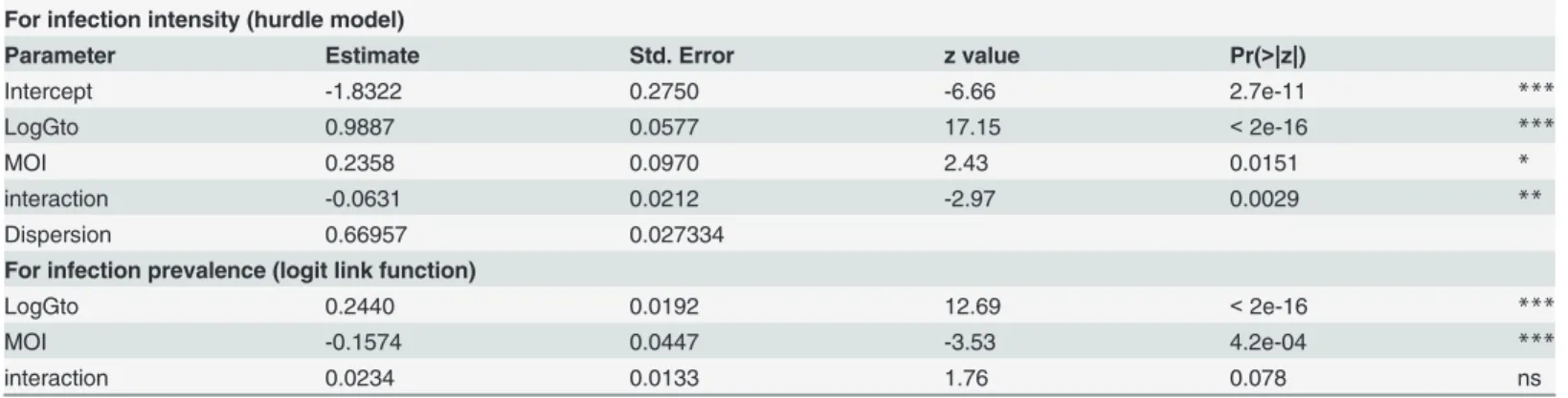
The estimates of the parameters of the hurdle negative binomial (HNB) model are shown in Table 1for positive count and zero components. Both gametocyte density and MOI significant-ly affected the oocyst number. The fit of the intensity of infection with gametocyte densities for the different MOIs is plotted inFig 1. In the HBN model, the correlations of oocyst counts with gametocyte density and MOI were positive (P<2e-16 andP= 0.0151, respectively;Table 1)
gametocyte density and MOI were highly significant (P<2e-16 andP= 4.2e-04, respectively),
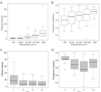
and the interaction was not significant (Table 1). The relation between gametocyte densities and the number of infected mosquitoes was positive, indicating that the higher gametocytemia the blood donor had, the higher the prevalence of infection (Fig 2B). In contrast, the effect of MOI was negative, which meant that infection prevalence was lower in multiclonal isolates (Fig 2D).
Table 1. Maximum likelihood estimates of the parameters in the Hurdle Negative Binomial model.
For infection intensity (hurdle model)
Parameter Estimate Std. Error z value Pr(>|z|)
Intercept -1.8322 0.2750 -6.66 2.7e-11 ***
LogGto 0.9887 0.0577 17.15 <2e-16 ***
MOI 0.2358 0.0970 2.43 0.0151 *
interaction -0.0631 0.0212 -2.97 0.0029 **
Dispersion 0.66957 0.027334
For infection prevalence (logit link function)
LogGto 0.2440 0.0192 12.69 <2e-16 ***
MOI -0.1574 0.0447 -3.53 4.2e-04 ***
interaction 0.0234 0.0133 1.76 0.078 ns
LogGto, logged-transformed gametocyte density;MOI, multipicity of infection;Interactionfor interaction between LogGto and MOI. The dispersion parameter that is accommodated in the HNB model reflects over-dispersion of parasites. Z value is a Student statistics. Significant codes
'***'<0.001 '**'<0.01
'*'<0.05; 'ns'0.05.
doi:10.1371/journal.pone.0123777.t001
Fig 1. A, fitted intensity of infection curve versus log-transformed of gametocytemia for the different MOIs in the HNB2 model.The analysis was performed from 125 feedings.
P
.
falciparum
genetic diversity throughout the development within the
mosquito vector
P.falciparum-positive salivary glands were pooled for 49 feedings, which led to 49 bulks from
547 sporozoite-positive salivary glands. The proportion of monoclonal infections was similar for theP.falciparumgametocyte isolates and the sporozoite populations, 14.3 and 8.2%, re-spectively (odds ratio: 1.86, 95% CI: 0.43–9.32,P= 0.524). The mean multiplicity of infection at the gametocyte and sporozoite stages varied from 3.57 ± 0.99 (range: 1–10) to 3.94 ± 0.88 (range: 1–8), respectively, and the difference was not significant (Wilcoxon matched pairs ranks test,V= -164,P= 0.155).
We observed different patterns of microsatellite polymorphism between the gametocyte and sporozoite populations for the same parasite donor. The fluorescence peak, which reflects the relative abundance of the alleles, varied between the income and outcome parasite stages, ga-metocytes and sporozoites, respectively, and most blood donors encountered changes in their allelic composition between the gametocyte and sporozoite populations (Fig 3). Indeed, for 87.8% (43 of 49) of the infections, we observed in sporozoite populations the presence of alleles not detected in the gametocyte population. Genotypes not detected at early stages of sporogony can reach detection level at a later stage, and this can reflect either an inaccurate assessment of the genotypes in the gametocyte samples or gametocyte genotypes present below the PCR threshold.
Fig 2. A and B, relation between gametocytes density in the parasite isolate and infection prevalence and infection intensity.Gametocyte densities were grouped into five classes, as indicated in the x axis. Infection prevalence is given as the mean proportion of infected mosquitoes, in percentage, and infection intensity as the median of oocyst counts perP.falciparum-positive gut. The boxplots were created from 125 feedings. The number of infected mosquitoes and the oocyst loads increase with the gametocyte density in the blood donor (hurdle model,P<2e-16 for both response variables).C and D, relation between multiplicity of infection in the parasite isolate and infection intensity and infection prevalence. Multiplicity of infection (MOI) is defined as the maximum number of alleles at the most polymorphic locus. Volunteers harboring MOI>4 were grouped. Infection prevalence is given as the mean proportion of infected mosquitoes, in percentage, and infection intensity represents the median of oocyst counts perP.falciparum
-positive gut for the given MOI class. The boxplots were created from 125 feedings.
Correlation of
P
.
falciparum
mating patterns with parasite transmission
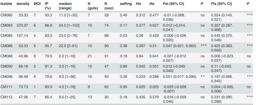
A total of 623 oocysts from 108 mosquito midguts infected on 9 blood donors were successfully analyzed with 6 microsatellite markers. As for the haploid forms ofP.falciparum, all 6 loci were highly polymorphic, with an average of 11.3 (±2.1) alleles per locus. The mean allelic rich-ness of oocyst populations within blood donors was 3.6 ± 1.9. The total observed and expected gene diversities over all 6 loci wereHo = 0.291 (±0.039) andHe = 0.404 (±0.045), respectively. However, significant differences betweenHo andHe were not observed for all gametocyte car-riers, indicating that inbreeding levels vary from one blood donor to another.
Fstatistics were computed to explore the genetic structure ofP.falciparumoocysts within and among mosquitoes fed on blood from the nine distinct gametocyte carrier volunteers. At the meta population level (all mosquitoes),P.falciparumdiploid stages exhibit a non-random distribution within the mosquito vector (Fst = 0.471, 95% CI: 0.452–0.487,P<10–4) and a
sig-nificant departure from panmixia (Fis = 0.280, 95% CI: 0.234–0.339,P<10–4). The genetic
differentiation among oocyst sub-populations (Fst) and the inbreeding coefficient (Fis) were computed at each microsatellite locus for the whole set of mosquito midguts (Fig 4A); the val-ues were similar to those previously described for populations ofP.falciparumfrom different areas [2,4]. The linkage disequilibrium was significant for all 15 pairs of loci, withP<10–3 for
each pair after Bonferroni correction. TheFst values per pair of samples (donors) varied from 0.162 to 0.933 and all pairwise values were highly significant.
At the donor level, a significant genetic differentiation between mosquitoes fed on the same donor was observed for only two gametocyte carriers: CM086 and CM096 (Fst = 0.047, 95% CI: 0.021–0.062; andFst = 0.051, 95% CI: 0.017–0.090, respectively,Table 2); genotypes were randomly distributed among mosquitoes for all the remaining blood donors. Departure from panmixia due to non-random mating of gametes within mosquito midguts was then estimated by theFis measures for each of the 9 blood donors. TheFis values ranged from 0.004 to 0.454 over the nine feedings, andFis was not significant for only 3 gametocyte carriers (CM088, CM111, and CM090) (Fig 4BandTable 2), which confirmed variations in inbreeding levels ac-cording to the blood donor.
Fig 3. Comparison ofP.falciparumallelic diversity in gametocyte (gto) and sporozoite (spz)
populations from the same isolates. A, donor CM066 at locus TA109;B, donor CM085 at locus PfPK2.The x-axis indicates the allele size at the given microsatellite locus; the y-axis represents arbitrary fluorescent units. Electrophoregrams show the parasite dynamics, from blood (gto) at the time of mosquito feeding to salivary glands (spz). Alleles not detected in the sexual stages are found in the sporozoites (A). The relative abundance of the different genotypes, determined by the height of the fluorescent units, varies between the gto and the spz populations (B), which reflects intense competitive interactions between parasite clones.
We computed a linear regression model to investigate correlations ofPlasmodiummating patterns with parameters of infection success. TheFiswas positively correlated to the medians of oocyst loads (R2= 0.840,P= 5.1e-4;Fig 5), indicating that infection intensity is higher when parasites do not mate randomly within the mosquito. We explored whether mating between clone relatives, as seen here by selfing (measured by identical genotypes at all 6 loci in the oo-cyst population), results from the parasite complexity, and indeed a linear regression model showed that selfing negatively correlates with MOI (R2= 0.786,P= 9.0e-4;S1 Fig).
Discussion
In this study, we have modeled the mosquito infection (infection intensity and infection preva-lence) according to gametocyte variables (gametocyte density and MOI) to provide insights into the transmission ofP.falciparumgenotypes in the field. Our results indicate that 1) the number of co-infecting clones in the gametocyte isolate impacts on the mosquito infection
Fig 4. Inbreeding statistics forP.falciparumoocysts recovered fromA.gambiaemidguts per microsatellite locus (A) and per blood donor (B).Fis gives a measure of the correlation of allelic types within oocysts relative to gene copies from different oocysts from the same mosquito.Fst measures the
correlation of the allelic type among different oocysts from the same mosquito relative to gene copies in oocysts from different mosquitoes fed on the same donor blood. The bars indicate the 95% CI for each value.
success, both in terms of prevalence and intensity of infection; and 2) sporogony within the mosquito vector has an important role in shaping the population structure ofP.falciparum.
We performed membrane feedings on blood from asymptomatic gametocyte carriers identi-fied in an area where malaria is hyper-endemic and the entomological inoculation rate exceeds 100 infected bites/person/year [18,19]. A large variability in the mosquito infection success was observed from one feeding to another. We then examined how the parameters from the game-tocyte isolates influence the mosquito infection, by scoring gamegame-tocyte densities
Table 2. Fstatistics for each of the nine experimental feedings.
Isolate density MOI IP (%)
median [range]
N (guts)
N (ooc)
selfing Ho He Fst (95% CI) P Fis (95% CI) P
CM080 33.33 7 93.3 11.0 [1–30] 7 28 0.46 0.312 0.477 -0.01 (-0.068.
0.036) ns 0.324 (0.145.0.521) *** CM083 370.37 6 88.9 24.0 [1–103] 10 74 0.17 0.377 0.627 0.012 (-0.014.
0.041) ns 0.357 (0.287.0.466) *** CM085 107.14 4 83.3 23.0 [2–76] 7 88 0.53 0.26 0.429 -0.006 (-0.026.
0.020) ns 0.445 (0.370.0.546) *** CM086 53.33 5 95.7 22.0 [2–61] 10 90 0.38 0.267 0.51 0.047 (0.021. 0.062) *** 0.425 (0.363.
0.503) ***
CM088 43.96 6 79.0 2.0 [1–16] 21 91 0.18 0.64 0.641 -0.001 (-0.012.
0.007) ns 0.006 (-0.023.0.037) ns CM090 68.18 2 81.3 3.0 [1–16] 15 47 0.89 0.042 0.051 0.012 (-0.040.
0.040) ns 0.11 (-0.043.0.247) ns CM096 98.48 6 78.6 9.0 [1–36] 16 93 0.39 0.233 0.286 0.051 (0.017. 0.090) ** 0.197 (0.066.
0.345) ***
CM111 73.73 1 83.3 4.5 [1–18] 9 82 0.95 0.025 0.025 -0.025 (-0.028.
-0.007) ns 0.004 (-0.005.0.006) ns CM112 47.06 7 85.4 9.0 [1–25] 13 30 0.18 0.435 0.579 -0.016 (-0.059.
0.046) ns 0.231 (0.090.0.398) ***
doi:10.1371/journal.pone.0123777.t002
Fig 5. Relationship between the median of the oocyst burden inP.falciparum-positive mosquitoes andFis.Each dot represents a blood donor. The oocyst loads are positively correlated to theFis values (P= 5.1e-4).
microscopically and genotyping gametocyte isolates using microsatellite markers. Our results show that not only gametocyte density but also MOI have an effect on infection outcomes. The gametocyte density strongly correlates with infection prevalence and intensity: mosquitoes fed on carriers with the highest gametocyte counts were more infected and carried more oocysts. The relationship between gametocyte density and transmission success has already been re-ported [20,21]; however, the influence of the multiplicity of infection ofP.falciparum gameto-cyte isolates on mosquito infection parameters has been poorly documented. We found in our studied area that the number of clones within natural gametocyte isolates negatively influenced the infection prevalence and the oocyst loads were higher in monoclonal infections, indicating that coinfections impact on the transmission success.
The transmissibility of multiple clones to mosquitoes, even when present at low density, has already been reported [20,22–24]. The current literature predicts that within-host competition between conspecific parasites results in a competitive advantage for virulent clones [25–27]. However, virulence in these studies refers to a strategy to optimize host exploitation, which is not the only parasite adaptation underlying competition in mixed infections [28,29]. Indeed, parasite plasticity and interactions with the host immunity also play an important role in deter-mining transmission success from coinfecting clones [26,30]. Accordingly, on the vector side, we previously reported that the immune responses mounted by the mosquito differed between monoclonal and multiclonal infections [31]. In the present study and in a previous one on a smaller set of feedings [31], we showed that mixed infections lead to a lower parasite burden. By contrast, a recent study reported that coinfection of two cultured strains of parasites did not affect the mosquito infection [32]. These findings may indicate that the reduced infection level we observed in mixed infections results from former parasite-parasite interactions within the human host and not from vector-parasite interactions. This would explain, in part, discrepan-cies between studies using wild isolates and those using laboratory strains ofP.falciparum [33,34]. Alternatively, the genetic diversity of the parasite may be an important factor to infect different species of sympatric mosquitoes and further studies will have to determine whether different vector species better transmit specificP.falciparumgenotypes [2,35]. b Increasing evi-dence also suggests that the mosquito genotype by parasite genotype and genotype by environ-ment interactions are a strong determinant of vector competence [36,37]. Our results highlight the great complexity of parasite-parasite and host-parasite interactionsin naturaand empha-size that both fundamental and field studies will be necessary to measure the importance of parasite and vector, and their biological and genetic features, in driving parasite transmission.
We found here that the overall genetic diversity of sporozoite populations was congruent with that of gametocyte isolates. The maintenance of genetic diversity throughout the sporogo-ny is consistent with previous studies that reported similar patterns of genetic diversity in ga-metocytes and infected mosquitoes, as seen in pooled oocysts within the midgut [23]. However, we observed in oocyst and sporozoite populations alleles that were not detected in the corresponding gametocyte samples, reflecting the imperfect detectability of minority clones [38]. In our study, the presence of new microsatellite alleles in oocysts and salivary glands of mosquitoes that fed on apparently mono-infected gametocyte carriers indicates that clones were present, but not detected, in the gametocyte population and, more importantly, that they were able to infect mosquitoes. Several studies have reported failure to detect minority clones in blood samples because the sensitivity of molecular methods is not optimal or the numerical-ly dominant clones may obscure the less abundant ones [22–24,38,39]. This limitation proba-bly underestimates the MOI in the gametocyte population and the correlation we found between the gametocyte complexity and the mosquito infection parameters may slightly differ
in natura. Nonetheless, our results thus confirm thatP.falciparumgametocyte genotypes with
The mating patterns ofP.falciparumdetermine the parasite population genetic structure, with important epidemiological consequences. Indeed, the dynamics of parasite transmission and the underlying spread of drug-resistant genotypes are directly dependent on the organ-ism’s mode of reproduction [41]. Non-random mating of gametes within the mosquito blood-meal (significantFis) was detected for a majority of feedings (6 of 9) despite random
distribution of gametocyte genotypes among mosquitoes (non-significantFst). Interestingly, oocyst burden wasFis-dependent, suggesting that the mating pattern ofP.falciparum influ-ences genotype transmission. Herein, we propose thatP.falciparumis capable of modulating inbreeding and outcrossing levels according to the genetic content of the gametocyte pool or to other environmental cues. This is in agreement with the recent hypothesis that malaria para-sites use kin discrimination to gauge the genetic diversity within the infection and adjust their sex-allocation in response to the presence of coinfecting genotypes [42]. The increased oocyst burden found in infections deviating from panmixia may reflect that mating between clone-mates gives rise to progenies with higher fitness, which is consistent with the higher infection intensity observed in monoclonal infections. However, from our oocyst genotyping data, it re-mains unclear how the genetic content of gametocyte isolates influences the outcrossing level. Further investigations aiming at measuring both the sex ratios and the densities of the different coinfecting genotypes are needed to better understand the transmission strategies in multiclo-nal infections.
Finally, these results are of importance for the understanding of vector-parasite interactions in the field, as we showed that the genetic composition of the gametocyte population affects the outcome of the infection in the mosquito vector. The current deployment of malaria control in-terventions should reduce the diversity of circulatingP.falciparumparasite strains, and lead to an increase of monoclonal infections that have better infectiousness for the mosquito vector. Our findings are then of great significance since they suggest an important epidemiological consequence of control interventions. This study shows the importance of monitoring and characterizing malaria infections to understand the changes in malaria epidemiology within the context of malaria control interventions, and to circumvent their unintended effect on vector transmission.
Supporting Information
S1 Fig. Relationship between MOI and selfing.Each dot represents a blood donor. MOI rep-resents the estimated number of clones per gametocyte carrier, Selfing is defined as mating be-tween two genetically identical gametes.
(TIF)
S1 Table. Characteristics of the gametocyte donors and parameters of transmission success in the Ngousso strain ofA.gambiae.
(DOC)
Acknowledgments
Author Contributions
Conceived and designed the experiments: IM DF AB. Performed the experiments: IM SN LA ZA MTT AC PHAA AB. Analyzed the data: IM WT FR AB. Contributed reagents/materials/ analysis tools: IM WT DF FR AB. Wrote the paper: IM AC FR AB.
References
1. Anderson TJ, Paul RE, Donnelly CA, Day KP. Do malaria parasites mate non-randomly in the mosquito midgut? Genet Res. 2000; 75(3):285–96. PMID:10893865
2. Annan Z, Durand P, Ayala FJ, Arnathau C, Awono-Ambene P, Simard F, et al. Population genetic struc-ture of Plasmodium falciparum in the two main African vectors, Anopheles gambiae and Anopheles funestus. Proc Natl Acad Sci U S A. 2007; 104(19):7987–92. PMID:17470800
3. Mzilahowa T, McCall PJ, Hastings IM. "Sexual" population structure and genetics of the malaria agent P. falciparum. PLoS One. 2007; 2(7):e613. PMID:17637829
4. Razakandrainibe FG, Durand P, Koella JC, De Meeus T, Rousset F, Ayala FJ, et al. "Clonal" population structure of the malaria agent Plasmodium falciparum in high-infection regions. Proc Natl Acad Sci U S A. 2005; 102(48):17388–93. PMID:16301534
5. Babiker HA, Schneider P, Reece SE. Gametocytes: insights gained during a decade of molecular moni-toring. Trends Parasitol. 2008; 24(11):525–30. doi:10.1016/j.pt.2008.08.001PMID:18801702
6. Nkhoma SC, Nair S, Cheeseman IH, Rohr-Allegrini C, Singlam S, Nosten F, et al. Close kinship within multiple-genotype malaria parasite infections. Proc Biol Sci. 2012. Epub 2012/03/09.
7. Harris C, Morlais I, Churcher TS, Awono-Ambene P, Gouagna LC, Dabire RK, et al. Plasmodium falcip-arum produce lower infection intensities in local versus foreign Anopheles gambiae populations. PLoS One. 2012; 7(1):e30849. Epub 2012/02/01. doi:10.1371/journal.pone.0030849PMID:22292059 8. Ribaut C, Berry A, Chevalley S, Reybier K, Morlais I, Parzy D, et al. Concentration and purification by
magnetic separation of the erythrocytic stages of all human Plasmodium species. Malar J. 2008; 7:45. doi:10.1186/1475-2875-7-45PMID:18321384
9. Anderson TJ, Haubold B, Williams JT, Estrada-Franco JG, Richardson L, Mollinedo R, et al. Microsatel-lite markers reveal a spectrum of population structures in the malaria parasite Plasmodium falciparum. Mol Biol Evol. 2000; 17(10):1467–82. PMID:11018154
10. Team RDC. R: A Language and Environment for Statistical Computing. Vienna, Austria: R Foundation for Statistical Computing; 2012. doi:10.1002/jcc.22917PMID:22278855
11. Mullahy J. Specification and testing of some modified count data models. J Econometrics 1986; 33:341–65.
12. Skaug H, Fournier D, Nielsen AN, Magnusson A, Bolker B. glmmADMB: generalized linear mixed mod-els using AD Model Builder. R package version 0.7.2.12. 2012.
13. Akaike H. Likehood of a model and information criteria. Journal of Econometrics. 1981; 16:2–23.
14. Goudet J. FSTAT (vers. 1.2): a computer program to calculate F-statistics. J Hered. 1995; 86:485–6.
15. Nei M. Estimation of Average Heterozygosity and Genetic Distance from a Small Number of Individuals. Genetics. 1978; 89(3):583–90. PMID:17248844
16. Weir BS, Cockerham CC. Estimating F-statistics for the analysis of population structure. Evolution. 1984; 38:1358–70.
17. Rousset F. Genepop'007: a complete reimplementation of the Genepop software for Windows and Linux. Mol Ecol Resources. 2008; 8:103–6. doi:10.1111/j.1471-8286.2007.01931.xPMID:21585727
18. Antonio-Nkondjio C, Awono-Ambene P, Toto JC, Meunier JY, Zebaze-Kemleu S, Nyambam R, et al. High malaria transmission intensity in a village close to Yaounde, the capital city of Cameroon. J Med Entomol. 2002; 39(2):350–5. PMID:11931035
19. Bonnet S, Gouagna LC, Paul RE, Safeukui I, Meunier JY, Boudin C. Estimation of malaria transmission from humans to mosquitoes in two neighbouring villages in south Cameroon: evaluation and compari-son of several indices. Trans R Soc Trop Med Hyg. 2003; 97(1):53–9. PMID:12886806
20. Bousema T, Dinglasan RR, Morlais I, Gouagna LC, van Warmerdam T, Awono-Ambene PH, et al. Mos-quito feeding assays to determine the infectiousness of naturally infected Plasmodium falciparum ga-metocyte carriers. PLoS One. 2012; 7(8):e42821. Epub 2012/09/01. doi:10.1371/journal.pone. 0042821PMID:22936993
22. Arez AP, Pinto J, Palsson K, Snounou G, Jaenson TG, do Rosario VE. Transmission of mixed Plasmo-dium species and PlasmoPlasmo-dium falciparum genotypes. Am J Trop Med Hyg. 2003; 68(2):161–8. PMID:
12641406
23. Nwakanma D, Kheir A, Sowa M, Dunyo S, Jawara M, Pinder M, et al. High gametocyte complexity and mosquito infectivity of Plasmodium falciparum in the Gambia. Int J Parasitol. 2008; 38(2):219–27.
PMID:17709108
24. Schneider P, Bousema T, Omar S, Gouagna L, Sawa P, Schallig H, et al. (Sub)microscopic Plasmodi-um falciparPlasmodi-um gametocytaemia in Kenyan children after treatment with sulphadoxine-pyrimethamine monotherapy or in combination with artesunate. Int J Parasitol. 2006; 36(4):403–8. PMID:16500657
25. Alizon S, de Roode JC, Michalakis Y. Multiple infections and the evolution of virulence. Ecol Lett. 2013; 16(4):556–67. Epub 2013/01/26. doi:10.1111/ele.12076PMID:23347009
26. de Roode JC, Pansini R, Cheesman SJ, Helinski ME, Huijben S, Wargo AR, et al. Virulence and com-petitive ability in genetically diverse malaria infections. Proc Natl Acad Sci U S A. 2005; 102(21):7624–
8. PMID:15894623
27. Wargo AR, de Roode JC, Huijben S, Drew DR, Read AF. Transmission stage investment of malaria parasites in response to in-host competition. Proc Biol Sci. 2007; 274(1625):2629–38. PMID:
17711832
28. Choisy M, de Roode JC. Mixed infections and the evolution of virulence: effects of resource competi-tion, parasite plasticity, and impaired host immunity. Am Nat. 2010; 175(5):E105–18. doi:10.1086/
651587PMID:20297955
29. Mackinnon MJ, Marsh K. The selection landscape of malaria parasites. Science. 2010; 328 (5980):866–71. doi:10.1126/science.1185410PMID:20466925
30. Raberg L, de Roode JC, Bell AS, Stamou P, Gray D, Read AF. The role of immune-mediated apparent competition in genetically diverse malaria infections. Am Nat. 2006; 168(1):41–53. PMID:16874614
31. Nsango SE, Abate L, Thoma M, Pompon J, Fraiture M, Rademacher A, et al. Genetic clonality of Plas-modium falciparum affects the outcome of infection in Anopheles gambiae. Int J Parasitol. 2012; 42 (6):589–95. Epub 2012/05/05. doi:10.1016/j.ijpara.2012.03.008PMID:22554991
32. Molina-Cruz A, Dejong RJ, Ortega C, Haile A, Abban E, Rodrigues J, et al. Some strains of Plasmodium falciparum, a human malaria parasite, evade the complement-like system of Anopheles gambiae mos-quitoes. Proc Natl Acad Sci U S A. 2012. Epub 2012/05/25.
33. Cohuet A, Osta MA, Morlais I, Awono-Ambene PH, Michel K, Simard F, et al. Anopheles and Plasmodi-um: from laboratory models to natural systems in the field. EMBO Rep. 2006; 7(12):1285–9. PMID:
17099691
34. Mendes AM, Schlegelmilch T, Cohuet A, Awono-Ambene P, De Iorio M, Fontenille D, et al. Conserved mosquito/parasite interactions affect development of Plasmodium falciparum in Africa. PLoS Pathog. 2008; 4(5):e1000069. doi:10.1371/journal.ppat.1000069PMID:18483558
35. Boissiere A, Gimonneau G, Tchioffo MT, Abate L, Bayibeki A, Awono-Ambene PH, et al. Application of a qPCR assay in the investigation of susceptibility to malaria infection of the M and S molecular forms of An. gambiae s.s. in Cameroon. PLoS One. 2013; 8(1):e54820. Epub 2013/01/26. doi:10.1371/ journal.pone.0054820PMID:23349974
36. Boissiere A, Tchioffo MT, Bachar D, Abate L, Marie A, Nsango SE, et al. Midgut Microbiota of the Malar-ia Mosquito Vector Anopheles gambMalar-iae and Interactions with Plasmodium falciparum Infection. PLoS Pathog. 2012; 8(5):e1002742. Epub 2012/06/14. doi:10.1371/journal.ppat.1002742PMID:22693451 37. Lefevre T, Vantaux A, Dabire KR, Mouline K, Cohuet A. Non-genetic determinants of mosquito
compe-tence for malaria parasites. PLoS Pathog. 2013; 9(6):e1003365. Epub 2013/07/03. doi:10.1371/ journal.ppat.1003365PMID:23818841
38. Wampfler R, Timinao L, Beck HP, Soulama I, Tiono AB, Siba P, et al. Novel Genotyping Tools for Inves-tigating Transmission Dynamics of Plasmodium falciparum. J Infect Dis. 2014; 210(8):1188–97. Epub
2014/04/29. doi:10.1093/infdis/jiu236PMID:24771862
39. Campino S, Auburn S, Kivinen K, Zongo I, Ouedraogo JB, Mangano V, et al. Population genetic analy-sis of Plasmodium falciparum parasites using a customized Illumina GoldenGate genotyping assay. PLoS One. 2011; 6(6):e20251. Epub 2011/06/16. doi:10.1371/journal.pone.0020251PMID:21673999 40. Ouedraogo AL, Bousema T, Schneider P, de Vlas SJ, Ilboudo-Sanogo E, Cuzin-Ouattara N, et al. Sub-stantial contribution of submicroscopical Plasmodium falciparum gametocyte carriage to the infectious reservoir in an area of seasonal transmission. PLoS One. 2009; 4(12):e8410. doi:10.1371/journal. pone.0008410PMID:20027314