Integrated Boost in Whole-Breast Irradiation
after Breast-Conserving Surgery: IMRT, IMRT
plus an Electron Boost and VMAT
Sangang Wu1☯, Youqun Lai1☯, Zhenyu He2☯, Yuan Zhou1, Shanyu Chen1, Mingming Dai1, Juan Zhou3, Qin Lin1*, Feng Chi2*
1Xiamen Cancer Center, Department of Radiation Oncology, the First Affiliated Hospital of Xiamen University, Xiamen, People’s Republic of China,2Sun Yat-sen University Cancer Center, State Key Laboratory of Oncology in South China, Department of Radiation Oncology, Collaborative Innovation Center of Cancer Medicine, Guangzhou, People’s Republic of China,3Xiamen Cancer Center, Department of Obstetrics and Gynecology, the First Affiliated Hospital of Xiamen University, Xiamen, People’s Republic of China
☯These authors contributed equally to this work.
*[email protected](QL);[email protected](FC)
Abstract
Objectives
To compare the target volume coverage and doses to organs at risks (OARs) using three techniques that simultaneous integrated boost (SIB) in whole-breast irradiation (WBI) after breast-conserving surgery, including intensity-modulated radiation therapy (IMRT), IMRT plus an electron boost (IMRT-EB), and volumetric-modulated arc therapy (VMAT).
Methods
A total of 10 patients with early-stage left-sided breast cancer after breast-conserving sur-gery were included in this study. IMRT, IMRT-EB and VMAT plans were generated for each patient.
Results
The conformity index (CI) of the planning target volumes evaluation (PTV-Eval) of VMAT was significantly superior to those of IMRT and IMRT-EB (P<0.05). The CI of the PTV Eval-boost of VMAT was better than that of IMRT (P= 0.018) and IMRT-EB (P<0.001), while the CI of the PTV Eval-boost of IMRT was better than that of IMRT-EB (P= 0.002).
The V5, V10 and Dmean in ipsilateral lung with VMAT were significantly higher than IMRT (P<0.05) and IMRT-EB (P<0.05). The Dmean, V5 and V10 in heart with VMAT were sig-nificantly greater than those of IMRT and IMRT-EB (P<0.05). There was no significant dif-ference in the OARs between IMRT and IMRT-EB (P>0.05).
OPEN ACCESS
Citation:Wu S, Lai Y, He Z, Zhou Y, Chen S, Dai M, et al. (2015) Dosimetric Comparison of the Simultaneous Integrated Boost in Whole-Breast Irradiation after Breast-Conserving Surgery: IMRT, IMRT plus an Electron Boost and VMAT. PLoS ONE 10(3): e0120811. doi:10.1371/journal.pone.0120811
Academic Editor:Qinghui Zhang, University of Nebraska Medical Center, UNITED STATES
Received:June 16, 2014
Accepted:February 6, 2015
Published:March 17, 2015
Copyright:© 2015 Wu et al. This is an open access article distributed under the terms of theCreative
Commons Attribution License, which permits
unrestricted use, distribution, and reproduction in any medium, provided the original author and source are credited.
Data Availability Statement:All relevant data are within the paper and its Supporting Information files.
Conclusions
Considered the target volume coverage and radiation dose delivered to the OARs (espe-cially the heart and lung), IMRT may be more suitable for the SIB in WBI than IMRT-EB and VMAT. Additional clinical studies with a larger sample size will be needed to assess the long-term feasibility and efficacy of SIB using different radiotherapy techniques.
Introduction
Whole-breast irradiation (WBI) has become an essential part of the multimodal breast-conserving treatment for breast cancer [1]. Additional tumor bed boost after WBI can further reduce the local recurrence rate in some patients at high risk of breast cancer [2–4]. However,
studies have found that the tumor bed boost would affect the cosmetic results [5,6], thereby al-tering the effectiveness of breast-conserving therapy. The long-term follow-up results of the 'boost versus no boost' trial conducted by the European Organisation for Research and Treat-ment of Cancer (EORTC) also found that tumor bed boost and use of photon beam boost were important factors affecting cosmetic results in patients [6]. But it was not the primary goal of this trial to investigate different outcome with different boost techniques [6]. In the last de-cades, a lot of attention has gone to the development of new techniques to reduce side effects. In case of breast irradiation this means late side effects on skin, heart and lungs.
The standard radiation therapy after breast-conserving surgery is sequential tumor bed boost for 60–66 Gy after the 5-week WBI for a total dose of 45–50 Gy, and the entire duration
is 6–7 weeks [7]. However, extended treatment time is resource-intensive, with resultant
psychosocial and economic implications. With the evolution of radiation physics, several tech-niques to deliver a tumor bed boost dose have become available. With the further understand-ing of the biology of breast cancer radiotherapy, the radiation dose of the breast cancer radiotherapy has changed, and recently, there have been an increasing number of studies on si-multaneous integrated boost (SIB) [8,9]. The rationale is a localized dose enhancement in the area at highest risk without prolonging treatment duration, thus not only providing improved patient comfort but also exploiting the higher sensitivity of breast tumor cells towards larger single doses, which has long been postulated in the linear quadratic model [10]. Daily SIB of 2.3 Gy can shorten the duration of radiation therapy [10], therefore, the inconvenience to the patient and cost to radiation oncology department is particularly attractive. However, more-advanced techniques are required to increase the uniformity of the radiation dose to improve the cosmetic outcomes and reduce the dose to the organs at risks (OARs) to reduce late radiation toxicities.
Currently, the main non-invasive tumor bed boost technique is photon or electron beam ir-radiation [4]; however, there is no strict standard for the irradiation technique used for the SIB. Compared with the conventional irradiation technique, intensity-modulated radiation therapy (IMRT) can perform boost in the tumor bed on the basis of daily radiotherapy, thereby shortening the duration of radiation therapy [11]. In the present study, we compared three techniques for the SIB: IMRT, IMRT plus an electron boost (IMRT-EB), and volumetric-modulated arc therapy (VMAT). We evaluated the target coverage and radiation dose delivery to the OARs to identify the most suitable technique for the SIB in the WBI after breast-conserving surgery.
Materials and Methods
Patients
This study included 10 patients with early-stage breast cancer who underwent radiation thera-py after breast-conserving surgery at the Department of Radiation Oncology, the First Affiliat-ed Hospital of Xiamen University from August 2013 to December 2013. The inclusion criteria were as follows: 1) female patient age of 18 years or older with left side breast cancer, who underwent breast-conserving surgery; 2) diagnosis of invasive cancer was confirmed by pathol-ogy; 3) axillary lymph node dissection was performed, or sentinel lymph node biopsy was nega-tive; 4) stage of I or II (TIN0M0, T2N0M0) according to the 2009 7th edition of the American Joint Committee on Cancer (AJCC)/Union for International Cancer Control (UICC) staging system; 5) microscopic margins were negative (>1cm); 6) complete immunohistochemistry
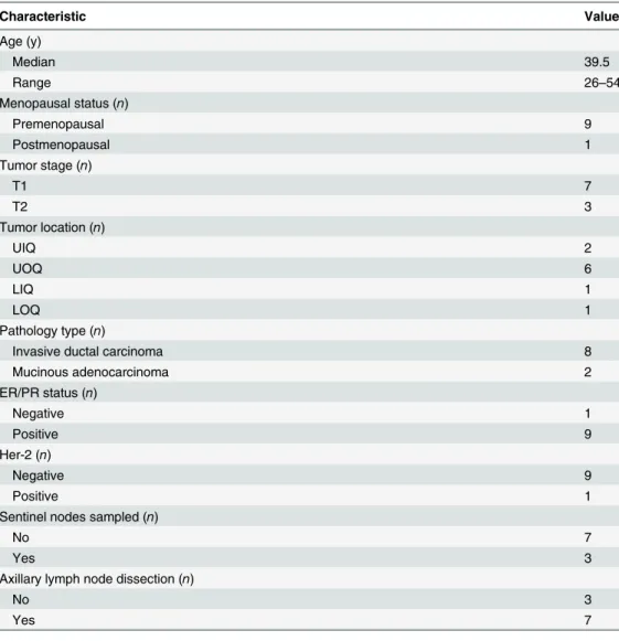
results including estrogen receptor (ER), progesterone receptor (PR), and human epidermal growth factor receptor 2 (Her2); 7) adjuvant chemotherapy or endocrine therapy was per-formed in accordance with standards and guidelines. The study was approved by the ethics committee of the First Affiliated Hospital of Xiamen University. All patients provided written consent for storage of their medical information in the hospital database and for research use of this information. Patient characteristics are listed inTable 1.
Delineation of the target volume and normal tissue
The delineation of the target volume and normal tissue was finally approved by three experi-enced radiation oncologist. Clinical target volume (CTV) for the whole breast was delineated according to the recommendations of the International Commission on Radiation Units (ICRU) report 83 [12]. The breast CTV included all visible breast parenchyma. The planning target volume of the breast (PTV-breast) added a 7- mm expansion in all directions around the CTV except for the skin surface, including the set-up margin and accounting for patient move-ment. The PTV for evaluation (PTV Eval-breast) was limited anteriorly to exclude the region outside of the patient and the first 5 mm of tissue under the skin, and was limited posteriorly to no deeper than the posterior surface of the ribs (to exclude lung).
Delineation of the lumpectomy (gross tumor volume, GTV): The GTV was delineated accord-ing to the metal mark placed duraccord-ing the surgery, the residual seroma after surgery, mammary gland interruption and density change on CT, and ultrasound findings of patients in the same fixed position including the distance from the tumor bed, i.e., the tumor cavity or interruption of the mammary gland to certain points on the skin, and the depth, width, and height of the tumor bed.
CTV boost included: GTV + 1 cm, limiting the CTV posteriorly to the anterior surface of the pectoralis major, and anterolaterally to 5 mm from skin, not crossing the midline. In gener-al, the pectoralis and/or serratus anterior muscles are excluded from the lumpectomy CTV un-less clinically warranted by the tumor pathology. The PTV boost was added to include a 7-mm expansion in all direction around the CTV boost. The PTV Eval-boost was limited anteriorly to exclude the region outside the patient and the first 5 mm of tissue under the skin, and was limited posteriorly to no deeper than the posterior surface of the ribs (to exclude lung).
Delineation of normal organs: The contralateral breast, lung, and heart were delineated on the CT image of each slice.Table 2shows the volume of target and normal tissue of the entire study population.
Radiation treatment planning
Table 2. The volume of target and normal tissue of 10 patients.
Target volume and normal tissue Volume
Mean (cm3) Median (cm3) Range (cm3)
PTV-Whole breast 473.10 482.03 256.12–731.02
CTV-Tumor bed 27.18 26.33 9.85–50.36
PTV-Boost 68.44 67.65 30.70–103.00
Ipsilateral lung 1086.00 1056.56 809.78–1499.45
Heart 524.50 547.26 364.1–613.32
Contralateral breast 467.51 450.99 298.02–633.56
CTV, clinical target volume; PTV, planning target volume.
doi:10.1371/journal.pone.0120811.t002
Table 1. Clinical characteristics of 10 patients.
Characteristic Value
Age (y)
Median 39.5
Range 26–54
Menopausal status (n)
Premenopausal 9
Postmenopausal 1
Tumor stage (n)
T1 7
T2 3
Tumor location (n)
UIQ 2
UOQ 6
LIQ 1
LOQ 1
Pathology type (n)
Invasive ductal carcinoma 8
Mucinous adenocarcinoma 2
ER/PR status (n)
Negative 1
Positive 9
Her-2 (n)
Negative 9
Positive 1
Sentinel nodes sampled (n)
No 7
Yes 3
Axillary lymph node dissection (n)
No 3
Yes 7
Abbreviations: UIQ = upper inner quadrant, UOQ = upper outer quadrant, LIQ = lower inner quadrant,
LOQ = lower outer quadrant.
PTV-boost by step and shot IMRT. An angle of 20–30° was used between the two beams in the
same direction, and the maximum number of segments was 50. IMRT-EB combined the two programs. The IMRT plan design is described above, but is emphasized only for PTV-breast. IMRT dose prescription was 28 fractions of 1.8 Gy. Electron boost plans used a single electron to each patient's PTV-boost. In order for the PTV-boost to be covered by the 95% isodose line, electron energy of 6, 9, 12 or 15 MeV was chosen according to the depth of the PTV-boost. The doses were then combined according to V95 including the 95% prescribed dose.
In VMAT, double ipsilateral partial arcs with a maximum individual length of 180° starting from the mid-sternum were adopted for this study. Collimator angles were individualized to patients, and ranged from 15° to 30°.
The evaluation criteria for the planned dose for the target area and the dose limitation of the OARs referred mainly to the IMRT-MC2 trial [13]. Radiation treatment of the whole breast in-cluded a total dose of 50.4 Gy and 1.8 Gy per fraction with 2.3 Gy per fraction with integrated boost to the tumor bed for a total dose of 64.4 Gy in 28 fractions. With regard to the organs at risk, less than 10% of the heart volume may receive>30 Gy, while less than 20% of the
ipsilat-eral lung may receive>20 Gy. The mean dose to the contralateral breast should be limited to
less than 5 Gy. Treatment planning was performed in Pinnacle (Version 9.2, Philips, USA) treatment planning system (TPS). All plans were normalized so that 95% of PTV-breast and PTV-boost received 95% of the prescribed dose (PD).
Calculation of conformity index (CI) and homogeneity index (HI)
CI = (VTref / VT) × (VTref / Vref), VTref, where VTref represents the target volume covered by isodose; VT is target volume; Vref is the total volume covered by 95% of isodose. CI range was 0–1, in which the conformity was better when the CI value was larger. HI = D5/D95,
where D5 represents the irradiation dose received by 5% of PTV-Eval, while D95 represents the irradiation dose received by 95% of PTV-Eval. The closer the HI value is to 1, the better the target uniformity will be.
Statistical analysis
One-way analysis of variance (ANOVA) test was used to compare dosimetric differences among plans using the three SIB techniques. All statistical tests were two-sided, and were per-formed using SPSS software (release 17.0, SPSS Inc., Chicago, IL, USA). Statistical significance was defined asP<0.05.
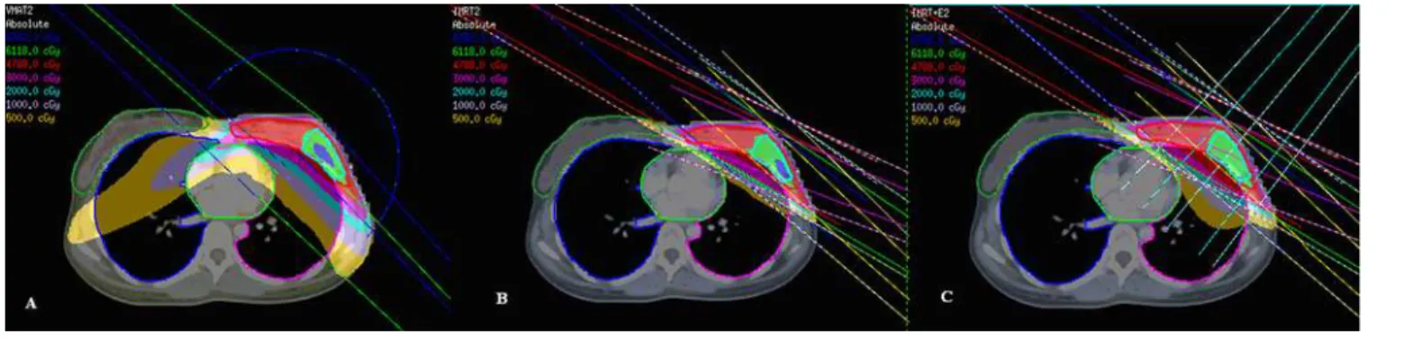
Fig 1. Beam arrangement, target coverage, and doses to normal organs in VMAT (A), IMRT (B), and IMRT-EB (C).
Results
Target volume coverage
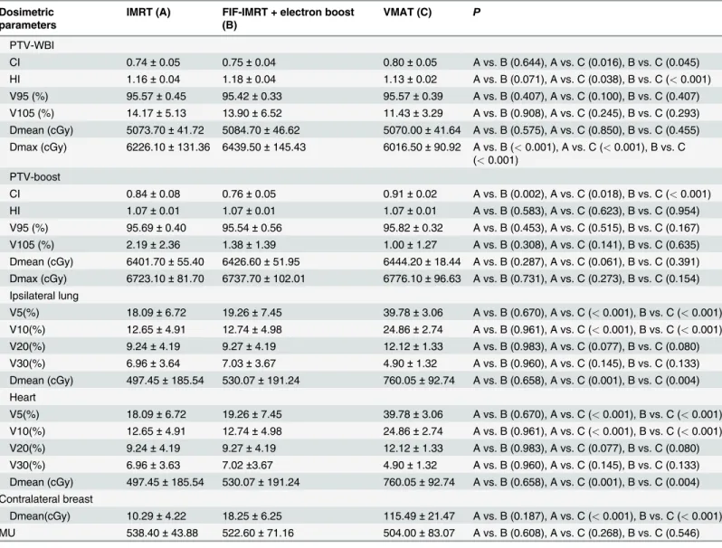
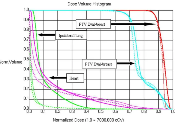
Table 3presents dosimetric parameters for IMRT, IMRT-EB and VMAT for the target volume coverage. Figs.1and2show the target volume coverage and doses to the normal tissue in the three treatment programs.
The CI and HI of the breast PTV-Eval of VMAT were significantly superior to those of IMRT and IMRT-EB (P<0.05), while the CI and HI of the breast PTV-EVAL were not
signifi-cantly different between the IMRT and IMRT-EB (P>0.05). There was no significant
differ-ence in the Dmean, V95, and V105 among the three radiotherapy techniques (P>0.05), but Table 3. Comparison of PTV and normal tissue for IMRT, IMRT plus an electron boost, and VMAT.
Dosimetric parameters
IMRT (A) FIF-IMRT + electron boost (B)
VMAT (C) P
PTV-WBI
CI 0.74±0.05 0.75±0.04 0.80±0.05 A vs. B (0.644), A vs. C (0.016), B vs. C (0.045)
HI 1.16±0.04 1.18±0.04 1.13±0.02 A vs. B (0.071), A vs. C (0.038), B vs. C (<0.001)
V95 (%) 95.57±0.45 95.42±0.33 95.57±0.39 A vs. B (0.407), A vs. C (0.100), B vs. C (0.407)
V105 (%) 14.17±5.13 13.90±6.52 11.43±3.29 A vs. B (0.908), A vs. C (0.245), B vs. C (0.293)
Dmean (cGy) 5073.70±41.72 5084.70±46.62 5070.00±41.64 A vs. B (0.575), A vs. C (0.850), B vs. C (0.455)
Dmax (cGy) 6226.10±131.36 6439.50±145.43 6016.50±90.92 A vs. B (<0.001), A vs. C (<0.001), B vs. C
(<0.001)
PTV-boost
CI 0.84±0.08 0.76±0.05 0.91±0.02 A vs. B (0.002), A vs. C (0.018), B vs. C (<0.001)
HI 1.07±0.01 1.07±0.01 1.07±0.01 A vs. B (0.583), A vs. C (0.623), B vs. C (0.954)
V95 (%) 95.69±0.40 95.54±0.56 95.82±0.32 A vs. B (0.453), A vs. C (0.515), B vs. C (0.167)
V105 (%) 2.19±2.36 1.38±1.39 1.00±1.27 A vs. B (0.308), A vs. C (0.141), B vs. C (0.635)
Dmean (cGy) 6401.70±55.40 6426.60±51.95 6444.20±18.44 A vs. B (0.287), A vs. C (0.061), B vs. C (0.391)
Dmax (cGy) 6723.10±81.70 6737.70±102.01 6776.10±96.63 A vs. B (0.731), A vs. C (0.273), B vs. C (0.154)
Ipsilateral lung
V5(%) 18.09±6.72 19.26±7.45 39.78±3.06 A vs. B (0.670), A vs. C (<0.001), B vs. C (<0.001)
V10(%) 12.65±4.91 12.74±4.98 24.86±2.74 A vs. B (0.961), A vs. C (<0.001), B vs. C (<0.001)
V20(%) 9.24±4.19 9.27±4.19 12.12±1.33 A vs. B (0.983), A vs. C (0.077), B vs. C (0.080)
V30(%) 6.96±3.64 7.03±3.67 4.90±1.32 A vs. B (0.960), A vs. C (0.145), B vs. C (0.133)
Dmean (cGy) 497.45±185.54 530.07±191.24 760.05±92.74 A vs. B (0.658), A vs. C (0.001), B vs. C (0.004)
Heart
V5(%) 18.09±6.72 19.26±7.45 39.78±3.06 A vs. B (0.670), A vs. C (<0.001), B vs. C (<0.001)
V10(%) 12.65±4.91 12.74±4.98 24.86±2.74 A vs. B (0.961), A vs. C (<0.001), B vs. C (<0.001)
V20(%) 9.24±4.19 9.27±4.19 12.12±1.33 A vs. B (0.983), A vs. C (0.077), B vs. C (0.080)
V30(%) 6.96±3.63 7.02±3.67 4.90±1.32 A vs. B (0.960), A vs. C (0.145), B vs. C (0.133)
Dmean (cGy) 497.45±185.54 530.07±191.24 760.05±92.74 A vs. B (0.658), A vs. C (0.001), B vs. C (0.004)
Contralateral breast
Dmean(cGy) 10.29±4.22 18.25±6.25 115.49±21.47 A vs. B (0.187), A vs. C (<0.001), B vs. C (<0.001)
MU 538.40±43.88 522.60±71.16 504.00±83.07 A vs. B (0.608), A vs. C (0.268), B vs. C (0.546)
CTV, clinical target volume; PTV, planning target volume; IMRT, intensity-modulated radiation therapy; VMAT, volumetric-modulated arc therapy; CI, conformity index; HI, homogeneity index (HI); MU, monitor unit.
the Dmax of the breast PTV-Eval of VMAT was significantly lower than that of IMRT
(P<0.001) and IMRT-EB (P<0.001). The Dmax of the breast PTV-Eval of IMRT was
signifi-cantly lower than that of the IMRT-EB (P<0.001).
The CI of the breast PTV-Eval-boost among the three radiotherapy techniques were signifi-cantly difference. The CI of the breast PTV-Eval-boost of VMAT was better than that of IMRT (P= 0.018) and IMRT-EB (P<0.001), and the CI of the PTV-Eval-boost of IMRT was better
than that of IMRT-EB (P= 0.002); however, there was no significant difference in the HI, Dmean, Dmax, V95 and V105 among the three radiotherapy techniques (P>0.05).
Doses to normal organs
Table 3presents the results of dosimetric comparison of ipsilateral lung and heart for 10 pa-tients with left-sided breast cancer. The V5 and V10 of ipsilateral lung in VMAT was signifi-cantly higher than that of IMRT (P<0.05) and IMRT-EB (P<0.05), and there was no
significant difference in the V5 and V10 between IMRT and IMRT-EB (P= 0.670).
There was no significant difference in the V20 and V30 of ipsilateral lung among the three radiotherapy techniques (P>0.05) (Figs.1and2). There was no significant difference in V5,
V10, V20, and V30 of ipsilateral lung between IMRT and IMRT-EB (P>0.05). The Dmean of
ipsilateral lung in VMAT was significantly higher than that of IMRT (P= 0.001) and IMRT-EB (P= 0.004), and there was no significant difference between IMRT and IMRT-EB (P= 0.658).
Fig 2. Dose volume histogram of the target volume coverage and normal organs in VMAT (medium solid), IMRT (medium dashed), and IMRT-EB (thin solid).
The V5 and V10 of heart in VMAT were significantly higher than that of IMRT and IMRT-EB (P<0.05), and there was no significant difference in the V5 or V10 between IMRT
and IMRT-EB (P>0.05). The V20 and V30 of heart in VMAT were not significantly different
from those of IMRT and IMRT-EB (P>0.05). The Dmean of heart in IMRT and IMRT-EB
was significantly less than that of VMAT (P<0.05). The V5, V10, V20, V30 and Dmean were
not significant difference between IMRT and IMRT-EB (P>0.05).
The Dmean of the contralateral breast in VMAT were significantly higher than that of IMRT and IMRT-EB (P<0.05), and there was no significant difference between IMRT and
IMRT-EB (P= 0.187). There was no significant difference in the MU among the three radio-therapy techniques (P>0.05).
Discussion
In the present study, we compared the target volume coverage and dose to the OARs of IMRT, IMRT-EB, and VMAT, the three techniques that SIB in WBI after breast-conserving surgery. The results showed that although VMAT had better target volume coverage, its extent of low-dose irradiation of the heart and lung was significantly greater than that of the other two tech-niques. The target volume coverage of IMRT was slightly lower than that of VMAT, but it was superior to that of IMRT-EB.
The CI and HI of target volume coverage are important indicators for the assessment of the radiotherapy techniques, and they might be important factors affecting the cosmetic effects of breast-conserving therapy. Early findings of EORTC 'boost versus no boost' randomized trial showed that tumor bed boost performed using the electron beam, photon beam, and interpola-tion technique did not cause significantly different cosmetic effects [14]; however, longer follow-up found that the photon beam irradiation was an important factor affecting the cos-metic results [6]. A possible reason may be related to the dosimetric characteristics of the pho-ton beam itself and the fact that radiotherapy technique was underdeveloped at that time. Thus, more volume of normal breast tissue underwent high-dose irradiation, thereby affecting the cosmetic results. The following dosimetric studies on the photon beam and electron beam boost found results opposite to those of the CI of the photon beam [15,16]. Currently, the elec-tron beam is a widely adopted tumor bed boost technique because its dosimetric characteristics are more suitable for superficial tumor bed; however, boost for deeper tumor bed requires in-creased electron beam energy, which will further increase the radiation dose to the heart, lung and normal breast tissue. There has been no standard for the SIB in WBI after breast conserv-ing surgery so far. Therefore, it is particularly important to explore a more precise conformal irradiation mode that is appropriate for the contemporary treatment program.
The dosimetric characteristics of IMRT and VMRT make radiotherapy with SIB feasible. At present, there are no studies on the integrated tumor bed boost techniques, IMRT, IMRT-EB, and VMAT in WBI after breast conserving surgery. In study on WBI, Jin et al. found that the CI of the VMAT target volume was lower than that of tangential field IMRT (P<0.05), and
the HI of the target volume was even worse (P<0.05). Therefore, VMAT was not
organs receiving high doses of radiation was reduced, accompanied by an increase in the vol-ume of normal organs receiving low doses of radiation [16].
In our study, the CI of target volume with VMAT was better than that of IMRT and IMRT-EB, and the CI of the target volume with IMRT was better than that of IMRT-EB; how-ever, there was no significant difference in the HI among the three techniques. Moreover, the range of the ipsilateral lung and heart receiving low doses of radiation in VMAT was greater than that in IMRT and IMRT-EB. The clinical results of SIB in whole-breast irradiation with different techniques is still lacking. The radiotherapy planning is based on CT images in mod-ern age, once the target volume coverage meets the requirements that will not affect local con-trol. Therefore, the technique that can minimize the doses to OARs has become the most appropriate choice. In present study, we found that IMRT is superior to the other two tech-niques when achieving comparable target volume coverage and doses to OARs, and it is a more appropriate method for the SIB.
Target volume movement due to respiration-induced organ motion is an important factor affecting the radiation dose received by target volume and OARs during breast IMRT. Zhang et al. has developed a model to estimate 3-dimensional motion in patients CT images for pre-dicting respiration-induced organ motion that has potential for improving the accuracy of dose calculation in radiotherapy [18]. In addition, the active breathing control (ABC) tech-nique can reduce or eliminate target volume caused by respiration, and more importantly, the ABC technique can decrease the radiation volume and dose to the lung and heart to reduce ra-diation injury. Remouchamps et al. reported that deep tangential field whole-breast IMRT was applied together with the ABC technique and the dose distribution in the IMRT target volume coverage became more homogeneous compared with conventional tangential field irradiation, and the radiation dose to the heart and lung was significantly reduced in the moderate deep in-spiration breath hold compared with the free breathe [19].
With the improved efficacy of breast cancer therapy, the likelihood of long-term survival of patients may be increased. Therefore, radiation toxicities during long-term follow-up are the subject of increasing attention. Henson et al. studied 558,871 patients with breast cancer who underwent postoperative radiotherapy, and they found that the 30-year radiotherapy-related mortality of lung cancer was higher than radiotherapy-related mortality during the first 20 years after radiotherapy (P= 0.002) [20]. VMAT may lead to more scattered radiation to the surrounding normal organs due to its own technical characteristics [21], especially to lung tis-sue. Dosimetric studies have shown that tangential field IMRT could markedly reduce the risk of lung cancer compared with multi-direction IMRT and VMAT [22]. The ipsilateral lung V5 and V10 of VMAT was significantly higher than that of IMRT and IMRT-EB, indicating that VMAT is associated with a higher possibility of radiation-induced lung cancer, but the results of long-term follow-up are still lacking.
The survival advantage after radiotherapy for breast cancer was probably offset by increased cardiovascular mortality in early series because of inferior radiotherapy techniques and choice of target volume such as internal mammary lymph nodes. Darby et al. analyzed patients treated for breast cancer between 1958 and 2001 and found that the probability of ischemic heart dis-ease started to incrdis-ease after several years post radiotherapy, and such incrdis-ease would last for at least 20 years [23]. Another study conducted long-term follow-up in 35,000 patients undergo-ing radiotherapy, and found that the incidence of cardiovascular disease increased remarkably after radiotherapy [24]. One study pointed out that cardiovascular mortality was not increased in patients who underwent radiotherapy after 1980 [25], but the effects of radiation therapy on the cardiovascular system will become apparent only after a long period of time, perhaps 30–40
long-term follow-up results of VMAT for the treatment of breast cancer [27]. Therefore, fur-ther studies are warranted to explore whefur-ther VMAT is suitable for the clinical treatment of patients with breast cancer.
Conclusions
In conclusion, our study results suggest that IMRT may be more suitable for the simultaneously integrated tumor bed boost in WBI after breast-conserving surgery than IMRT-EB and VMAT when ensuring appropriate target coverage and considering doses to OARs, especially the heart and lung. However, since the conclusion of the present study is based on a dosimetry study and the sample size is small. Additional clinical studies with a larger sample size will be needed to assess the long-term feasibility and efficacy of SIB using different radiotherapy techniques.
Supporting Information
S1 Dataset. The data underlying the findings in present study. (XLS)
Author Contributions
Conceived and designed the experiments: SGW YQL ZYH QL FC. Performed the experiments: YZ SYC MMD. Analyzed the data: SGW JZ. Contributed reagents/materials/analysis tools: SYC JZ. Wrote the paper: SGW YQL ZYH.
References
1. Clarke M, Collins R, Darby S, Davies C, Elphinstone P, Evans E, et al. (2005) Effects of radiotherapy and of differences in the extent of surgery for early breast cancer on local recurrence and 15-year sur-vival: an overview of the randomised trials. Lancet 366:2087–106. PMID:16360786
2. Bartelink H, Horiot JC, Poortmans PM, Struikmans H, Van den Bogaert W, Fourquet A, et al. (2007) Im-pact of a higher radiation dose on local control and survival in breast-conserving therapy of early breast cancer: 10-year results of the randomized boost versus no boost EORTC 22881–10882 trial. J Clin
Oncol 25:3259–3265. PMID:17577015
3. Senkus E, Kyriakides S, Penault-Llorca F, Poortmans P, Thompson A, Zackrisson S, et al. (2013) Pri-mary breast cancer: ESMO Clinical Practice Guidelines for diagnosis, treatment and follow-up. Ann Oncol 24(suppl 6): vi7–23. doi:10.1093/annonc/mdt284PMID:23970019
4. National Comprehensive Cancer Network (NCCN) Clinical Practice Guidelines in Oncology, Breast Cancer. Version 3, 2014.http://www.nccn.org/professionals/physician_gls/pdf/breast.pdf(Accessed 26 April 2014).
5. Poortmans PM, Collette L, Horiot JC, Van den Bogaert WF, Fourquet A, Kuten A, et al. (2009) Impact of the boost dose of 10 Gy versus 26 Gy in patients with early stage breast cancer after a microscopically incomplete lumpectomy: 10-year results of the randomised EORTC boost trial. Radiother Oncol 90:80–85. doi:10.1016/j.radonc.2008.07.011PMID:18707785
6. Immink JM, Putter H, Bartelink H, Cardoso JS, Cardoso MJ, van der Hulst-Vijgen MH, et al. (2012) Long-term cosmetic changes after breast-conserving treatment of patients with stage I-II breast cancer and included in the EORTC 'boost versus no boost' trial. Ann Oncol 23:2591–2598. PMID:22499858
7. American College of Radiology. (2007) Practice guideline for the breast conservation therapy in the management of invasive breast carcinoma. J Am Coll Surg 205:362–376. PMID:17660085
8. Liem X, Chira C, Fourquet A, Campana F, Peurien D, Fournier-Bidoz N, et al. (2014) [Preliminary re-sults of whole breast helical tomotherapy with simultaneous integrated boost in the adjuvant treatment of breast cancer] [Article in French]. Cancer Radiother 18:15–22. doi:10.1016/j.canrad.2013.07.149 PMID:24316350
9. Alford SL, Prassas GN, Vogelesang CR, Leggett HJ, Hamilton CS. (2013) Adjuvant breast radiotherapy using a simultaneous integrated boost: clinical and dosimetric perspectives. J Med Imaging Radiat Oncol 57:222–229. doi:10.1111/j.1754-9485.2012.02473.xPMID:23551785
adjuvant radiotherapy? Statement of the German and the Austrian Societies of Radiooncology (DEGRO/ÖGRO). Strahlenther Onkol 189:193–196. doi:10.1007/s00066-012-0300-3PMID:
23358687
11. Thilmann C, Zabel A, Grosser KH, Hoess A, Wannenmacher M, Debus J. (2001) Intensity-modulated radiotherapy with an integrated boost to the macroscopic tumor volume in the treatment of high-grade gliomas. Int J Cancer 96:341–349. PMID:11745504
12. ICRU Report 83. (2010) Prescribing, Recording, and Reporting Photon-Beam Intensity-Modulated Ra-diation Therapy (IMRT). International Commission on RaRa-diation Units and Measurements, Bethesda. 13. Askoxylakis V, Jensen AD, Häfner MF, Fetzner L, Sterzing F, Heil J, et al. (2011) Simultaneous
inte-grated boost for adjuvant treatment of breast cancer—intensity modulated vs. conventional radiothera-py: the IMRT-MC2 trial. BMC Cancer 11:249. doi:10.1186/1471-2407-11-249PMID:21676232
14. Poortmans P, Bartelink H, Horiot JC, Struikmans H, Van den Bogaert W, Fourquet A, et al. (2004) The influence of the boost technique on local control in breast conserving treatment in the EORTC 'boost versus no boost' randomised trial. Radiother Oncol 72:25–33. PMID:15236871
15. V Van Parijs H, Reynders T, Heuninckx K, Verellen D, Storme G, De Ridder M. (2014) Breast conserv-ing treatment for breast cancer: dosimetric comparison of different non-invasive techniques for addition-al boost delivery. Radiat Oncol 9:36. doi:10.1186/1748-717X-9-36PMID:24467916
16. Park SH, Kim JC. (2013) Comparison of electron and x-ray beams for tumor bed boost irradiation in breast-conserving treatment. J Breast Cancer 16:300–307. doi:10.4048/jbc.2013.16.3.300PMID: 24155759
17. Jin GH, Chen LX, Deng XW, Liu XW, Huang Y, Huang XB. (2013) A comparative dosimetric study for treating left-sided breast cancer for small breast size using five different radiotherapy techniques: con-ventional tangential field, filed-in-filed, tangential-IMRT, multi-beam IMRT and VMAT. Radiat Oncol 8:89. doi:10.1186/1748-717X-8-89PMID:23587298
18. Zhang Q, Pevsner A, Hertanto A, Hu YC, Rosenzweig KE, Ling CC, et al. (2003) A patient-specific re-spiratory model of anatomical motion for radiation treatment planning. Med Phys 34:4772–81.
19. Remouchamps VM, Vicini FA, Sharpe MB, Kestin LL, Martinez AA, Wong JW. (2003) Significant reduc-tions in heart and lung doses using deep inspiration breath hold with active breathing control and intensity-modulated radiation therapy for patients treated with locoregional breast irradiation. Int J Radiat Oncol Biol Phys 55:392–406. PMID:12527053
20. Henson KE, McGale P, Taylor C, Darby SC. (2013) Radiation-related mortality from heart disease and lung cancer more than 20 years after radiotherapy for breast cancer. Br J Cancer 108:179–182. doi:
10.1038/bjc.2012.575PMID:23257897
21. Otto K. (2008) Volumetric modulated arc therapy: IMRT in a single gantry arc. Med Phys 35:310–317. PMID:18293586
22. Abo-Madyan Y, Aziz MH, Aly MM, Schneider F, Sperk E, Clausen S, et al. (2014) Second cancer risk after 3D-CRT, IMRT and VMAT for breast cancer. Radiother Oncol (in press).
23. Darby SC, Ewertz M, McGale P, Bennet AM, Blom-Goldman U, Brønnum D, et al. (2013) Risk of
ische-mic heart disease in women after radiotherapy for breast cancer. N Engl J Med 368:987–998. doi:10. 1056/NEJMoa1209825PMID:23484825
24. McGale P, Darby SC, Hall P, Adolfsson J, Bengtsson NO, Bennet AM, et al. (2011) Incidence of heart disease in 35,000 women treated with radiotherapy for breast cancer in Denmark and Sweden. Radio-ther Oncol 100:167–175. doi:10.1016/j.radonc.2011.06.016PMID:21752480
25. Patt DA, Goodwin JS, Kuo YF, Freeman JL, Zhang DD, Buchholz TA, et al. (2005) Cardiac morbidity of adjuvant radiotherapy for breast cancer. J Clin Oncol 23:7475–7482. PMID:16157933
26. Kodama K, Fujiwara S, Yamada M, Kasagi F, Shimizu Y, Shigematsu I. (1996) Profiles of non-cancer diseases in atomic bomb survivors. World Health Stat Q 49:7–16. PMID:8896251