Three-dimensional conformal breast
irradiation in the prone position
1Department of Radiation Oncology, Medical Faculty, Ankara University,
Cebeci Hospital, Dikimevi, Ankara, Turkey
2Department of Radiation Oncology, Medical Faculty, Kocaeli University,
Sopali Çiftligi, Derince, Izmit, Turkey C. Kurtman1,
M. Nalça Andrieu1,
A. Hiçsönmez1 and
B. Çelebio—lu2
Abstract
The prone position can be used for the planning of adjuvant radio-therapy after conservative breast surgery in order to deliver less irradiation to lung and cardiac tissue. In the present study, we com-pared the results of three-dimensional conformal radiotherapy plan-ning for five patients irradiated in the supine and prone position. Tumor stage was T1N0M0 in four patients and T1N1M0 in one. All patients had been previously submitted to conservative breast surgery. Breast size was large in three patients and moderate in the other two. Irradiation in the prone position was performed using an immobiliza-tion foam pad with a hole cut into it to accommodate the breast so that it would hang down away from the chest wall. Dose-volume histo-grams showed that mean irradiation doses reaching the ipsilateral lung were 8.3 ± 3.6 Gy with the patient in the supine position and 1.4 ± 1.0 Gy with the patient in the prone position (P = 0.043). The values for the contralateral lung were 1.3 ± 0.7 and 0.3 ± 0.1 Gy (P = 0.043) and the values for cardiac tissue were 4.6 ± 1.6 and 3.0 ± 1.7 Gy (P = 0.079), respectively. Thus, the dose-volume histograms demonstrated that lung tissue irradiation was significantly lower with the patient in the prone position than in the supine position. Large-breasted women appeared to benefit most from irradiation in the prone position. Prone position breast irradiation appears to be a simple and effective alterna-tive to the conventional supine position for patients with large breasts, since they are subjected to lower pulmonary doses which may cause less pulmonary side effects in the future.
Correspondence
C. Kurtman
Department of Radiation Oncology Medical Faculty, Ankara University Cebeci Hospital, Dikimevi Ankara
06100 Turkey Fax: +90-312-319-7884
E-mail: [email protected] or [email protected]
Received October 14, 2002 Accepted August 7, 2003
Key words
•Prone position
•Breast cancer
•Radiotherapy
Introduction
The management of primary breast can-cer with conservative surgery and radiation therapy is a widely accepted alternative to mastectomy (1,2). However, standard tan-gential breast radiotherapy not only treats portions of the chest wall, but also exposes lung and heart tissue to radiation (3,4).
Radiation pneumonitis following conser-vative surgery and radiation therapy for breast
to result in unacceptable dose inhomogene-ity or in significant doses delivered to nor-mal tissue (7). In the present study, we describe the results of three-dimensional con-formal radiotherapy (3D-CRT) in the prone position.
Material and Methods
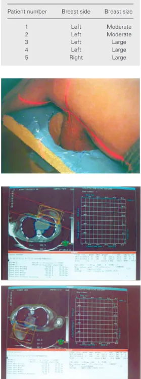
When the patient is irradiated in the prone position the treated breast hangs down away from the chest wall and the radiation dose to normal tissue is minimized. We studied five patients with a diagnosis of infiltrative duc-tal carcinoma of the breast subjected to con-servative surgery and referred to our radio-therapy clinic. Tumor stage was T1, N0, M0 in four patients and T1, N1, M0 in one. Patient characteristics are listed in Table 1. The patients were positioned on an immobi-lization foam pad both in the supine position and in the prone position. The ipsilateral arm was placed above the head and the contralat-eral arm on the immobilization foam pad. A breast ring was fixed on the breast to hold it in an upright position. A hole cut into the pad under the breast permitted the breast to hang down during treatment in the prone position (Figure 1).
Computed tomography images were ob-tained with a Picker® I.Q. T/C computed tomography apparatus (Picker International, Inc., Cleveland, OH, USA) both in the su-pine and prone positions. The target volume, body outline, left lung tissue, right lung tis-sue and cardiac tistis-sue were drawn, the target isocenter was fixed, and virtual simulation was performed with a Picker Voxel Q vir-tual simulation workstation. Beam’s eye view, isodose distribution, inhomogeneity correction, multileaf collimators and dose volume histogram were obtained with a Varian® Cad-plan treatment-modeling workstation (Varian Associates Inc., On-cology Systems, Palo Alto, CA, USA). The median and lateral borders of the breast tissue determined clinically and with beam’s eye view were included in the fields using tangential parallel-opposed photon beams. 3D-CRT planning was done for each position. Patient treatment was planned using a 6-mV photon power Varian® Clinac 2300 C/D apparatus, with one daily session of 2 Gy, five sessions per week and a Figure 1. A hole was cut into the
foam pad under the breast that allowed the breast to hang down with the patient in the prone po-sition.
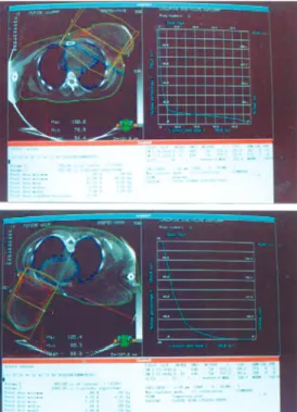
Figure 2. Case 1. Dose-volume histogram and isodose distribu-tion in the target volume in the supine position (top) and in the prone position (bottom).
Table 1. Characteristics of breasts of patients with primary breast cancer.
Patient number Breast side Breast size
1 Left Moderate
2 Left Moderate
3 Left Large
4 Left Large
total dose of 50 Gy over five weeks. Wedges were used to enhance the best isodose distri-bution and the treatment plans, and dose volume histograms obtained for the supine position and the prone position were com-pared.
The Wilcoxon test was applied to com-pare the results obtained in the two posi-tions.
Results
The plans were performed using the isocentric technique and the isodose distri-butions were observed for the five cases in the supine position and in the prone position. The isodose distributions were optimized with wedges on the transverse plane. Dose volume histograms were constructed for the target volume, left lung tissue volume, right lung tissue volume and cardiac tissue vol-ume in each case for both the supine and the prone positions (Figures 2 and 3).
The isodoses covering target volumes at the base and on the lateral edge of the breast and the mean target doses for both the supine and the prone positions are listed in Table 2.
The mean doses delivered to lung and cardiac tissues in each case in the supine and prone positions are shown in Table 3. The ipsilateral lung was the left one in four pa-tients and the right one in the fifth patient (Table 3).
The mean doses delivered to normal tis-sues in the supine position compared to the prone position are listed in Table 4. The mean doses to the ipsilateral lung were 8.3 ± 3.6 Gy for the supine position and 1.4 ± 1.0 Gy for the prone position (P = 0.043). The values for the contralateral lung were 1.3 ± 0.7 and 0.3 ± 0.1 Gy (P = 0.043) and the values for cardiac tissue were 4.6 ± 1.6 and 3.0 ± 1.7 Gy (P = 0.079), respectively. The results showed that the mean doses delivered to the lungs were significantly lower in the prone position than in the supine position.
Figure 3. Case 3. Dose-volume histogram and isodose distribu-tion for the ipsilateral left lung tissue volume in the supine sition (top) and in the prone po-sition (bottom).
Table 2. Isodose percentage in the target volume and mean target dose for each patient in the prone and supine positions.
Isodose percentage in the target volume Mean target dose (Gy)
At the base At the lateral edge of the breast of the breast
Patient 1
Supine 82.8 108.5 50.0 (99.9 ± 3.0%) Prone 77.8 103.5 48.8 (97.6 ± 2.8%)
Patient 2
Supine 96.6 111.2 48.4 (96.8 ± 4.9%) Prone 94.7 106.1 49.1 (98.3 ± 3.5%)
Patient 3
Supine 75.0 108.8 47.5 (95.0 ± 4.3%) Prone 67.1 109.0 49.5 (99.0 ± 5.2%)
Patient 4
Supine 93.8 109.2 51.1 (102.2 ± 4.7%) Prone 96.0 111.9 49.2 (98.5 ± 5.5%)
Patient 5
Supine 95.8 117.1 50.3 (100.7 ± 3.7%) Prone 95.6 115.3 51.3 (102.6 ± 5.9%)
Discussion
The management of primary breast can-cer with conservative surgery and radiation therapy is a widely accepted alternative to mastectomy (1). There is an increasing inter-est in the late effects of therapy on the
sur-vival of patients treated for breast cancer (8-11). The long-term complications following conservative surgery and radiation therapy for early stage breast cancer are low, and changes in radiation technique may reduce their occurrence (12).
Lingos et al. (5) assessed a large number of patients regarding the factors contributing to radiation pneumonitis. They concluded that radiation pneumonitis following con-servative surgery and radiation therapy for breast cancer was a rare complication and more likely to occur in patients treated with both a 3-field technique and chemotherapy. Three percent of patients treated with a 3-field technique and with chemotherapy de-veloped radiation pneumonitis compared to 0.5% for all other patients (P = 0.0001). When patients were treated with combined chemoradiotherapy and the 3-field technique, the incidence of radiation pneumonitis was 8.8% for concurrent chemotherapy compared with 1.3% for patients who received sequen-tial chemotherapy and radiation therapy (P = 0.002). Over the limited range of volumes treated, irradiated lung volume was not asso-ciated with an increased risk of radiation pneumonitis. On the other hand, the risk of radiation pneumonitis appeared to be related to the volume of lung irradiated in two other studies (13,14). The use of computed tomog-raphy in tangential breast irradiation pro-vides a detailed picture of the dose distribu-tions throughout the breast volume and sur-rounding normal tissue. Three-dimensional treatment planning allows dose escalation to the target volume without significantly in-creasing the dose received by surrounding normal tissue (15). The full scale computed tomography scan with a true three-dimen-sional dose algorithm is more accurate than the three-slice model (16).
Gyenes et al. (17) reported that the com-puted tomography-based three-dimensional treatment planning system might be con-formed to reduce the irradiated cardiac vol-ume. In the cited study, most of the patients Table 4. Mean radiation dose delivered to normal tissue in the supine and prone
positions for the patient group as a whole.
Mean radiation dose (Gy)
Supine Prone
Ipsilateral lung tissue 8.3 ± 3.60 1.4 ± 1.05* Cardiac tissue 4.6 ± 1.60 3.0 ± 1.70 Contralateral lung tissue 1.3 ± 0.70 0.3 ± 0.10*
*P < 0.05 compared to the supine position (Wilcoxon test).
Table 3. Mean radiation dose delivered to normal tissue in each patient in the supine and prone positions.
Mean radiation dose (covering isodose)
Supine Prone
Patient 1
Ipsilateral lung 7.6 (15.2 ± 29.8%) 3.2 (6.4 ± 16.5%) Contralateral lung 0.8 (1.6 ± 1.2%) 0.5 (1.0 ± 0.8%) Heart 5.5 (11.1 ± 18.8%) 4.8 (9.7 ± 17.3%)
Patient 2
Ipsilateral lung 5.3 (10.6 ± 19%) 0.9 (1.9 ± 2.3%) Contralateral lung 0.8 (1.7 ± 0.5%) 0.3 (0.6 ± 0.6%) Heart 4.9 (8.1 ± 11.2%) 3.2 (6.4 ± 9.1%)
Patient 3
Ipsilateral lung 5.8 (11.6 ± 22.6%) 0.5 (1.1 ± 0.8%) Contralateral lung 0.6 (1.2 ± 0.6%) 0.1 (0.3 ± 0.3%) Heart 6.3 (12.6 ± 19.5%) 1.7 (3.5 ± 2.8%)
Patient 4
Ipsilateral lung 8.8 (17.7 ± 28.7%) 0.9 (1.8 ± 3.4%) Contralateral lung 1.1 (2.2 ± 2.0%) 0.4 (0.9 ± 0.6%) Heart 3.9 (7.8 ± 8.2%) 4.4 (8.8 ± 12.2%)
Patient 5
Ipsilateral lung 14.2 (28.4 ± 36.0%) 1.3 (2.6 ± 4.5%) Contralateral lung 2.1 (4.3 ± 2.3%) 0.1 (0.2 ± 0.1%) Heart 2.1 (4.3 ± 2.3%) 0.7 (1.5 ± 0.6%)
with left-sided T1, N0, M0 breast cancer did not receive irradiation to a substantial car-diac volume (17).
A dose to the contralateral breast due to primary breast irradiation is possible as a function of the technique used for the pri-mary treated breast. Several factors contri-bute significantly to the opposite breast dose and the situation can be improved by good techniques (18).
Gray et al. (19) documented a slightly yet measurably inferior cosmetic result in large-breasted or heavy women. However, the dif-ference between the large-breasted group and the others was not great and should not exclude these women from consideration for breast conservation techniques.
Cross et al. (20) described a technique for the conservative irradiation of women with very large breasts. Patients were treated in a modified lateral decubitus technique which offered breast-conserving therapy to women with large breasts, without poor cosmesis.
Merchant and McCormick (7) reported that prone position breast irradiation appears to be a simple and effective alternative to irradiation of the breast in the conventional supine position when the supine position is likely to result in unacceptable dose inhomo-geneity or significant doses delivered to nor-mal tissues. Large-breasted women appeared to benefit most from this treatment.
Bieri et al. (21) determined the effects of treatment techniques, such as supine and prone positioning, on the absorbed dose in organs at a distance from the irradiated breast. Peripheral doses delivered to the abdomen,
pelvic organs, bone marrow and lung were significantly higher for supine than for prone tangential breast irradiation.
Recently, Mahe et al. (22) used the prone position technique for breast irradiation af-ter conservative surgery with the help of a special Plexiglas platform in 35 patients with large pendulous breasts. The feasibility was excellent for positioning, dosimetric studies, and immediate tolerance in more than 90% of the patients. This system had the major advantage of ensuring a similar and repro-ducible position for computed tomography scanning and treatment.
In the present study, 3D-CRT planning was used to deliver a lower dose to normal tissue in the prone position compared to the supine position. In the prone position, the ipsilateral breast tissue was allowed to hang down away from the chest wall and the contralateral breast tissue was pressed to a lateral position. The mean doses reaching the ipsilateral lung were 8.3 ± 3.6 Gy for the supine position and 1.4 ± 1.0 Gy for the prone position. The values for the contralat-eral lung were 1.3 ± 0.7 and 0.3 ± 0.1 Gy and the values for cardiac tissue were 4.6 ± 1.6 and 3.0 ± 1.7 Gy, respectively. The doses delivered to the ipsilateral lung tissue (P = 0.043), cardiac tissue (P = 0.079) and con-tralateral lung tissue (P = 0.043) were lower in the prone position than in the supine posi-tion. Large-breasted women appeared to ben-efit most from the planning in the prone position. This result will be better evaluated in the future by increasing the number of patients, also permitting us to reach a con-clusion based on clinical follow-up data.
References
1. Fisher B, Bauer M, Margolese R et al. (1985). I. Five-year results of a randomized clinical trial comparing total mastectomy and segmen-tal mastectomy with or without radiation in the treatment of breast cancer. New England Journal of Medicine, 12: 665-673.
2. Vitucci C, Tirelli C, Graziano F & Santoro E (2000). Results of conser-vative surgery for limited sized infiltrating breast cancer: analysis of 962 tested patients: 24 years of experience. Journal of Surgical
Oncology, 74: 108-115.
3. Cross P, Joseph DJ, Cant J, Cooper SG & Deham JW (1992). Tangential breast irradiation: Simple improvements. International Journal of Radiation Oncology, Biology, Physics, 23: 433-442. 4. Foo ML, McCullough EC, Foote RL, Pisansky TM & Shaw EG (1993).
International Journal of Radiation Oncology, Biology, Physics, 27: 403-417.
5. Lingos TI, Recht A, Vicini F, Abner A, Silver B & Harris JR (1991). Radiation pneumonitis in breast cancer patients treated with con-servative surgery and radiation therapy. International Journal of Radiation Oncology, Biology, Physics, 21: 355-360.
6. Gez E, Sulkes A, Isacson R, Catane R & Weshler Z (1985). Radiation pneumonitis: a complication resulting from combined radiation and chemotherapy for early breast cancer. Journal of Surgical Oncology, 30: 116-119.
7. Merchant TE & McCormick B (1994). Prone position breast irradia-tion. International Journal of Radiation Oncology, Biology, Physics, 30: 197-203.
8. Haybittle JL, Brinkley D, Houghton J, A’Hern RP & Baum MM (1989). Postoperative radiotherapy and late mortality: evidence from the cancer research campaign trial for early breast cancer. British Medical Journal, 298: 1611-1614.
9. Kelly CA, Wang X, Chu JCH & Hartsell WF (1996). Dose to contralat-eral breast: A comparison of four primary breast irradiation tech-niques. International Journal of Radiation Oncology, Biology, Phys-ics, 34: 727-732.
10. Majeski J, Austin RM & Fitzgerald RH (2000). Cutaneous angiosar-coma in an irradiated breast after conservation therapy for cancer: association with chronic lympheudema. Journal of Surgical Oncolo-gy, 74: 208-213.
11. Varsos G & Yahalom J (1991). Lactation following conservation surgery and radiotherapy for breast cancer. Journal of Surgical Oncology, 46: 141-144.
12. Pierce SM, Recht A, Lingos TI, Abner A, Vicini F, Silver B, Herzog A & Harris JR (1992). Long-term radiation complications following conservative surgery (CS) and radiation therapy (RT) in patients with early stage breast cancer. International Journal of Radiation Oncolo-gy, BioloOncolo-gy, Physics, 23: 915-923.
13. Rotstein S, Lax I & Svane G (1990). Influence of radiation therapy on the lung tissue in breast cancer patients: CT-assessed density changes and associated symptoms. International Journal of
Radia-tion Oncology, Biology, Physics, 18: 173-180.
14. Groth S, Zaric A, Sorensen PB & Larsen J (1986). Regional lung function impairment following post-operative radiotherapy for breast cancer using direct or tangential field techniques. British Journal of Radiology, 59: 445-451.
15. Chin LM, Cheng CW, Siddon RL, Rice RK, Mijnheer BJ & Harris JR (1989). Three dimensional photon dose distributions with and with-out lung corrections for tangential breast intact treatments. Interna-tional Journal of Radiation Oncology, Biology, Physics, 17: 1327-1335.
16. Cheng CW, Das IJ & Stea B (1994). The effect of the number of computed tomographic slices on dose distributions and evaluation of treatment planning systems for radiation therapy on intact breast. International Journal of Radiation Oncology, Biology, Physics, 30: 183-195.
17. Gyenes G, Gagliardi G, Lax I, Fornander T & Rutqvist LE (1997). Evaluation of irradiated heart volumes in stage I breast cancer pa-tients treated with postoperative adjuvant radiotherapy. Journal of Clinical Oncology, 15: 1348-1353.
18. Frass BA, Robenson PL & Lichter AS (1985). Dose to the contralat-eral breast due to primary breast irradiation. International Journal of Radiation Oncology, Biology, Physics, 11: 485-497.
19. Gray JR, McCormick B, Cox L & Yahalom J (1991). Primary breast irradiation in large-breasted or heavy women: Analysis of cosmetic outcome. International Journal of Radiation Oncology, Biology, Phys-ics, 21: 347-354.
20. Cross MA, Elson HR & Aron BS (1989). Breast conservation radia-tion therapy technique for women with large breasts. International Journal of Radiation Oncology, Biology, Physics, 17: 199-203. 21. Bieri S, Russo M, Rouzaud M & Kurtz JM (1997). Influence of
modifications in breast irradiation technique on dose outside the treatment volume. International Journal of Radiation Oncology, Bi-ology, Physics, 38: 117-125.