Dengue severity in the elderly in Puerto Rico
Enid J. García-Rivera
1and José G. Rigau-Pérez
1Objective. Severe dengue affects all age groups in the Americas, but little detailed infor-mation is available about this disease in the elderly. The objective of this article is to describe the disease in this age group.
Methods. We reviewed suspected dengue-case investigation forms submitted with diagnos-tic samples as well as clinical reports from infection control nurses in Puerto Rico, for the pe-riod of 1994 through 1999.
Results. We assigned the laboratory-positive case-patients to four age groups: infants (1 year: 554), youth (2 to 18 years: 6 857), adults (19 to 64 years: 9 433), and elderly (≥65 years: 822). Regardless of infecting serotype, the elderly were more likely to have been hospi-talized (48% vs. 33%) (P<0.01) and were less likely to show hemorrhage (26% vs. 33%) (P<0.01). On multivariate analysis, controlling for gender and the presence of hemorrhage, the elderly had a higher risk for hospitalization and death than did the youths and the adults.
Conclusions. The elderly appear to be more likely than youth and younger adults to develop severe illness when infected with the dengue virus, in a pattern similar to that of infants. The clinical evaluation of elderly patients with dengue must include a careful assessment of in-creased capillary permeability and occult hemorrhage in order to avoid complications from de-layed identification and treatment of severe dengue infection. These findings are of increasing importance for dengue epidemiology and medical care in view of the expanding nature of dengue and dengue hemorrhagic fever in a world that also has a growing number and propor-tion of elderly persons.
Dengue, aged, Puerto Rico.
ABSTRACT
Dengue is an acute viral disease transmitted by Aedes mosquitoes, with a global distribution in tropical and subtropical areas. Most dengue infec-tions are asymptomatic, and disease manifestations may range from mini-mal symptoms to death. The
syn-drome known as dengue fever (DF) produces fever of acute onset, severe headache, myalgia or arthralgia, nau-sea or vomiting, and rash. On the other hand, dengue hemorrhagic fever (DHF) and dengue shock syndrome (DSS) are life-threatening illnesses associated with fever, hemorrhage, thrombocytopenia, and increased vas-cular permeability (1).
For the last three decades, dengue infection has been reemerging as an important cause of illness in the world, and most Central American and Ca-ribbean countries have had notable epidemics of DHF (2). With this
resur-the disease has increased, and it has been transmitted to a broader spec-trum of the population. Severe dengue infection has been most frequently associated with younger age groups, on the basis of excellent studies con-ducted mostly in Southeast Asia (3– 12). In contrast, in the Americas, se-vere dengue infection has been re-ported among all age groups (13–20). The elderly (aged ≥ 65 years) have not been the subject of earlier studies of risk factors or clinical manifestations of dengue infection.
Aging is associated with atypical symptoms and higher rates of illness
Key words
1 United States of America, Centers for Disease
bility to infections in general. This may be related to exogenous or endoge-nous factors such as environmental conditions, the presence of comorbid disease, and physiologic or immuno-logic changes (21). Whether the elderly differ from other age groups in clini-cal manifestations, disease severity, or risk factors associated with severe dengue is not known.
An excellent opportunity to evalu-ate such differences came from the lab-oratory-based surveillance system for dengue maintained by the Puerto Rico Department of Health in collaboration with the Dengue Branch, a unit of the Centers for Disease Control and Pre-vention (CDC) of the United States of America that is located in San Juan, Puerto Rico. A commonwealth associ-ated with the United States, the island of Puerto Rico is located in the Carib-bean and has an area of 3 454 square miles (9 104 km2). In year 2000 the population was 3 808 610 and 11% were aged 65 years or older (22). Puerto Rico is divided into 78 mu-nicipios. Each municipio has an urban or semiurban nucleus (city or town) and may include both urban and rural areas.
In Puerto Rico, dengue is endemic and intermittently epidemic. The most recent islandwide outbreaks occurred in 1994 and 1998 (23). Each year, the dengue surveillance system receives reports for dengue patients in all age groups and from all municipios. The purpose of this study was to charac-terize, using laboratory-based surveil-lance information, the clinical manifes-tations of dengue infection and the risk factors associated with severe dengue infection in the elderly, in comparison to other age groups in Puerto Rico.
METHODS
Surveillance data
We analyzed surveillance data for case-patients in Puerto Rico with onset between 1 January 1994 and 15 Octo-ber 1999. The CDC Dengue Branch re-ceives blood specimens from clinics,
hospitals, and laboratories throughout Puerto Rico. The samples are accom-panied by a dengue case investigation form (DCIF), which includes demo-graphic and clinical information, in-cluding whether the patient had de-veloped hemorrhagic manifestations or was hospitalized at the time the blood specimen was drawn. Reports from hospital infection control nurses (ICNs), who voluntarily submit de-tailed clinical information from inpa-tients with suspected dengue, were also analyzed.
Laboratory data
Serum samples collected less than 6 days after the onset of symptoms (acute-phase samples) were processed for virus isolation in either C6/36 mos-quito cell cultures or inoculated into
Toxorhynchites amboinensisor Aedes ae-gyptimosquitoes (24). Dengue viruses were identified by the use of serotype-specific monoclonal antibodies in an indirect fluorescent antibody test on virus-infected cell cultures or tissue from inoculated mosquitoes. Serum specimens collected 6 days or more after the onset of illness (convalescent-phase samples) were tested for anti-dengue immunoglobulin M (IgM) by the IgM antibody-capture enzyme-linked immunosorbent assay (MAC-ELISA) (25). If a serum specimen gave positive results for the virus, it was further evaluated with the immuno-globulin G (IgG) ELISA to determine whether the infection was primary or secondary (26, 27).
Case classification
Patients with clinical manifestations compatible with dengue whose serum specimens were referred for labora-tory diagnosis to the CDC Dengue Branch were considered to have a sus-pected case of dengue. Confirmation of current dengue infection was based on the following criteria: 1) dengue virus isolation from serum or autopsy tissue samples (virus-positive cases) or 2) seroconversion from negative
to positive or a fourfold or greater change in anti-dengue antibody titers in paired serum samples. Probable dengue case-patients were those indi-viduals in whom a single serum sam-ple was positive for anti-dengue IgM or showed anti-dengue IgG antibody titer by ELISA ≥163 840. For this study, confirmed and probable case-patients were considered together as laboratory-positive case-patients. Single speci-mens negative for virus or for anti-dengue IgM antibody, if collected 5 or fewer days from the onset of symp-toms, were considered nondiagnostic, and the case was categorized as inde-terminate. In the 1994 and 1998 epi-demics, priority for testing was given to samples from more severely ill pa-tients, regardless of age, or from mu-nicipalities where an increase in in-cidence had not been previously detected. Samples that were not pro-cessed because of the criteria for test-ing applied durtest-ing the 1994 and 1998 epidemics were considered nondiag-nostic, and the case was also catego-rized as indeterminate. In specimens collected 6 or more days after the onset of symptoms, the absence of IgM was considered to rule out the diagnosis of dengue, and the case-patient was con-sidered negative for dengue.
Suspected dengue case-patients were classified on the basis of age (by age group), laboratory diagnosis (by serologic or virologic testing), im-mune response (primary vs. secondary dengue infection), and severity (DF vs. DHF/DSS).
All patients were assigned to one of four age groups: infants (age ≤ 1 year), youths (2 to 18 years), adults (19 to 64 years), or the elderly (≥ 65 years). Age was defined as the age at onset of symptoms, stated on the DCIF or the ICN reports. Case-patients in whom age was unknown were excluded from the analysis. The comparisons be-tween the age groups were limited to laboratory-positive case-patients.
de-fined as having the virus isolated from an acute-phase serum sample with no detectable anti-dengue IgG antibody by IgG ELISA. A patient with a current secondary case was defined as having the virus isolated from an acute-phase serum sample with an anti-dengue IgG antibody titer by IgG ELISA (27).
Only the ICN reports provided suf-ficient information to allow for clini-cal classification of cases. Laboratory-positive hospitalized case-patients were classified as having DF, DHF, or DSS by using the World Health Organiza-tion (WHO) case definiOrganiza-tions (1).
Statistical methods
In this retrospective study, we used DCIF data to compare the proportion of patients who exhibited hemorrhage, who were hospitalized, or who died in each age group. Using ICN reports, we evaluated the presence of symp-toms, the results of clinical testing, and the disease classification (DF vs. DHF/DSS) for each group among laboratory-positive hospitalized dengue case-patients. Only case-patients for which the information for a defined variable was present were included in the analysis. Statistical significance of comparisons was ascertained using chi-square, two-tailed Fisher’s exact test, or single factor analysis of variance
(Kruskal-Wallis test). Post hoc analysis (multiple comparisons for percentages or means) was performed by using the Tukey test to compare each value with every other value, and the Dunnett pro-cedure was used to compare the results from the elderly versus those from each of the other age groups (28).
Using stratified univariate analysis, we compared the association between each risk factor (gender, history of hemorrhage, immunologic status, and virus serotype) and each outcome (proportions of hospitalized and fatal cases) in each age group. For multi-variate analysis, logistic regression models were used to identify indepen-dent factors associated with hospital-ization and death. Models were tested for interaction and confounders. Epi Info version 6.04b software (29) and Computer Programs for Epidemiolo-gists (PEPI) version 3.01 software (30) were used for all statistical analyses.
RESULTS
From 1 January 1994 to 15 October 1999, specimens from 59 669 patients with suspected dengue in Puerto Rico were referred for laboratory diagnosis to CDC’s Dengue Branch. Among them, 2 271 (3.8%) were from infants, 24 395 (40.9%) were from youth, 29 920 (50.1%) were from adults, and 3 083
(5.2%) were from elderly patients. From 1994 to 1999, the proportion of suspected dengue cases in Puerto Rico in persons aged 65 years or older in-creased from 4.4% to 7.3%. A defini-tive laboratory diagnosis was made in 22 743 of the suspected cases (38.1%). There were 17 666 (29.6%) laboratory-positive cases (14 512 [82%] by sero-logic and 3 154 [18%] by virosero-logic methods), 5 077 (8.5%) specimens were classified as negative, and 36 926 (61.9%) were indeterminate. By age group, the proportions of laboratory-positive results were: 24.4% (554 cases) in infants, 28.1% (6 857 cases) in youths, 31.5% (9 433 cases) in adults, and 26.6% (822 cases) in the elderly. By dengue serotype, of the 3 154 viral isolations, 761 of them (24.1%) were DEN-1, 1 294 (41.0%) were DEN-2, 163 (5.2%) were DEN-3, and 936 (29.7%) were DEN-4.
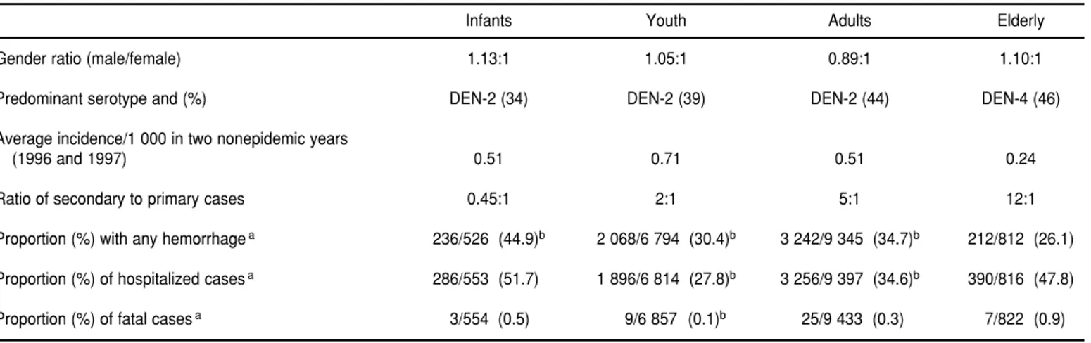
As shown in Table 1, males made up a slight majority of the laboratory-positive dengue case-patients in every age group except the adults. Of the 44 patients with a laboratory-positive case who died during the study pe-riod, 22 (50%) were male. DEN-4 was the predominant serotype among the elderly. The elderly showed the lowest incidence rate in a comparison with data for other age groups in the two nonepidemic years of 1996 and 1997. Among the 3 154 virus-positive case-patients, 2 379 of them (75.4%) had a
TABLE 1. General characteristics of laboratory-positive dengue cases, by age group, Puerto Rico, 1994–1999
Infants Youth Adults Elderly
Gender ratio (male/female) 1.13:1 1.05:1 0.89:1 1.10:1
Predominant serotype and (%) DEN-2 (34) DEN-2 (39) DEN-2 (44) DEN-4 (46)
Average incidence/1 000 in two nonepidemic years
(1996 and 1997) 0.51 0.71 0.51 0.24
Ratio of secondary to primary cases 0.45:1 2:1 5:1 12:1
Proportion (%) with any hemorrhagea 236/526 (44.9)b 2 068/6 794 (30.4)b 3 242/9 345 (34.7)b 212/812 (26.1)
Proportion (%) of hospitalized casesa 286/553 (51.7)b 1 896/6 814 (27.8)b 3 256/9 397 (34.6)b 390/816 (47.8)
secondary type immune response. As expected, the ratio of secondary to pri-mary infection increased with age, from 0.45:1 in infants to 12:1 in the elderly. The elderly also had a statis-tically significant lower frequency of hemorrhage and a statistically signifi-cant higher proportion of hospitalized patients than did either the youth or adult case-patients (Table 1). Regard-less of infecting serotype, the elderly were more likely to have been hospi-talized (48% vs. 33%) (P < 0.01) and were less likely to show hemorrhage (26% vs. 33%) (P< 0.01). The elderly had the highest case fatality rate (0.9%), and the youth had the lowest (0.1%); the difference was statistically significant (P<0.05).
Hospitalized laboratory-positive case-patients
Among the 1 757 laboratory-positive hospitalized case-patients from whom
age information was available, no sta-tistically significant differences were found among the age groups in terms of gender distribution (not shown), disease classification, or case fatality ratio (Table 2). Overall, 1 559 (88.7%) were classified as having dengue fever and 198 (11.3%) as having DHF or DSS. No cases of DSS were reported among the elderly.
Also as shown in Table 2, rash, he-patomegaly, and mucocutaneous hem-orrhage were observed less fre-quently in the elderly, but the elderly also experienced bleeding in the lower gastrointestinal tract and microhema-turia (defined as a finding of more than five red blood cells per high power field, or a positive chemical test for blood in urine) more often than did the other age groups. Microhematuria was the most common hemorrhagic manifestation in all age groups except infants, and it was reported in 68% of the elderly with any hemorrhage. Excessive capillary permeability,
mea-sured as the presence of hemoconcen-tration (hematocrit increased by 20% or more, or decreased as much after in-travenous fluid therapy), or other ob-jective evidence of increased capillary permeability (such as hypoproteine-mia, pleural effusions, or other effu-sions), was also detected significantly more frequently in the elderly than in youths and adults. Among hospital-ized patients, no clinically significant differences between the groups were found in mean maximum and mini-mum hematocrit, minimini-mum blood pressure, platelet count, albumin lev-els, or liver enzyme levels (data not shown).
Risk analysis
When considering only laboratory-positive case-patients, male gender in-creased the risk for hospitalization for infants (odds ratio (OR) = 1.62, 95% confidence interval (CI) = 1.14–2.30)
TABLE 2. Clinical characteristics for laboratory-positive hospitalized dengue cases, by age group, Puerto Rico, 1994–1999a
Infants Youths Adults Elderly
Characteristic No. % No. % No. % No. %
Constitutional symptoms
Fever 19 90.5 562 98.8 984 98.1 118 97.5
Rash 15 71.5b 245 49.4b 346 40.0b 27 25.7
Pleural or abdominal effusions 2 10.5 13 3.0 9 1.2 2 2.2
Hepatomegaly 2 10.5b 22 4.9b 30 3.8b 0 0.0
Hemorrhagic manifestations
Petechiae, purpura, or ecchymoses 5 25.0 103 22.0 158 19.0 16 15.4
Epistaxis 2 10.0 33 7.2 58 7.0 4 3.8
Gum bleeding 1 5.0 24 5.2 73 8.8b 3 2.9
Vaginal bleeding (women) 0 0.0 10 2.2 33 4.2b 0 0.0
Hematuria 0 0.0 13 2.8 32 4.0 3 2.9
Lower gastrointestinal bleeding 1 5.9 12 2.9 19 2.6 8 8.1
Hematemesis 2 10.0 27 5.8 43 5.2 3 2.9
Hemoptysis 0 0.0 9 1.8 26 3.2 1 1.0
Microhematuria 1 8.3 98 22.8b 293 37.5 38 40.0
Bleeding at puncture site 0 0.0 28 6.2 70 8.7 5 4.9
Other bleeding 1 5.3 3 0.7 15 1.9 1 1.0
Excessive capillary leakage 6 28.6 104 18.0b 229 22.2b 42 33.3
Severe outcome
Shock 1 5.3 6 1.3 10 1.3 0 0.0
Death 1 8.3 6 1.9 10 1.8 3 4.3
Disease classification
Dengue fever 17 81.0 523 90.6 910 88.1 109 86.2
Dengue hemorrhagic fever 3 14.3 51 8.8 119 11.5 17 13.5
Dengue shock syndrome 1 4.8 3 0.5 4 0.4 0 0.0
and youths (OR = 1.13; 95% CI = 1.01– 1.26), and hemorrhage increased the risk of hospitalization in all groups except the elderly. Neither male gen-der nor history of hemorrhage signifi-cantly increased the risk of death for patients in any of the age groups.
When we looked at age subgroups among the elderly (65–69, 70–79, 80–89, and 90 years or older), the fre-quency of reported hemorrhage, hos-pitalization, or disease outcome was similar. An increased risk for hemor-rhage in comparison to other age groups was seen only in males 80–89 years old (OR = 4.31; 95% CI = 1.46– 13.13). However, neither gender, hem-orrhage, nor secondary infection in-creased the risk for hospitalization in any of the elderly age subgroups (data not shown).
Multiple regression analysis among the laboratory-positive patients showed that hemorrhage and male sex were independent risk factors for hospital-ization (Table 3). Hemorrhage was also an independent risk factor for death. After adjustment for gender and the presence of hemorrhage, the elderly and infants had similar risks of hospitalization and death. The elderly had 2.4 times the risk of hospitaliza-tion as did youths, and 1.7 times the risk of adults. Also, the risk of death in the elderly was 6.8 times that of youths and 3.4 times that of adults.
DISCUSSION
Although dengue infection in adults has been described in prior publica-tions (11, 14–20, 31–36), this study pro-vides the first detailed analysis of clin-ical manifestations of dengue in the elderly (aged 65 years or older). Three markers of disease severity were eval-uated in this study: the presence of hemorrhage, hospitalization, and fatal outcome. Even when the clinical pre-sentations of dengue fever and severe dengue infection in this age group are similar to the disease presentations de-scribed in clinical studies in other age groups, subtle differences were found. Hemorrhages were reported less
fre-hospitalized more often and had a higher case fatality ratio than did youths and adults. These findings are of increasing importance for dengue epidemiology and medical care, in view of the expanding nature of dengue and DHF in a world that is facing an aging population.
Many of the clinical manifestations of dengue fever found in our study of the elderly are consistent with previ-ous clinical descriptions in adults. Rash was less commonly found in the elderly (probably because of the rela-tive frequency of secondary infections in this segment of the population (33)), and the percentage of DHF patients with hepatomegaly decreased with age (none of the elderly patients was observed to have hepatomegaly). He-patic weight declines with age, and in vivo ultrasound studies have shown that liver volume is 17% to 28% lower in those over age 65 than in those under age 40, which could be a con-tributing factor for this finding (37, 38).
Another finding to be kept in mind in the clinical evaluation of an elderly patient is that fewer hemorrhagic man-ifestations, especially mucosal and cu-taneous hemorrhages, were reported in this age group. However, hemor-rhage is a marker for severe disease and even though the aged report fewer hemorrhagic manifestations (overall), microhematuria and occult gastroin-testinal bleeding occurred more fre-quently than did other hemorrhagic manifestations. Therefore, if obvious bleeding is not evident, it should be
patients in Puerto Rico (14), in our study the most frequent hemorrhagic manifestation in all age groups except infants was microscopic hematuria. It was present in 40% of the elderly case-patients who exhibited hemorrhage when they sought treatment and was the only hemorrhagic manifestation in 68% of the 17 elderly case-patients with DHF. We also found a higher proportion of elderly patients with in-creased vascular permeability. The pathophysiological mechanism be-hind this is not clear, but the higher frequency of this important contribu-tor to DHF (39–42) could be an impor-tant factor in the severity of dengue in the elderly.
Our study was based on surveil-lance data, which by its nature may have led to preferential reporting of severe cases and incomplete recording of important items such as hospitaliza-tion status and disease outcome. Dif-ferential reporting of symptoms by age, a potential explanation for our findings, could not be assessed with the data available, but less frequent re-porting of hemorrhage based solely on the age of the patient would not be expected. If differential reporting were present, it would be related more to the source of reporting (e.g., physi-cians, nurses, or ICNs) and, therefore, should be equally distributed among all age groups.
We found that the elderly with dengue were hospitalized as often as infants and more frequently than youths and adults. This might be
ex-TABLE 3. Multivariate analysis showing odds ratio (OR) and 95% confidence interval (CI) for risk for hospitalization and death among laboratory-positive dengue cases in Puerto Rico, 1994–1999
Hospitalization Death
Groups compareda OR 95% CI OR 95% CI
Elderly vs. infants 0.91 0.73–1.13 1.80 0.46–7.04
Elderly vs. youth 2.43b 2.10–2.82 6.78b 2.51–18.3
Elderly vs. adults 1.71b 1.48–1.98 3.45b 1.49–8.05
Hemorrhage vs. no-hemorrhage 1.51b 1.42–1.62 2.06b 1.14–3.74
Males vs. females 1.09b 1.03–1.17 1.07 0.59–1.94
1. World Health Organization. Dengue haemor-rhagic fever. Diagnosis, treatment, prevention and control. 2nd ed. Geneva: WHO; 1997. 2. Pinheiro FP, Corber SJ. Global situation of
dengue and dengue haemorrhagic fever, and its emergence in the Americas. World Health Stat Q 1997;50(3–4):161–169.
3. Thein S, Aung MM, Aye M, Zaw A, Aye K, Aye KM, et al. Risk factors in dengue shock syndrome. Am J Trop Med Hyg 1997;56:566– 572.
4. Burke DS, Nisalak A, Johnson DE, Scott RM. A prospective study of dengue infections in Bangkok. Am J Trop Med Hyg 1988;38:172– 180.
5. Sangkawibha N, Rojanasuphot S, Ahandrik S. Risk factors in dengue shock syndrome: a prospective study in Rayong, Thailand. Am J Epidemiol 1984;120:653–669.
6. George R, Lum LC. Clinical spectrum of dengue. In: Gubler DJ, Kuno G, eds. Dengue and dengue hemorrhagic syndrome. Walling-ford, United Kingdom: CAB International; 1997. Pp. 89–113.
7. Nelson E. Hemorrhagic fever in children in Thailand. Trop Pediatr 1960;56:101–107. 8. Sumarmo, Wulur H, Jahja E, Gubler DJ,
Suharyono W, Sorensen K. Clinical observa-tions on virologically confirmed fatal dengue
infections in Jakarta, Indonesia. Bull World Health Org 1983;61:693–701.
9. Halstead S, Nimmannitya S, Margiotta M. Dengue and chinkungunya virus infection in man in Thailand, 1962–1964. Am J Trop Med Hyg 1969;18:972–983.
10. Eram S, Setyabudi Y, Sadono I, Sutrisno S, Gubler DJ, Sulianti J. Epidemic dengue hem-orrhagic fever in rural Indonesia. Am J Trop Med Hyg 1979;28:711–716.
11. Hayes C, Manaloto C, Gonzalez A, Ranoa P. Dengue infections in the Philippines: clinical and virological finding on 517 hospitalized pa-tients. Am J Trop Med Hyg 1988;39:110–116. 12. Songco R, Hayes C, Leus CD, Manaloto COR.
Dengue fever/dengue hemorrhagic fever in Filipino children: clinical experience during the 1983–1984 epidemic. Southeast Asian J Trop Med Publ Hlth 1987;18:284–290. 13. Gubler DJ. Dengue and dengue hemorrhagic
fever: its history and resurgence as a global public health problem. In: Gubler DJ, Kuno G, eds. Dengue and dengue hemorrhagic fever. Wallingford, United Kingdom: CAB Interna-tional; 1997. Pp. 1–22.
14. Rigau-Pérez JG, Puerto Rico Association of Epi-demiologists. Clinical manifestations of dengue hemorrhagic fever in Puerto Rico 1990–1991. Rev Panam Salud Publica 1997;1(5):381–388.
15. Miagostovich MP, Ramos RG, Nicol AF, Nogueira RMR, Cuzzi-Maya T, Oliveira AV, et al. Retrospective study on dengue fatal cases. Clin Neuropath 1997;16(4):204–208. 16. Gomez Dantes H, Koopman JS, Addy CL,
Zarate ML, Vaca Marin MA, Longini IM, et al. Dengue epidemics on the Pacific coast of Mexico. Int J Epid 1988;17(1):178–186. 17. Travassos da Rosa A, Vasconcelos P,
Travas-sos da Rosa ES, Rodrigues SG, Mondet B, Cruz A, et al. Dengue epidemic in Belém, Pará, Brazil 1996–97. Emerg Inf Dis 2000;6(3): 298–301.
18. Rosso F, Restrepo de Meza MT, Alzate A, Muñóz J, Moreno CH. Dengue hemorrágico en el Hospital Universitario del Valle, 1990– 1992. Colombia Médica 1994;25(1):10–14. 19. Rodríguez Gómez JH, Calderón Moncloa JC.
Dengue clásico: aspectos epidemiológicos en el Hospital de Apoyo Integrado Tarapoto-1990. Acta Médica Peruana 1992;16(3):187–193. 20. Díaz A, Kourí G, Guzmán MG, Lobaina L,
Bravo J, Ruiz A, et al. Description of the clinical picture of dengue hemorrhagic fever/dengue shock syndrome (DHF/DSS) in adults. Bull Pan Am Health Organ 1988;22(2): 133–144. 21. Bell R, High K. Alterations of immune defense
mechanisms in the elderly: the role of nutri-tion. Infect Med 1997;14:415–424.
REFERENCES infants and elderly as “fragile” since
they are more likely to develop com-plications during any disease process and therefore require closer observa-tion. This would lead to less-severe cases being hospitalized more often and also to an increase in the propor-tion of patients hospitalized due to a concern (age) unrelated to dengue. However, if age were the reason for differential rates of hospitalization, we would expect a lower case fatality rate among the elderly, because milder cases would inflate the denominator. Nevertheless, in our study we found the opposite, that is, a higher case fa-tality rate among the elderly. These considerations highlight the need for prospective clinical studies that in-clude the elderly.
Dengue complications are enhanced in well-nourished children with good immune response (43). The nutritional and immune status of the elderly was not assessed in this study, but it is clear that even when the elderly may in gen-eral have diminished immunity, they
still present with severe dengue. This requires further evaluation, in consid-eration of the multifactorial etiology of severe dengue infection. Another rele-vant contributor to the findings in this study might be concurrent disease, since aging is associated with the pres-ence of chronic conditions and in-creased susceptibility to infectious disease. Previous studies have docu-mented rare occurrences of severe dengue and co-infections or the pres-ence of chronic diseases (44–46). Sur-veillance data such as those we ana-lyzed do not provide information about coexisting diseases. However, a previous study of all 57 DHF patients documented in Puerto Rico in 1990 and 1991 (aged 0 to 86 years) found no evi-dence of comorbidity among them, suggesting that DHF diagnosis would rarely be attributed to comorbidity (14).
In summary, the elderly in Puerto Rico often develop severe illness when infected with the dengue virus. They show higher rates of hospitalization, DHF, and death than do infected
youth and younger adults, in a pattern similar to that of infants. We applied the WHO case definition for DHF strictly, so DHF diagnosis was not applied differentially. An elderly case may present with increased vascular permeability and may require hospi-talization (two markers of disease se-verity), even in the absence of evident hemorrhage. The clinical evaluation of elderly patients with suspected dengue must include a thorough cli-nical examination. An assessment of the occurrence of mild hemorrhage, including the presence of microhema-turia, occult blood in stools, and in-creased capillary permeability is very important to avoid complications from delayed identification and treatment of severe dengue infection.
Acknowledgments. The authors
22. United States of America, Census Bureau. Census 2000 data for Puerto Rico. Available from: http://www.census.gov/census2000/ states/pr.html [Internet site]. Accessed 15 Jan-uary 2003.
23. Rigau-Perez JG, Ayala-Lopez A, García-Rivera EJ, Hudson SM, Vorndam V, Reiter P, et al. The reappearance of dengue-3 and sub-sequent dengue-4 and dengue-1 epidemic in Puerto Rico in 1998. Am J Trop Med Hyg 2002;67:355–362.
24. Gubler DJ, Kuno G, Sather GE, Velez M, Oliver A. Mosquito cell cultures and specific monoclonal antibodies in surveillance for dengue viruses. Am J Trop Med Hyg 1984;33: 158–165.
25. Burke DS, Nisalak A, Ussery MA. Antibody capture immunoassay detection of Japanese encephalitis virus immunoglobulin M and G antibodies in cerebrospinal fluid. J Clin Mi-crobiol 1982;15:1034–1042.
26. Chungue E, Marché G, Pichart R, Boutin JP, Roux J. Comparison of immunoglobulin G enzyme-linked immunosorbent assay (IgG-ELISA) and hemagglutination inhibition (HI) test for the detection of dengue antibodies: prevalence of dengue IgG-ELISA antibodies in Tahiti. Trans R Soc Trop Med Hyg 1989;83: 708–711.
27. Miagostovich MP, Nogueira RMR, dos Santos FB, Schartmayr HG, Araujo ESM, Vorndam V. Evaluation of an IgG enzyme-linked immuno-sorbent assay for dengue diagnosis. J Clin Virol 1999;14:183–189.
28. Zar JH. Biostatistical analysis. 2nd ed. Engle-wood Cliffs, New Jersey, United States of America: Prentice-Hall; 1984.
29. Dean AG, Dean JA, Coulombier D, Brendel KA, Smith DC, Burton AH, et al. Epi Info,
ver-sion 6: a word processing, database, and sta-tistics program for epidemiology on micro-computers. Atlanta, Georgia, United States: Centers for Disease Control and Prevention; 1994.
30. Abramson JH, Gahlinger PM. Computer pro-grams for epidemiologists (PEPI). Version 3.01. Available from: http://www.usd-inc.com/ pepi.html [Internet site]. Accessed November 1999.
31. Wali JP, Biswas A, Handa R, Aggarwal P, Wig N, Dwivedi SN. Dengue hemorrhagic fever in adults: a prospective study of 110 cases. Trop Doct 1999;29:27–30.
32. Kuberski T, Rosen L, Reed D, Mataika J. Clin-ical and laboratory observations on patients with primary and secondary dengue type in-fectious with hemorrhagic manifestations in Fiji. Am J Trop Med Hyg 1977;26:775–783. 33. Cobra C, Rigau-Pérez J, Kuno G, Vorndam V.
Symptoms of dengue fever in relation to host immunologic response and virus serotype, Puerto Rico, 1990–1991. Am J Epidemiol 1995; 142:1204–1211.
34. Guzman MG, Kouri GP, Bravo J, Soler M, Vazquez S, Morier L. Dengue hemorrhagic fever in Cuba, 1981: a retrospective seroepide-miologic study. Am J Trop Med Hyg 1990;42: 179–184.
35. Macridi NG. L’épidémie de dengue a Athè-nes. Rev Hyg Med Preventive 1929;51(4):241– 267.
36. Guzmán MG, Alvarez M, Rodríguez R, Rosario D, Vázquez S, Valdés L, et al. Fatal dengue hemorrhagic fever in Cuba, 1997. Int J Infect Dis 1999;3(3):130–135.
37. Merck & Co. The aging liver. In: The Merck manual of geriatrics. 2nd ed. Merck & Co.; 1995. Available from: http://www.merck.
com/pubs/mm_geriatrics/59x.htm [Internet site]. Accessed 28 June 2000.
38. Merck & Co. Normal aging changes. In: The Merck manual of geriatrics. 2nd ed. Merck & Co.; 1995. Available from: http://www. merck.com/pubs/mm_geriatrics/33x.htm [Internet site]. Accessed 28 June 2000. 39. Sen P, Middleton J, George P, Gombert M, Lee
Douglas, Louria D. Host defense abnormali-ties and infections in older persons. Infect Urol 1995;8(1):23–29.
40. Kalayanarooj S, Vanghn DW, Nimmannitya S. Early clinical and laboratory indicators of acute dengue illness. J Infect Dis 1997;176: 313–321.
41. Monath TP. Early indicators in acute dengue infection. Lancet 1997;350:1719–1720. 42. Kliks SC, Nisalak A, Brand W, Wahl L, Burke
DS. Antibody-dependent enhancement of dengue virus growth in human monocytes as a risk factor for dengue hemorrhagic fever. Am J Trop Med Hyg 1989;40:444–451. 43. Thisyakorn U, Nimmannitya S. Nutritional
status of children with dengue hemorrhagic fever. Clin Inf Dis 1993;16:295–297.
44. Bravo JR, Guzman MG, Kouri GP. Why dengue hemorrhagic fever in Cuba? Individual risk factors for dengue hemorrhagic fever/dengue shock syndrome (DHF/DSS).Trans R Soc Trop Med Hyg 1987;81:816–820.
45. Goh KT. Changing epidemiology of dengue in Singapore. Lancet 1995;346:1098.
46. Pancharoen C, Thisyakorn U. Coinfections in dengue patients. Pediatr Infect Dis J 1998;17: 81–82.
Manuscript received 9 April 2002. Revised version ac-cepted for publication on 15 November 2002.
Objetivos. Las formas graves de dengue afectan a todos los grupos de edad en las
Américas. Sin embargo, es escasa la información detallada sobre esta enfermedad en adultos mayores. El objetivo de este trabajo es describir esta enfermedad en personas de edad avanzada.
Métodos. Se revisaron los formularios usados para investigar los casos sospechados
de dengue que fueron enviados con muestras para el diagnóstico, así como los formes clínicos confeccionados por personal de enfermería dedicado al control de in-fecciones en Puerto Rico en el período entre 1994 y 1999.
Resultados. Los casos positivos según las pruebas de laboratorio fueron asignados a cuatro grupos de edad: niños (≤1 año: 554 casos), jóvenes (de 2 a 18 años: 6 857 casos), adultos (de 19 a 64 años: 9 433 casos) y adultos mayores (≥65 años: 822 casos). Inde-pendientemente del serotipo infectante, los adultos mayores fueron los más propensos a haber sido hospitalizados (48% vs. 33%) (P< 0,01) y los menos propensos a las he-morragias (26% vs. 33%) (P< 0,01). Según el análisis de múltiples variables, con datos controlados según el sexo y la presencia de hemorragia, los adultos mayores mostraron un mayor riesgo de ser hospitalizados y de morir que los jóvenes y los adultos.
Conclusiones. En comparación con los jóvenes y los adultos, los adultos mayores se
muestran más propensos a desarrollar formas graves de dengue al ser infectados por el virus causal, siendo el patrón similar al de los niños. La evaluación clínica de los adultos mayores con dengue debe incluir una minuciosa evaluación del aumento de la permeabilidad capilar y de posibles hemorragias ocultas, con el fin de evitar com-plicaciones por la demora en identificar y tratar los casos graves de dengue. En vista de la propagación del dengue y de su forma hemorrágica, estos resultados son de una importancia creciente en lo referente a la epidemiología y la atención médica de los casos de dengue, en un mundo donde también van en aumento el número de adultos mayores y la proporción de la población que ellos representan.
RESUMEN