J. Evid. Based Med. Healthc., pISSN- 2349-2562, eISSN- 2349-2570/ Vol. 3/Issue 57/July 18, 2016 Page 3011
THROMBOCYTOPENIA AT A TERTIARY CARE CENTRE
Ravindra Kumar Sudarsi1, Markandeya Rao G. K2, Nanditha Nallamanddi3, Giridhar Gorrepotu4
1Associate Professor, Department of Medicine, Osmania Medical College/General Hospital, Hyderabad. 2Professor, Department of Medicine, Osmania Medical College/General Hospital, Hyderabad.
3Resident, Department of Medicine, Kamineni Institute of Medical Sciences, Hyderabad. 4Resident, Department of Medicine, Sri Venkat Sai Medial College, Hyderabad.
ABSTRACT
BACKGROUND AND OBJECTIVES
The aetiologies of thrombocytopenia are diverse. Various studies on thrombocytopenia done in the past have related to specific aetiologies. This study attempts to determine the common aetiologies of thrombocytopenia and bleeding manifestations in adult patients admitted under Department of Medicine.
METHODS
Patients older than 18 years of age who were first time found to have thrombocytopenia at admission under Department of Medicine between 1st October 2014 and 31st September 2015 were followed up during their stay in hospital and the diagnosis made, bleeding manifestations, and requirement of platelet transfusions were recorded.
RESULTS
200 patients were included in the study. Dengue/dengue-like fever was the diagnosis made in 30% of patients followed by malaria (22%), undiagnosed aetiology (9%), HELLP (6%), snake bite and sepsis 5% each, ITP and megaloblastic anaemia 4.5% each, haematological malignancies and CTD 3% each, and other rare causes formed the rest.
Bleeding secondary to thrombocytopenia was seen in 36 patients. 22.2% of them were diagnosed as dengue, 19.4% had malaria, 16.7% had ITP, 11.1% of patients with HELLP, 8.3% each had malignancy and undiagnosed viral infections, 5.6% each had CTD and MDS, and 2.8% had aplastic anaemia.
Dengue fever was responsible for thrombocytopenia-related bleeding in 8 cases, ITP in 6 cases, malaria in 7 cases, HELLP in 4 cases, malignancy and undiagnosed aetiology 3 cases each, CTD and MDS 2 cases each, and aplastic anaemia in 1 case.
The common bleeding manifestations were GIT (Melena), petechial rash, and menorrhagia occurring in 33.3%, 22.2% and 13.8% of total number of patients respectively.
30.5% of those who had bleeding secondary to thrombocytopenia had platelet count <5000/μL. 68.7% of those with platelet count <5000/μL had bleeding manifestation.
36.6% of those who had bleeding secondary to thrombocytopenia had platelet count 5,000-10,000/μL. 65.0% of those with platelet count 5,000-10,000/μL had bleeding manifestation.
46 patients were given platelet transfusions, 65.2% of these cases were transfused because of bleeding and 34.78% were prophylactic.
INTERPRETATION AND CONCLUSION
Dengue fever was the commonest cause of newly found thrombocytopenia in adult patients admitted under Department of Medicine. The commonest bleeding manifestation secondary to thrombocytopenia was GIT bleed. Though, Dengue and Malaria were the most common aetiology diagnosed in patients with bleeding secondary to thrombocytopenia, the proportion of aplastic anaemia, MDS, and ITP patients who had bleeding manifestation was higher than the proportion of Dengue and Malaria patients who developed bleeding. Major proportion of bleeding manifestation occurred in patients with platelet count <5000/μL. There was no definitive trigger value of platelet for platelet transfusion and transfusion because of bleeding was greater than prophylactic transfusion.
KEYWORDS Thrombocytopenia.
HOW TO CITE THIS ARTICLE: Sudarsi RK, Rao MGK, Nallamanddi N,et al. Thrombocytopenia at a tertiary care centre. J. Evid. Based Med. Healthc. 2016; 3(57), 3011-3019. DOI: 10.18410/jebmh/2016/656
INTRODUCTION: Nature has designed a complex, but ingenious system to maintain blood in the vascular system fluid and free from clots, yet allow the rapid formation of solid plug to close ruptures as other forms of injury to blood vessels. This process is referred to as normal haemostasis.1
Financial or Other, Competing Interest: None. Submission 11-06-2016, Peer Review 24-06-2016, Acceptance 30-06-2016, Published 18-07-2016. Corresponding Author:
Dr. Ravindra Kumar Sudarsi,
#A-70, Vengal Rao Nagar, Hyderabad-500038. E-mail: [email protected]
J. Evid. Based Med. Healthc., pISSN- 2349-2562, eISSN- 2349-2570/ Vol. 3/Issue 57/July 18, 2016 Page 3012 Thrombosis denotes the formation of a clotted mass of
blood within the noninterrupted vascular system. It represents to a considerable extent, a pathologic extension of normal haemostasis. Platelets play a central role in normal haemostasis and therefore also in thrombosis. Despite their lack of nucleus, head, and heart, these tiny structures (about 2 micrometer in diameter) are amazingly versatile. In the haemostatic cascade, platelets undergo 3 important reactions: (1) adhesion and shape change, (2) secretion (relapse reaction), and (3) aggregation, collectively referred to as platelet activation.1
Thrombocytopenia is characterised by bleeding most often from small vessels. This bleeding can manifest as petechiae over the skin, haemorrhages from mucosa of
gastrointestinal or genitourinary tract. Intracranial
haemorrhage is a dangerous consequence in
thrombocytopenic patients.
In 1910, W.W. Duke demonstrated the relation of blood platelets to haemorrhagic disease and for the first time demonstrated that haemorrhagic disease caused by thrombocytopenia could be relieved by platelet transfusion.2
Platelet transfusions are given prophylactically or therapeutically to thrombocytopenia patients and to patients undergoing invasive procedures.
Platelets are very important in haemostatic mechanism. The causes of thrombocytopenia are varied and they can be associated with bleeding, which at times can be life threatening (e.g. intracranial bleed). Timely recognition and treatment of underlying condition, platelet transfusions are required to prevent fatal outcomes.
OBJECTIVES: This study aims to determine the following; 1. The relative frequency of different disease conditions
presenting as newly-found thrombocytopenia in adult patients getting admitted under Department of Medicine at an Indian tertiary care hospital.
2. The proportion of patients who had bleeding
manifestations.
3. The different bleeding manifestations in order of their commonness of occurring.
4. The percentage of patients requiring interventions like platelet substitutes, steroids, splenectomy.
5. To determine whether low platelet count or presence
of bleeding manifestation was considered more often as indicator for platelet transfusions.
METHODS:
Source of Data: Adult patients admitted under Department of Medicine who were detected to have thrombocytopenia at admission. The trend of the platelet count post admission, bleeding manifestations and requirement of platelet transfusions in these were noted during their hospital stay.
Method of Collection of Data:
Inclusion Criteria: Patients presenting with thrombocytopenia to Department of Medicine during the period between 1st October 2014 and 31st September 2015 (12 months) satisfying the following inclusion criteria were included in the study.
a) Age >18 years.
b) Platelet count <1,00,000/μL (at admission).
Exclusion Criteria: a) Age <18 years.
b) Patients who were earlier diagnosed to have
conditions that are known to cause thrombocytopenia (e.g. known cases of haematological malignancies, aplastic anaemia, MDS, ITP).
c) Patients who have received chemotherapy for
neoplasms in previous admissions subsequently presenting as thrombocytopenia.
d) Patients who have already received platelet
transfusion prior to admission.
All patients included in the study were followed up during their course in the hospital. The trend of their platelet counts was recorded. The decision as to when the platelet counts were repeated was that of the treating physician.
The bleeding manifestations patients presented with or developed during their course in hospital were recorded.
The diagnosis made in each of these cases was noted down.
The proportion of study patients requiring interventions to improve platelet count like platelet transfusion, steroids and biologicals, and the reason for such intervention was recorded.
Proportion of patients receiving platelet transfusions because of presence of a bleeding manifestation and proportion requiring prophylactic platelet transfusion were determined.
RESULTS:
Aetiology: During the study period of 12 months (October 2014 to September 2015), 200 patients satisfied the inclusion criteria and were included in the study. There were 124 males (mean age: 32.4 yrs.) and 76 females (mean age: 25.5 yrs.) in the study population. The commonest aetiology responsible for newly-diagnosed thrombocytopenia in adult patients admitted under Department of Medicine was found to be dengue. 60 patients were diagnosed to have dengue making up 30 percent of the total number of cases.
Malaria was a close second diagnosis with 44 cases (22%). Of the patients with malaria, Plasmodium falciparum was the cause for thrombocytopenia in 29 patients (66% of the total number of malaria cases) followed by Plasmodium vivax with 11 patients and 4 patients were found to have mixed infection.
Undiagnosed aetiology was next most common cause of thrombocytopenia where the patient presented with thrombocytopenia and dengue, malaria, salmonella, HIV, HBsAg serology was negative constituting 18 cases [9% of the total cases].
HELLP syndrome was fourth most common cause of thrombocytopenia making 12 cases [6%].
J. Evid. Based Med. Healthc., pISSN- 2349-2562, eISSN- 2349-2570/ Vol. 3/Issue 57/July 18, 2016 Page 3013 ITP was next most common cause making up 9 cases
of total [4.5%].
Megaloblastic anaemia was next most common cause of thrombocytopenia making 8 cases of total [4%].
Haematological malignancies and connective tissue disorders constitute 6 cases each contributing 3% of cases each. Among malignancies, 2 cases are multiple myeloma, 2 cases are AML, 1 case each of non-Hodgkin’s lymphoma and CLL. Among connective tissue disorders, 4 cases are SLE and 2 cases are mixed connective tissue disorder.
HIV constitutes 5 cases [2.5%], Salmonella constitutes 4 cases [2%], and hepatitis constitutes 2 cases [1%].
Myelodysplastic syndrome 2 cases [1%], aplastic anaemia 1 case [0.5%].
Portal hypertension with hypersplenism 2 cases [1%].
DISCUSSION: A total number of 200 patients were included in the study during the study period of 12 months from October 2014 to September 2015. Viral fever was the most common aetiology found in adult patients who were found to have thrombocytopenia for the first time and were admitted under Department of Medicine. 30% of the patients were diagnosed to have dengue.
Fever-associated thrombocytopenia accounted for 143 cases. There were 67.8% males and 32.2% females in this
group. This ratio was similar to a study done by Nair PS et al3 at a New Delhi tertiary care hospital between March 2002
to June 2003 where 109 patients with fever-associated thrombocytopenia were studied. There were 69.7% males and 30.3% females in that study.
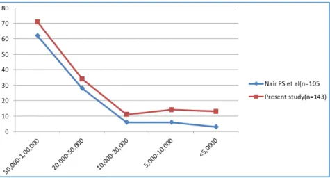
Platelet count range (cells/μL)
Nair PS, St. Stephen’s Hospital, New
Delhi, 2003
Present study
50,000-1,00,000 62(56.8%) 71(49.6%)
20,000-50,000 28(25.7%) 34(23.8%)
10,000-20,000 6(9.2%) 11(7.7%)
5,000-10,000 6(5.5%) 14(9.8%)
<5,000 3(2.7%) 13(9.1%)
Table 1: Number of Patients with Fever-Associated Thrombocytopenia in Different Platelet Ranges
There were more patients in the below <5000/μL platelet count group in the present study compared to the study done in New Delhi (9.1% vs. 2.7%). The percentages of patients below 20,000/μL platelet count were 38% in the present study and 17.4% in the study mentioned above.
Chart 1: Comparison of Platelet Counts in the Present Study with That of the Study Done by Nair PS et al, New Delhi, 2003
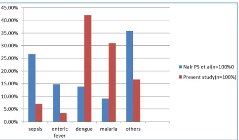
Sepsis was the commonest cause of fever-associated thrombocytopenia in the St. Stephen’s study followed by enteric fever and dengue fever. In contrast to this data from Northern India, dengue was the leading cause of fever-associated thrombocytopenia followed by malaria and sepsis. Only five (2.5%) confirmed cases of enteric fever had platelet count <1,00,000/μL in the present study whereas 14% of cases had enteric fever in the St. Stephen's study. Enteric fever presenting with thrombocytopenia hence appears to be less common in Southern India where the present study was conducted, compared to that in the North.
Aetiology of thrombocytopenia
Nair PS et al, New Delhi,
2003
Present study
Sepsis 29(26.65%) 10(7%)
Salmonella 16(14.7%) 5(3.4%)
Dengue 15(13.8%) 60(42%)
Malaria 10(9.2%) 44(31%)
Others 39(35.8%) 24(16.6%)
Total 109 143
J. Evid. Based Med. Healthc., pISSN- 2349-2562, eISSN- 2349-2570/ Vol. 3/Issue 57/July 18, 2016 Page 3014
Chart 2: Comparison of Aetiologies of Fever-Associated Thrombocytopenia
Between present study and study done by Nair PS et al New Delhi, 2003, the commonest bleeding manifestation secondary to thrombocytopenia was GIT bleed, which was seen in 33.3% of cases followed by petechial rash in 22.2%. 13.8% of women had menorrhagia.
The frequency of bleeding manifestation in fever-associated thrombocytopenia was less in the present study compared to the study done in St. Stephen's, New Delhi.
12.6% of those with fever-associated thrombocytopenia had bleeding manifestation in the present study whereas 41.3% had bleeding manifestation in the St. Stephen's study. Petechial rash and GI bleed were the most common bleeding manifestation in the St. Stephen's study (9.2% each). Petechial rash was not high in the list in the present study among patients with fever-associated thrombocytopenia (2.8%).
Bleeding manifestation No. of patients Percentage of patients
Nair’s PS et al Present study Nair’s PS et al Present study
Petechial rash 10 4 9.2% 2.8%
GI bleed 10 6 9.2% 4.2%
Epistaxis 7 2 6.4% 1.4%
Gum bleed 6 1 5.5% 0.7%
Haematuria 5 2 4.6% 1.4%
Haemoptysis 3 0 2.8% 0%
Others including menorrhagia 4 3 3.7% 2.15
45/109 18/143 41.3% 12.6%
Table 3: Comparison of Bleeding Manifestations Between Present
Study and Study Done by Nair’s P S et al New Delhi 2003
However, GI bleed (4.2%) was most frequent bleeding manifestation in this study also.
Bleeding manifestations in patients studied for suspected dengue fever in a study one at Hawaii in 2001-2002 (1644 cases) found petechiae to be the commonest bleeding manifestation followed by menorrhagia and epistaxis. Though, viral exanthema was reported. There was no report of petechiae in patients with dengue fever in the present study. Petechiae were seen commonly in cases of ITP. Bleeding manifestation because of thrombocytopenia secondary to viral fever were seen only in 7 of the 60 cases of viral fever included in the study with two each having menorrhagia and melena, one patient had a petechial rash, one had epistaxis, and one had haematuria. There were no cases with haematemesis or gum bleed in patients with dengue fever. The fact that the sample size of cases of viral
fever was small compared to the Hawaii study is a limitation in the comparison.
Bleeding manifestation
Paul V Effler et al, Hawai4 2001-2002
(% 0f patients)
Present study (% of patients)
Petechiae 8% 1.7%
Menorrhagia 5.5% 3.3%
Epistaxis 4.4% 1.7%
Gum bleed 4.5% 0%
Melena 3.1% 3.3%
Haematuria 3.1% 1.7%
Haematemesis 1.3% 0%
J. Evid. Based Med. Healthc., pISSN- 2349-2562, eISSN- 2349-2570/ Vol. 3/Issue 57/July 18, 2016 Page 3015 Plasmodium falciparum and Plasmodium vivax are
endemic infections in India and are associated with mild haematological abnormalities. Severe thrombocytopenia is common in isolated falciparum and mixed falciparum/vivax malaria, but is very rare in isolated P. vivax infection. In the present study, 27.3% of cases of Plasmodium vivax malaria had platelet count <40000/μL whereas a study done by Myong-don, et al4 in South Korea in 1996-1999 on 101
patients of Plasmodium vivax had only 5% fall into this group. Lower platelet counts secondary to isolated Plasmodium vivax malaria appears to be commoner in the Indian study. It is however to be noted that none had platelet count less than 20,000/μL. In Horstmann's series,(5)
the lowest count in 39 cases of vivax malaria was 44 x 109/L.
Profound thrombocytopenia secondary to isolated vivax malaria have been reported from India, one with an initial platelet count 5 x 10⁹/L(6) and another 8 x 10⁹/L(7 ), these
being first and second cases of profound thrombocytopenia secondary to vivax infection being reported in literature. 54.5% of cases of vivax malaria fell into the 40 x 109 to 80
x 109 category whereas 45.4% into this category in the
South Korean study. Thrombocytopenia secondary to isolated vivax infection probably tends to be more severe in India.
Platelet count (/μL)
Myoung-don OH et al, Seoul, South korea1996-99
Present study
<40,000 5(5%) 3(27.3%)
40,000-80,000 46(45.4%) 6(54.5%)
>80,000 29(49.6%) 218.2%)
Total 101 11
Table 5: Comparison of Platelet Count Range in Plasmodium Vivax Malaria
Definitive studies (e.g. well-designed, prospective, randomised clinical trials) are not available either historically or at present to support evidence-based decisions regarding a trigger level of platelet count that indicates prophylactic platelet transfusion.(8) Instead, retrospective reviews and
anecdotal reports provide observational data to assist in best guess clinical practices. Reasonable clinical practice until more definitive data become available is to transfuse enough platelets per each transfusion to maintain the blood platelet count >10 x 109/L in stable non-bleeding patients, >20 x
109/L in unstable non-bleeding patients, and >50 x 109/L in
bleeding patients or in those undergoing invasive procedures.(9)
In the present study, 16 out of 46 patients who received platelet transfusion were transfused prophylactically. No definitive trigger value of platelet count for prophylactic transfusion was observed. However, 81.3% of those who received prophylactic platelet transfusions had a platelet count less than 20,000/μL. 18.7% of the prophylactic platelet transfusions were however given to patients with platelet count >20,000/μL. No uniform guidelines for prophylactic transfusion therapy were hence found to be followed.
This study had several limitations. As the study was conducted only in patients admitted under Department of Medicine, it did not include surgical cases. Obstetric and gynaecological causes of thrombocytopenia, which were referred to our hospital were studied and patients with portal hypertension, hypersplenism who were admitted directly under Department of Gastroenterology were not included in the study. Drug-induced thrombocytopenia was also not evaluated. Most of the haematological malignancies were admitted directly in the cancer hospital and hence not evaluated thoroughly.(10)
J. Evid. Based Med. Healthc., pISSN- 2349-2562, eISSN- 2349-2570/ Vol. 3/Issue 57/July 18, 2016 Page 3016
Disease Number
of Cases
Percentage of cases
HIV 5 2.5
Salmonella 4 2
MDS 2 1
Aplastic anaemia 1 0.5
Portal hypertension with
hypersplenism 2 1
Table 6: Miscellaneous Causes of Thrombocytopenia
Chart 4: Haematological and Lymphoid Malignancies Responsible for Thrombocytopenia
Chart 5: Infectious Causes of Thrombocytopenia
Chart 6: Type of Malaria in Patients With Thrombocytopenia
Bleeding Manifestations: Of the total 200 patients with platelet count <1,00,000/μL, bleeding manifestations were seen in a total of 48 patients (24%).
2 patients with portal hypertension had variceal bleed. 2 cases of HELLP syndrome, 4 cases of snake bite, 4 cases of sepsis also had PT and APTT raised and were in DIC. Bleeding in these 12 cases [6%] was not attributed to thrombocytopenia.
In remaining 36 patients [18%], bleeding was attributed to thrombocytopenia. The diagnosis in these
patients was ITP [6 patients 16.7%], dengue fever [7 cases 19.4%], malignancies 3 cases [8.3%], malaria 7 cases each [19.4%], HELLP syndrome 4 cases [11.1%], undiagnosed aetiology 2 cases [5.8%], sepsis 3 cases [8.3%], enteric fever 1 case [2.8%], HIV 1 case [2.8%], MDS and CTD 2 cases each [5.6%], and aplastic anaemia 1 case [2.8%].
Though the maximum number of cases with bleeding was higher in dengue fever than other causes, the total number of patients also were higher in the dengue fever group, the frequency of bleeding manifestation was higher in aplastic anaemia [100% had bleeding], MDS [100% had bleeding], and ITP [66.7% had bleeding] compared to 11.7% in dengue [7 out of 60 cases of dengue had bleeding].
Thrombocytopenia secondary to malignancy was responsible for bleeding in 3 cases [3 out of 6 cases] constituting 50% of cases making this also most common cause of frequency of bleeding in thrombocytopenia patients.
4 cases of malaria, 4 cases of HELLP, 2 cases of undiagnosed aetiology, 2 cases of MDS, 1 case of aplastic anaemia, 2 cases of connective tissue disorders had bleeding manifestations.
Bleeding manifestations secondary to
thrombocytopenia were seen most commonly with platelet count <5000/μL. 11 out of 16 cases, i.e. 68.7% of patients with platelet count of <5000/μL had bleeding. 65% of patients with platelet count of 5000-10,000/μL (13 out of 20 patients) and 22.7% of patients with platelet count between 10,000-20,000/μL (5 out of 22 cases) had bleeding. There were 40 patients between 20,000 to 50,000/μL group. 4 cases (10%) of them had bleeding. The occurrence of bleeding was least in 50,000-1,00,000 group with 3 cases of bleeding out of 102 patients in this group (2.94%).
30 out of 36 cases that had bleeding attributable to thrombocytopenia were transfused platelets. 1 case of dengue, 1 case of malaria, 2 cases of undiagnosed aetiology had minimal GI bleed and bleeding subsided spontaneously and hence did not necessitate platelet transfusion. They also had platelet count >20,000 at presentation. Malaria patient with bleeding improved spontaneously with treatment of anti-malaria without platelet transfusion. 1 patient of AML who was bleeding succumbed to death before platelets were given. 1 patient of mixed connective tissue disorder had membranoproliferative glomerulonephritis and had massive haematemesis and succumbed to death.
J. Evid. Based Med. Healthc., pISSN- 2349-2562, eISSN- 2349-2570/ Vol. 3/Issue 57/July 18, 2016 Page 3017
Disease Number
of Cases
Proportion of cases
Dengue 7 19.4
Malaria 4 11.1
ITP 6 16.7
HELLP 4 11.1
Malignancy 3 8.3
Undiagnosed Aetiology 2 5.8
Sepsis 3 8.3
Enteric fever 1 2.8
HIV 1 2.8
MDS 2 5.6
CTD 2 5.6
Aplastic Anaemia 1 2.8
Total 36 100%
Table 7: Aetiologies Responsible for Bleeding Manifestations Secondary to Thrombocytopenia
and Number of Patients Under Each Category
Chart 8: Proportion of Patients Bleeding in Each Group
Disease
Total No. of cases
No. of cases
bleed Proportion
Aplastic
anaemia 1 1 100%
MDS 2 2 100%
ITP 9 6 66.7%
Malignancy 6 3 50%
HELLP 12 4 33.3%
CTD 6 2 33.3%
Undiagnosed
Aetiology 18 2 11.1%
Malaria 44 4 9.0%
Dengue 60 7 11.7%
Sepsis 10 3 30.0%
Enteric fever 4 1 25.0%
HIV 5 1 20.0%
Total = 36
Table 8: Percentage of Patients Bleeding Among Each Group of Disease
Groups:
Platelet count
Total no. of Patients
No. of pt’s with bleeding Manifestations
Percentage
<5000/μL 16 11 68.7%
5000-10,000/μL 20 13 65%
10,000-20,000/μL 22 05 22.7%
20,000-50,000/μL 40 04 10%
50,000-1,00,000/μL 102 03 2.94%
Total 200 36
Table 9: Patients with Bleeding Manifestation Under Different Platelet Range
The commonest bleeding manifestation secondary to thrombocytopenia was GIT bleed, which was seen in 12 patients. Petechial rash was found in 8 patients, menorrhagia was seen in 5 patients, 4 patients had epistaxis, 4 had haematuria, 2 patients had gum bleed, and 1 had IC bleed secondary to thrombocytopenia.
Bleeding manifestations
No. of
patients Percentage
Melena [GI bleed] 12 33.3%
Petechial rash 08 22.2%
Menorrhagia 05 13.8%
Haematuria 04 11.1%
Epistaxis 04 11.1%
Gum bleed 02 5.5%
IC bleed 01 2.8%
Table 10: Types of Bleeding Manifestation and Number of Cases in Each Type
Note: Some patients had more than one bleeding manifestation, so the most persistent bleeding manifestation was taken into account.
Interventions:
Platelet Transfusions: 46 patients were given platelet transfusions. 30 of these patients had bleeding manifestations and they were given platelet transfusions
therapeutically. 16 cases received transfusions
prophylactically.
So, bleeding manifestations was the indication for platelet transfusion in 65.2% of patients the rest 34.78% being prophylactic.
J. Evid. Based Med. Healthc., pISSN- 2349-2562, eISSN- 2349-2570/ Vol. 3/Issue 57/July 18, 2016 Page 3018 Steroids: 20 patients had required to be put on steroids. All
patients with ITP [9 cases], aplastic anaemia [1 case], connective tissue disorder [6 cases], and sepsis [4 out of 10 cases] were put on steroids as part of their management plan. Steroids were also required as part of sepsis management in 4 patients.
As the study population included cases who were detected for the first time to have thrombocytopenia, the role of splenectomy was not considered in any one of them. All ITP patients were put on steroids and it was too early to decide on surgical management in any one of these cases.
Biologicals: 1 case of ITP did not respond to steroids and she was started on rituximab. Her platelets improved with rituximab. Indication for Platelet Transfusion Platelet count No. of Cases Indication of Platelet Transfusion
Therapeutic 30 Bleeding
Prophylactic 16
<5000/μL 5 High risk of
bleeding
5000-10,000/μL 6 High risk of bleeding
10,000-20,000/μL 2
Need to undergo bone
marrow examination
20,000-50,000/μL 2
Need to undergo bone
marrow examination
50,000-1,00,000/μL 1 comorbidities Elderly with Table 11: Indications for Platelet Transfusions
Aetiology No. of Patients
ITP 9
Aplastic anaemia 1
Connective tissue disorder 6
Sepsis 4
Table 12: Patients in Whom Steroids Were Used and Their Indications
CONCLUSION:
This study shows that dengue fever is the commonest
diagnosis made in adult patients who are newly detected to have thrombocytopenia at admission to the medicine ward.
Patients with febrile illness with thrombocytopenia are high likely to be suffering from dengue fever. Malaria and other undiagnosed aetiology are also high in the list of diseases presenting as thrombocytopenia.
The severity of thrombocytopenia secondary to
isolated vivax malaria tends to be more common in India.
One fifth of patients with platelet count less than 1,00,000/μL tend to have bleeding manifestation commonest being GIT bleed, petechial rash, and menorrhagia.
Majority of the bleeding occurs with platelet count less than 10,000/μL. Though more number of
patients in infectious group had bleeding
manifestations, the number of patients is also high in this group. Though, the number of patients in non-infectious group was less, but almost all had bleeding manifestations.
The proportion of patients receiving prophylactic platelet transfusion was less than patients receiving transfusion because of presence of a bleeding manifestation.
There was no definitive trigger value of platelet count below which platelet transfusions were ordered though majority of the prophylactic transfusions were done when platelet count was less than 10,000/μL.
SUMMARY:
Over a period of 12 months between September 2014
and October 2015, 200 adult patients admitted under Department of Medicine and with platelet count less than 1,00,000/μL were included in the study. There were 124 males and 76 females with mean age 30 yrs. in both males and females.
Dengue fever with 30% of the total number of cases
was the commonest cause of thrombocytopenia.
Malaria and undiagnosed aetiology each with 22% and 9% respectively of total number of patients were a close second.
HELLP (6%), Snake bite (5%), Sepsis (5%), and ITP
(4.5%) were the other common causes.
Plasmodium falciparum malaria accounted for 66% of
the malaria cases. Plasmodium vivax was solely the cause for thrombocytopenia in 25% of patients with malaria. 9% had mixed infection.
27.3% of patients with Plasmodium vivax malaria had
platelet count <40,000/μL, but none had
<20,000/μL.
Bleeding manifestations were seen in 18% of the study population. 8% of these patients had platelet count <5000/μL. 68.7% of those with platelet count <5000/μL had bleeding.
GI bleed (33.3%), petechial rash (22.2%), and
menorrhagia (13.8%) were the frequent
haemorrhagic manifestations secondary to
thrombocytopenia.
Infectious causes like dengue and malaria was the leading aetiology for bleeding accounting for 41.7% of patients with haemorrhagic manifestation. 100% of patients in aplastic anaemia and MDS group 66.7% of patients in ITP had bleeding manifestations.
J. Evid. Based Med. Healthc., pISSN- 2349-2562, eISSN- 2349-2570/ Vol. 3/Issue 57/July 18, 2016 Page 3019
46 patients were given platelet transfusions. 65.2%
of patients had bleeding and hence had required platelet transfusions, the rest were prophylactic.
81.3% of those who received platelet transfusion prophylactically had platelet count <20,000/μL. No definitive trigger value of platelet count for prophylactic transfusion was observed.
REFERENCES
1. Cotran, Kumar, Robbins SL. Pathological basis of disease. 5th edn. Philadelphia: WB Saunders company
1994:99-102.
2. Duke WW. The relation of blood platelets to
haemorrhagic disease: description of a method for determining the bleeding time and coagulation time and report of three cases of haemorrhagic disease relieved by transfusion. JAMA 1910;55(14):1185-1192.
3. Naik PS, Jain A, Khandari U, et al. study of fever-associated thrombocytopenia. JAPI 2003;51:1173. 4. Effler PV, Pang L, Kitsutani P, et al. Dengue fever,
Hawaii, 2001-2002. Emerg Infect Dis
2005;11(5):742-749.
5. Oh MD, Shin H, Shin D, et al. Clinical features of vivax malaria. Am J Trop Med Hyg 2001;65(2):143-146.
6. Horstmann RD, Dietrich M, Bienzle U, et al.
Malaria-induced thrombocytopenia. Blut 1981;42(3):157-164.
7. Kakar A, Bhoi S, Prakash V, et al. Profound
thrombocytopenia in Plasmodium vivax malaria. Diagn Microbiol Infect Dis 1999;35(3):243-244.
8. Arvand M, Bhakdi S, Dahlback B, et al.
Staphylococcus aureus alpha-toxin attack on human platelets promotes assembly of the prothrombinase complex. J Biol Chem 1990;265(24):14377-14381.
9. Makkar RP, Mukhopadhyay S, Monga A, et al.
Plasmodium vivax malaria presenting with severe thrombocytopenia. Braz J Infect Dis 2002;6(5):263-5.
10. Stephan F, Hollande J, Richard O, et al.