Novembro, 2014
Inês Neves Santos Silva Rodrigo
Licenciatura em Ciências BiomédicasUnraveling intrinsic mechanisms of
Nerve Regeneration
Dissertação para obtenção do Grau de Mestre em Engenharia Biomédica
Orientador: Dra. Mónica Sousa, Investigador Principal, IBMC
III
Unraveling intrinsic mechanisms of Nerve RegenerationCopyright © Inês das Neves Santos da Silva Rodrigo, Faculdade de Ciências e Tecnologia, Universidade Nova de Lisboa.
A Faculdade de Ciências e Tecnologia e a Universidade Nova de Lisboa têm o direito, perpétuo e sem limites geográficos, de arquivar e publicar esta dissertação através de exemplares impressos
V
I have not failed. I've just found 10000 ways that won't work.VII
Agradecimentos
Quando chegamos a esta meta final, é impossível não recordar as pessoas que de alguma forma marcaram o meu percurso pela FCT e também pelo grupo Nerve Regeneration.
Quero começar por agradeçer à Dra. Mónica Sousa, não só por me ter dado a oportunidade de fazer parte do grupo Nerve Regeneration, mas também pelo apoio, formação e conselhos oferecidos durante o ano que estive no Porto.
Ao Professor Dr. Mário Secca, que visionou este curso do qual orgulhosamente faço parte. Por todo carinho e apoio que mostrou aos estudantes do curso, mas também por me incentivar e apoiar na procura do tema de tese de mestrado, um muito obrigado.
A todo o grupo Nerve Regeneration, que me acolheu e foi como uma família durante a minha estadia no Porto. Ao Dr. Fernando, pelos ensinamentos, conselhos e por me incentivar sempre a querer fazer melhor. À Marlene e ao Tiago, por me porem sempre um sorriso na cara e tanto se preocuparem comigo. Às minhas colegas de mestrado, a Carla e a Rita Costa, à Joana, à Catarina, à Rita Malheiro, à Vera, à Jessica, à Filipa, ao Francisco e ao Sérgio, guardo, entre conversas e risadas, muitas lições e boas memórias. À Dra. Márcia Liz, pelos conselhos e ensinamentos, e ao Dr. Pedro Brites por partilhar conosco o seu conhecimento e as suas ideias visionárias.
Aos meus amigos de sempre. À Catarina e à Ana Raquel, entre tantas vivências, discussões, lágrimas e risadas, a minha vida não era tão boa se não vos tivesse conhecido. Ao Diogo, pela paciência, mas acima de tudo pelo amigo que mostraste ser. Ao João. À Filipa, por fazer sempre questão de me recordar das boas coisas que já nos aconteceram.
Não podia acabar sem agradecer à minha família que de tantas formas me apoiaram. Um especial obrigado aos meus pais, pois para além de serem o meu maior apoio, fazem-me sempre acreditar que para tudo há uma solução.
IX
Abstract
Unlike injury to the peripheral nervous system (PNS), where injured neurons can trigger a regenerative program that leads to axonal elongation and in some cases proper reinnervation, after injury to the central nervous system (CNS) neurons fail to produce the same response. The regenerative program includes the activation of several injury signals that will lead to the expression of genes associated with axonal regeneration. As a consequence, the spawned somatic response will ensure the supply of molecular components required for axonal elongation.
The capacity of some neurons to trigger a regenerative response has led to investigate the mechanisms underlying neuronal regeneration. Thus, non-regenerative models (like injury to the CNS) and regenerative models (such as injury to the PNS) were used to understand the differences underlying those two responses to injury. To do so, the regenerative properties of dorsal root ganglion (DRG) neurons were addressed. This particular type of neurons possesses two branches, a central axon, that has a limited capacity to regenerate; and a peripheral axon, where regeneration can occur over long distances.
In the first paradigm used to understand the neuronal regeneration mechanisms, we evaluated the activation of injury signals in a non-regenerative model. Injury signals include the positive injury signals, which are described as being enhancers of axonal regeneration by activating several transcription factors. The currently known positive injury signals are ERK, JNK and STAT3. To evaluate whether the lack of regeneration following injury to the central branch of DRG neurons was due to inactivation of these signals, activation of the transcription factors pELK-1, p-c-jun (downstream targets of ERK and JNK, respectively) and pSTAT3 were examined. Results have shown no impairment in the activation of these signals. As a consequence, we further proceed with evaluation of other candidates that could participate in axonal regeneration failure. By comparing the protein profiles that were triggered following either injury to the central branch of DRG neurons or injury to their peripheral branch, we were able to identify high levels of GSK3-β, ROCKII and HSP-40 after injury to the central branch of DRG neurons. While in vitro knockdown of HSP-40 in DRG neurons showed to be toxic for the cells, evaluation of pCRMP2 (a GSK3-β downstream target) and pMLC (a ROCKII downstream target), which are known to impair axonal regeneration, revealed high levels of both proteins following injury to the central branch when comparing with injury to their peripheral one. Altogether, these results suggest that activation of positive injury signals is not sufficient to elicit axonal regeneration; HSP-40 is likely to participate in the cell survival program; whereas GSK3-β and ROCKII activity may condition the regenerative capacity following injury to the nervous system.
In the second paradigm chosen, the Conditioning lesion was used as a regenerative model to evaluate some aspects of axonal transport and to identify the protein profile elicited by the nuclear changes under an axonal regeneration program. A particular characteristic of DRG neurons is that an injury to their peripheral branch - the conditioning lesion - prior to an injury to their central branch, besides increasing axonal transport, elicits regeneration of both branches. Since the signaling molecule
X
cAMP has been described as the central mediator of the Conditioning lesion regenerative effect, we asked whether by increasing cAMP levels through administration of rolipram, we could mimic the increased axonal transport observed following a conditioning lesion. Our results have shown that increasing cAMP levels did not elicit increased axonal transport. On the other hand, evaluation of the protein profile prompted by the nuclear response of DRG neurons following conditioning injury has allowed the identification of several proteins that where upregulated and being anterogradely transported to the injury site when comparing with uninjured DRG neurons. Amongst them, GNB-2 was identified as a potential enhancer of axonal regeneration.
Overall, this thesis elucidates some of the intrinsic mechanisms that determine whether following injury axonal regeneration occurs or fails.
XI
Resumo
Ao contrário do que o que acontece após lesão no sistema nervoso periférico (SNP), onde os neurónios lesionados são capazes the activar um programa regenerativo capaz de induzir crescimento axonal e em certos casos correcta reinervação; após lesão no sistema nervoso central (SNC), os neurónios são incapazes de reproduzir a mesma resposta. O programa regenerativo inclui a activação de diversos sinais indicadores de lesão que induzem a expressão de genes associados à regeneração axonal. Como consequência, a resposta somática gerada vai providenciar os componentes necessários ao crescimento axonal.
A capacidade de alguns neurónios activarem um programa regenerativo, levou-nos a investigar os mecanismos associados à regeneração neuronal. Desta forma, modelos não-regenerativos (como lesão no SNC) e regenerativos (como lesão no SNP) foram usados para compreender as differenças inerentes a estas duas respostas. Neste sentido, fizemos uso das propriedades regenerativas dos neurónios dos ganglios da raiz dorsal. Este tipo peculiar de neurónios possui dois ramos, um central que possui uma capacidade limitada de regenerar, e um periférico, cuja regeneração axonal pode ocorrer por longas distâncias.
No primeiro paradigma utilizado para compreender os mecanismos de regeneração nervosa, foi proposto avaliar a activação de sinais indicadores de lesão num modelo não-regenerativo. Sinais indicadores de lesão incluem os sinais de lesão positivos, que estão descritos como sendo potenciadores da regeneração axonal através da sua capacidade de activar diversos factores de transcrição. Actualmente, os sinais de lesão positivos conhecidos são as proteinas ERK, JNK and STAT3. Com o objectivo de avaliar as causas responsáveis pela limitada capacidade regenerativa do ramo central dos neuronios dos ganglios da raiz dorsal, a activação dos factores de transcrição pELK-1, p-c-jun (respectivos alvos da actividade de ERK e JNK) e pSTAT3 foi verificada. Os resultados revelaram nenhuma limitação na sua activação. Como consequência, procedeu-se à avaliação de outros candidatos que poderiam de alguma forma contribuir para a ausência de regeneração axonal. Através da comparação dos perfis proteicos activados após lesão no ramo central ou no ramo periférico dos neurónios dos ganglios da raiz dorsal, foi possivel identificar elevados níveis das seguintes proteinas, GSK3-β, ROCKII and HSP-40, após lesão nos ramos centrais deste tipo de neurónios. Enquanto que diminuição in vitro dos níveis de HSP-40 em neurónios de ganglios da raiz dorsal mostrou ser tóxica para as células, a avaliação das fosforilações de CRMP2 e MLC (alvos da activadade da GSK3-β e ROCKII, respectivamente) revelou níveis altos destes substratos após lesão no ramo central dos neurónios dos ganglios da raiz dorsal em comparação com lesão nos seus ramos periféricos. Em conclusão, estes resultados mostraram que a activação de sinais de lesão positivos não é suficiente para induzir regeneração axonal; que a proteína HSP-40 poderá estar envolvida no programa de sobrevivência da célula; e que a actividade das proteínas GSK3-β e ROCKII condiciona a capacidade regenerativa dos neurónios após lesão do sistema nervoso.
No segundo paradigma utilizado, a lesão condicionada foi usada como modelo regenerativo para avaliar alguns aspectos do transporte axonal e para identificar o perfil proteico instigado pelas
XII
modificações nucleares num programa de regeneração axonal. Uma característica dos neurónios dos ganglios da raiz dorsal é que a realização de uma lesão no seu ramo periférico - a lesão condicionante – antes de se proceder a uma lesão no seu ramo central, para além de induzir um maior transporte axonal, permite a regeneração de ambos os ramos. Uma vez que a molécula sinalizadora cAMP tem sido descrita como o mediador central do efeito regenerativo da lesão condicionante, colocou-se a questão se, aumentando os níveis de cAMP através da administração de rolipram, se poderia reproduzir um aumento no transporte axonal observado após lesão condicionante. Os resultados mostraram que um aumento nos níveis de cAMP, não induz um aumento no transporte axonal. Por outro lado, avaliação do perfil proteico activado pela resposta nuclear à lesão condicionante permitiu a identificação de várias proteínas que, para além de se encontrarem expressas em elevados níveis em comparação com neurónios não lesionados, estavam a ser transportadas do soma para o local de lesão. De entre essas proteínas, a molécula GNB-2 foi identificada como potencial promotor da regeneração axonal.
Em suma, esta Tese elucida alguns dos mecanismos intrinsicos aos neurónios que determinam se após lesão dos sistema nervoso regeneração ocorre ou não.
XIII
Contents
AGRADECIMENTOS ... VII ABSTRACT ... IX RESUMO... XI LIST OF FIGURES ... XV LIST OF TABLES ... XVII ABBREVIATIONS ... XIXPROLOGUE ... 1
CHAPTER 0 - INTRODUCTION ... 3
1. ORGANIZATION AND FUNCTION OF THE NERVOUS SYSTEM ... 7
2. THE NERVOUS TISSUE ... 9 2.1 Neurons ... 9 2.1.1 Intrinsic mechanisms ... 11 2.1.1.1 Protein synthesis ... 11 2.1.1.2 Axonal transport ... 11 2.2 Glial cells ... 12
3. INJURY TO THE NERVOUS SYSTEM ... 15
3.1 Injury Models to study Axonal Regeneration ... 16
3.2 Extrinsic Mechanisms that modulate Axonal Regeneration ... 18
3.2.1 Wallerian degeneration in PNS ... 18
3.2.2 Wallerian degeneration in CNS ... 19
3.3 Intrinsic Mechanisms that modulate Axonal Regeneration ... 21
3.3.1 Local response to injury ... 22
3.3.1.1 Growth cone assembling ... 22
3.3.1.2 Injury signaling mechanisms ... 25
3.3.1.2.1 Depolarization ... 25
3.3.1.2.2 Negative Injury signals ... 27
3.3.1.2.3 Positive injury signals ... 27
3.3.2 Somatic response to injury ... 29
3.3.3 Axonal elongation ... 31
CHAPTER 1 – INTRINSIC CHANGES FOLLOWING DORSAL ROOT INJURY – WHY DOES AXONAL REGENERATION FAIL? ... 33
PRELIMINARY RESULTS ... 37
Local activation of positive injury signals following DRI ... 38
Local activation of a specific protein profile following DRI... 40
RESEARCH GOALS ... 43
MATERIALS AND METHODS ... 45
RESULTS AND DISCUSSION ... 49
XIV
Local activation of a specific protein profile following DRI... 50
CHAPTER 2 – INTRINSIC CHANGES FOLLOWING A CONDITIONING INJURY – WHAT MAKES AXONAL REGENERATION POSSIBLE? ... 55
PRELIMINARY RESULTS ... 59
cAMP as the central mediator of the conditioning effect? ... 60
Novel modulators of axonal regeneration ? ... 60
RESEARCH GOALS ... 65
MATERIALS AND METHODS ... 67
RESULTS AND DISCUSSION ... 69
cAMP as the central mediator of the conditioning effect ? ... 69
Novel modulators of axonal regeneration ? ... 70
EPILOGUE ... 73
REFERENCES ... 75
XV
List of figures
Figure 1 | Human Spinal cord ... 8
Figure 2 | Neuron ... 9
Figure 3| Types of neurons ... 10
Figure 4| Types of neurons that participate in the reflex arc ... 10
Figure 5 | Axonal transport ... 12
Figure 6 | Glial cells found in the nervous system ... 13
Figure 7 | Injury models to study axonal regeneration ... 17
Figure 8 | Wallerian degeneration in PNS and axon regeneration after peripheral nerve injury ... 19
Figure 9 | Representation of CNS injury site and respective formation of the glial scar ... 20
Figure 10 |Activation of the regeneration program following injury ... 21
Figure 11 | Growth cone structure and components ... 23
Figure 12 | Extracellular cues determine the elongation pathway of the axon ... 23
Figure 13 | RhoA/ROCKII pathway in growth cone ... 24
Figure 14 | Injury signaling mechanisms ... 25
Figure 15 | Activation of cAMP in regeneration-competent neurons ... 26
Figure 16 | Retrograde transport of pSTAT3 ... 28
Figure 17 | Retrograde transport of pERK ... 29
Figure 18 | Nuclear changes following nerve injury ... 30
Figure 19 | Epigenetic changes following nerve injury ... 31
Figure 1.1 | DRI does not prompt an efficient axonal regeneration ... 37
Figure 1.2 | DRI fails to efficiently upregulate RAG expression ... 38
Figure 1.3 | Injury-ligation paradigm ... 38
Figure 1.4 | DRI induces local activation of positive injury signals ... 39
Figure 1.5 |DRI elicits the retrograde transport of positive injury signals to DRGs ... 39
Figure 1.6 | pSTAT3 is translocated to nucleus of DRG neurons following DRI ... 40
Figure 1.7 | HSP-40, ROCKII and GSK3-β are increased in the DRG following DRI ... 42
Figure 1.8 | The transcription factor pELK-1 is translocated to the nucleus following DRI ... 49
Figure 1.9 | The transcription factor p-c-jun is translocated to the nucleus following DRI ... 49
Figure 1.10 |DRI increases the phosphorylation of MLC ... 51
Figure 1.11 | GSK3- β phosphorylation is increased following DRI ... 52
Figure 1.12 | Phosphorylation of CRMP2 is increased in DRG following DRI ... 52
Figure 1.13 | HSP-40 knock down is toxic for the cells ... 53
XVI
Figure 2.2 | Representative scheme of the radiolabeling assay used to assess the protein profile
transported from the soma to the spinal cord ... 61 Figure 2.3 | Conditioning increases the synthesis and transport of proteins to the central branch of DRGs
... 62 Figure 2.4 |A conditioning lesion upregulates protein levels ... 62 Figure 2.5 | Treatment with rolipram in cultured DRG neurons did not elicit an increase in the velocity of
transport of lysosomes and synaptophysin-positive vesicles ... 69 Figure 2.6 | Validation of knock down efficiency in Cath.-a-differentiated (CAD) cells ... 70 Figure 2.7 | In vitro knockdown of GNB-2 exhibits a significant decrease in the length of the longest
neurite ... 71 Figure 2.8 | GNB-2 knock down impairs neurite outgrowth ... 71
XVII
List of tables
Table 1.1 | Axoplasmic proteins differentially regulated as assessed by both Kinexus analysis and
immunoblot validation after DRI when compared with SNI ... 41 Table 1.2 | List of primary antibodies used for immunobloting ... 46
Table 2.1 | Primers used for knock down validation ... 68
Table S1 | Target proteins for which a significant variation was found by microarray analysis of dynein-immunoprecipitated axoplasm from DRI and SNI samples ... 85 Table S2 | Protein identification by MALDI-TOF/TOF of 2D gel spots of spinal cord extracts ... 87
XIX
Abbreviations
AAD Axonal acute degeneration
AC Adenyl cyclase
AMP Adenosino-5'-monophosphate
ANX Annexin
ARG Arginase
ATF-3 Activating transcription factor 3
ATP Adenosine-5'-triphosphate
BNB Blood-nerve-barrier
BSA Albumin bovine serum
BSCB Blood-spinal cord-barrier
CAD Cath.-a-differentiated cells
cAMP Cyclic adenosine monophosphate
Cdc42 Cell division control protein 42 homolog
CL Conditioning lesion
CNS Central nervous system
CREB cAMP response element-binding protein
CRMP Collapsin response mediator protein
CSPG Chondroitin sulphate proteoglycans
DAB 3,3´-diaminobenzidine
DAPI 4',6-diamidino-2-phenylindole
db-Camp Dibutyryl cyclic adenosine monophosphate
XX
DMSO Dimethyl sulfoxide
DNA Deoxyribonucleic Acid
DREZ Dorsal root entry zone
DRG Dorsal root ganglion
DRI Dorsal root injury
ECM Extracellular matrix
Elk-1 E twenty six like transcription factor 1
ERK Extracellular signal regulated kinase
FBS Fetal bovine serum
GAP-43 Growth associated protein 43
GCPR G protein-coupled receptors
GDC Granular disintegration of axonal cytoskeleton
GNB-2 Guanine nucleotide binding protein beta 2 subunit
GSK3-β Glycogen synthase kinase 3 beta
H3 Histone 3
HAT Histone acetyltransferase
HDAC Histone deacetylase
HEK293T Human Embryonic Kidney 293T cells
HPRT Hypoxanthine-guanine phosphorobosyltransferase
HSP Heat shock protein
JNK c-Jun N-terminal kinase
MAPKK/MEK Mitogen-activated protein kinase kinase
MLC Myosin light chain
mRNA RNA messenger
XXI
NPY Neuropeptide Y
P/S Penicillin/Streptomycin
PBS Phosphate buffered saline
PDE Phosphosdiesterase
PFA Paraformaldehyde
PFAC Histone acetyltransferase p300/CBP-associated factor
PLL Poly-L-Lysine
PNS Peripheral nervous system
Rac Ras-related C3 botulinum toxin
RAG Regeneration-associated genes
RhoA Ras homolog gene family, member A
RhoGDI Rho GDP dissociation inhibitor
ROCK Rho-associated kinase
RT Room temperature
RT-qPCR Real time-quantitative polymerase chain reaction
SCa Slow component a
SCb Slow component b
SCI Spinal cord injury
SCs Schwann cells
Sirt2 Sirtuin 2
SMAD Mothers against decapentaplegic homolog
SNI Sciatic nerve injury
STAT3 Signal transducer and activator of transcription 3
1
Prologue
In the first year of college, when asked what a biomedical engineer does, professor Pedro Vieira answered by saying something like “The biomedical engineer is the bridge between those who create the equipment and those who use it. He/she is the one that translates for engine language the requests in the biomedicine field, thus allowing the development of new technologies that will provide new therapies, new findings”. Few years later, in the Cell and Tissue Engineering course, I had the opportunity to get in contact with the nerve regeneration field and all the possibilities that it offered. From pharmaceutical therapies, electric stimulation, scaffolds production to stem cell transplantation, there is an all-world of study trying to unravel mechanisms capable of improving function of the nervous system following injury. Being myself such passionate about the complexity of the nervous system, I knew that I would like to embark through this road. But how could a biomedical engineer focus its efforts to develop materials capable of improving nerve regeneration without getting in contact with the field? After almost 8 months of looking and several e-mails (and I must confess, of meditation too), I received a message from Dr. Mónica Sousa inviting me for get together and allowing me to perform this thesis work.
Saying this, before I present the structure of this thesis, there is one thing that should be taken in account: some injured neurons are capable of triggering intrinsic mechanisms that lead to axonal regeneration (such as neurons from the peripheral nervous system), while others fail to do it (like central nervous system neurons) (Liu et al 2011). Many have been the efforts to understand the mechanisms underlying these different capacities and, although much has been achieved, reports have failed to completely explain those mechanisms and much as yet to be unraveled.
In this sense, the goal of this thesis was to dissect some of the intrinsic factors that determine whether injured neurons switch to a non-regeneration or a regeneration-competent state. As a consequence, this work is divided in three chapters and an epilogue. While, in the first chapter, Chapter 0, some of the concepts that I acquired during the last year will be discussed, in the following two chapters, I will present the two approaches used to study the biological mechanisms of axonal regeneration. It is important to phrase that, although the two approaches have the same goal (to understand how axonal regeneration can be improved), they are focused in different features of axonal regeneration: whereas the first one, which will be addressed in Chapter I, took advantage of a non-regenerative model to understand axonal regeneration failure; the second, which will be discussed in Chapter II, used a regenerative model to understand intrinsic factors that elicit successful regeneration. Not less important, since my thesis work is part of a continuum investigation that started at the Nerve Regeneration lab, in the beginning of these two chapters I will also present some of the preliminary results that have allowed me to complete this thesis. Finally, in the epilogue, I will draw some general conclusions that resulted from the experience that I had during this last year.
3
Chapter 0
Chapter 1 - Introduction
Introduction
5
The main goal of this chapter is to introduce some theoretical concepts that will put in context the work produced in this thesis. As a consequence, a brief description of the function and organization of the nervous system, as well as the constituents of the nervous tissue will be introduced. Then, some of the
findings made in the nerve regeneration field will be dissected, giving special emphasis to the intrinsic mechanisms that modulate axonal regeneration, since it is the focus of this thesis.
7
1. Organization and function of the Nervous System
In mammals, including humans and rodents, the nervous system can be classified into two structures, namely the central nervous system (CNS) which comprises the brain and the spinal cord; and the peripheral nervous system (PNS) that encounters cranial and spinal nerves. Both peripheral and central nervous systems work in a dependent manner, sensing and executing somatic and autonomous tasks (Tortora & Derrickson 2011).
The CNS is known for being the main source of stimuli processing, like memories and thoughts. It is also responsible for the majority of the signals that encode muscle contraction and gland secretion; and also for the analysis of some of the sensory inputs. The PNS, on the other hand, covers all nervous tissue that doesn’t belong to the CNS, including nerve fibers that have thousands of motor and sensory axons; ganglions which are clusters that contain cell bodies from sensory neurons; and sensory receptors, specialized neuronal cells that convert external stimuli into electrical messages. Because it is not in the interest of this thesis, the anatomy and physiology of the brain and cranial nerves will not be addressed, and only the spinal cord and spinal nerves will be covered (Tortora & Derrickson 2011).
Mammals’ spinal cord can be classified in four main regions: cervical (c), thoracic (T), lumbar (L) and sacral (S). Depending on the species, each domain is divided in different segments. For instance, while in humans the cervical region is divided in 8 segments, thoracic in 12, lumbar in 5 and sacral also in 5 (Fig.1), in rodents, cervical, thoracic, lumbar and sacral have, respectively, 5, 13, 6 and 4 segments. Each segment connects with one pair of nerves, through the dorsal (sensory) and ventral (motor) roots. Also associated to each segment, there are two ganglions - the dorsal root ganglions (DRGs) - that precede the dorsal roots (Tortora & Derrickson 2011, Watson et al 2008).
Sensory information travels along the spinal nerve into the dorsal root and enters the spinal cord through the dorsal root entry zone (DREZ), as it is seen in figure 1. Depending on the signal’s nature, it can be processed in the spinal cord or be transmitted to the brain along ascending tracks. Of note, each dorsal root contributes with about one third of its sensory neurons to the formation of these ascending paths, the dorsal column fibers. The other two thirds establish a synapse after entering the spinal cord to give origin to the spinothalamic tract. If the signal is processed in the brain, the message is then conducted along descending tracks to the corresponding ventral root; if analyzed in the spinal cord, the signal is immediately redirected into ventral root and finally to its target, in a process called reflex arc (Tortora & Derrickson 2011).
This well-established organization is crucial for a good and complete performance of body functions. For instances, severe aggressions to the spinal cord or nerves can interrupt the communication between the brain and the body, which results in most cases in loss of sensation and movement below the injury site. This means that an injury at lumbar level can result for example in paraplegia and bladder dysfunction, while an injury at sciatic nerve is more likely to end in member paralysis or chronic pain (Bradbury & McMahon 2006, Taylor et al 2010).
8
Figure 1 | Human Spinal cord
Communication between the body and the brain is made through the spinal cord which is segmented into four main groups: cervical (C), thoracic (T), lumbar (L) and sacral (S). Each nerve connects with a specific fragment of one of these 4 groups, through the ganglion and dorsal (sensory) root; and through the ventral (motor) root. Some of the sensory components of nerves are aligned in ascending tracks (colored in red), while motor neurons that come from the brain are lined up in descending tracks (colored in blue). The spinal cord is wrapped by vertebras that protect the nervous tissue from external aggressions. Adapted from (Bradbury & McMahon 2006)
DREZ (Dorsal)
DRG
9
2. The nervous tissue
The nervous tissue is a heterogeneous combination of cells that work together in order to keep the nervous system functional. Depending on their role and location in the nervous system these cells can be classified as neurons or neuroglia (also called glial cells). While neurons are responsible for conducting and processing external and internal stimuli, neuroglia has a more supportive and protective role, since it serves as scaffold, helps in electrochemical transmission and also provides nutrients to neurons (Tortora & Derrickson 2011). Knowing the histology of the nervous system is a crucial step to understand physiological changes, since each cell may contribute, either in a positive or negative manner, to nervous system survival. Therefore, in this section neurons’ structure and function will be discussed, as well as some aspects of neuroglia.
2.1 Neurons
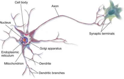
The simplest description of a neuron is that it is a polarized cell that possesses three well-established regions, namely a cell body (which contains the nucleus, membrane organelles, vesicles, endoplasmic reticulum and the Golgi apparatus), an axon that ends at the synaptic terminals, and branched structures called dendrites (Fig.2). While dendrites are responsible for receiving the action potentials, axons conduct them to the synaptic terminals, where stimulation of the adjacent cell is established through the release of neurotransmitters into the synaptic gap (Tortora & Derrickson 2011).
Figure 2 | Neuron
In the simplest representation of a neuron, neurons possess three regions, namely a cell body, that contains the nucleus, vesicles and membrane organelles, endoplasmic reticulum and Golgi apparatus; dendrites, which receive and transmit the action potentials; and an axon, that conducts the action potentials to their synaptic terminals. In synaptic terminals neurotransmitters are release to the synaptic gap, thus stimulating the adjacent cell. Adapted from (Communications 2013)
10
However, in the majority of the cases, neurons assume more complex shapes. Depending on their polarization they can be classified into four categories: unipolar, bipolar, pseudo-unipolar and multipolar (Fig.3) (Bear & Rintoul 2013).
Figure 3| Types of neurons
Neurons assume different sizes and shapes. Depending on their polarization they can be classified in a. unipolar; b. bipolar; c. multipolar and d. pseudounipolar. Adapted from (Bear & Rintoul 2013)
The particular structure of each neuron allows proper connection between them and thus efficient electrochemical transmission. For instance, in the reflex arc process, sensory input induces the stimulation of the dorsal root ganglion neurons, which are pseudounipolar neurons. These cells have two axons responsible for afferent transmission to interneurons, present in the spinal cord, which in turn will stimulate motor neurons. Both interneurons and motor neurons are multipolar neurons, which mean that they receive the stimuli in dendrites and transmit it along their axons (Fig.4) (Reece et al 2011, Tortora & Derrickson 2011).
Figure 4| Types of neurons that participate in the reflex arc
In the arc reflex, the dorsal root ganglion (DRG) neurons (blue) are responsible for the afferent transmission, interneurons (green) integrate the information and motorneurons (red) allow efferent transmission. This capability is due to the unique structure of the neurons that participate in this process. While DRG neurons are pseudounipolar neurons, which means that they have two axons; interneurons and motorneurons are multipolar neurons which mean that they receive the stimuli through dendrites and transmit it through their axon (Reece et al 2011).
Efficient communication between neurons and, consequently competent performance of the nervous system, requires neuronal survival that depends on proper function of internal mechanisms.
11
2.1.1 Intrinsic mechanisms
Besides the unique ability of transmitting electrochemical messages, a lot happens within these cells, namely in soma which is the center of the genomic and metabolic changes that are essential for neuron survival and maintenance. However, cell metabolism is not restricted to the cell body, and both axons and dendrites require constantly support (Stahl 2008). Here, some neuron-specific features that allow cell survival and maintenance are addressed.
2.1.1.1
Protein synthesis
The major macromolecular components of neurons, as well as of any other cell, are proteins. Besides ensuring the unique structure of neurons, they are also crucial for neuronal function, since they participate in innumerous intracellular signaling mechanisms that allow proper response to extracellular stimulus, cell survival, and information storage and processing (Fallon & Taylor 2013). For years it was though that protein synthesis was exclusive of neuronal soma, although with the emergence of new evidences, it is now known that protein synthesis machinery is present in dendrites and axons during development, and also in mature peripheral axons (Twiss & Fainzilber 2009). Indeed, due to the extension of dendrites and axons, which in some cases stand several centimeters from the cell body, the presence of mRNA and translational machinery in different dendritic and axonal compartments allows a quick response to local requests by regulating local protein composition (Steward & Schuman 2003).
2.1.1.2
Axonal transport
An important feature of neuronal cells is their capacity to transport components along extended distances, which is fundamental for neuronal function and survival (Hirokawa et al 2010). This particular characteristic is due to a specialized microtubule and actin cytoskeleton that allows polarized transport by molecular motors (Kapitein & Hoogenraad 2011). Within dendrites and axons, longitudinal filaments of microtubules serve as road to molecular motors, such as dynein and kinesin, which are respectively responsible for retrograde and anterograde transport of molecules and organelles. While retrograde transport refers to the movement of cargoes that travel to the cell body, anterograde transport refers to the movement from the cell body (Fig. 5).
12
Figure 5 | Axonal transport
Axonal transport allows the transport of molecules and organelles through long distances. It can be classified in retrograde and anterograde transport. While retrograde transport is responsible for the movement of cargoes to the cell body. Anterograde transport corresponds to the movement from the cell body. (Pasinelli & Brown 2006)
In addition, in synaptic regions and in the tip of growing axons, the cytoskeleton is mainly composed by actin filaments and the transport of cargoes is coordinated by myosins (Kapitein & Hoogenraad 2011).
2.2 Glial cells
Glial cells are cells responsible for supporting neurons to perform their activities. Besides serving as physical support, they are responsible for modulating the environment that surrounds neurons (Bear & Rintoul 2013).
One of the main differences between the CNS and PNS is the neuroglia composition. While in PNS neuroglia is composed by Schwann and satellite cells, in CNS the majority of its components are astrocytes, oligodendrocytes, microglia and ependymal cells (Tortora & Derrickson 2011)(Fig. 6). Both Schwann cells, in PNS, and oligodendrocytes, in CNS, are responsible for axonal myelination, which not only offers physical support but also allows the rapid transmission of action potentials. Schwann cells also offer trophic support so that growing neurons reach their target and are also implicated in neuronal survival. Also in PNS, satellite cells regulate changes between neuronal soma and interstitial fluid. In CNS,
13
while astrocytes conduct nutrients and other substances from blood to neurons, microglia is responsible for the elimination of cell debris and pathogens through phagocytosis (Jessen 2004).
Figure 6 | Glial cells found in the nervous system
Glia cells provide support to neurons and regulate the external environment. a. Neuroglia from the peripheral nervous system includes Schwann cells and satellite cells; b. In central nervous system, astrocytes, oligodendrocytes, microglia and ependymal cells are the main glial cells (Bear & Rintoul 2013).
15
3. Injury to the nervous system
Axonal regeneration is possible
Following nervous system injury, neurons undergo a sequence of morphologic and metabolic changes that in most cases results in neuronal death and loss of nerve function (Bradbury & McMahon 2006). However, some neurons can trigger a regenerative program and axonal regeneration occurs (Liu et al 2011). Cruikshank was the first to suggest that damaged nerves could regenerate and regain their function, but it was Ramon y Cajal’s work in nervous system degeneration and regeneration that supported those previous results (Ochs 1977). Since then, much has been done in order to understand the mechanisms underlying nerve regeneration, especially regarding the PNS which, unlike the CNS is able to trigger a regenerative program that leads to axonal elongation and in some cases reestablishment of neuronal connections (Huebner & Strittmatter 2009).
It is not new that the PNS and CNS have different responses when injured. In fact, in 1928, Ramon y Cajal highlighted these differences in a publication where several studies in neurons either from CNS and PNS were performed (Ramon y Cajal & May 1928). In one of those studies, it was shown that sensory central branches of DRGs once injured, either within the spinal cord or between the cord and the ganglion, behaved much likely to the peripheral nerves but regeneration was usually frustrated. Moreover, by showing that injured sensory roots stop growing when in contact with CNS tissue, he proposed that regeneration failure in CNS could be due to mechanical obstacles or lack of trophic factors. Five decades later, David and Aguayo demonstrated that injured CNS axons could regenerate into transplanted peripheral grafts, supporting the idea that interactions between axons and cellular environment modulate the different regenerative capacities of peripheral and central nervous system (David & Aguayo 1981).
The emergence of evidences that intrinsic signaling is also important for axonal regeneration, led Ambron and colleagues to raise the question whether a lesion could trigger intrinsic mechanisms that contribute to the neuronal response to injury. Indeed, by performing several studies in Aplysia they showed that the ability of injured neurons to regenerate their axons was also dependent on intracellular signaling (Ambron & Walters 1996). In addition, in vitro assays using dorsal root ganglia neurons from animals with either peripheral or central nervous system injury, revealed different growing patterns. While DRG neurons with previous injury on their central branch displayed short and branched neurites (projections that arise from the cell body), DRG neurons injured on their peripheral branches were able to extend long neurites, thus emphasizing that injury to the PNS and CNS elicit different responses and consequently different regeneration capacities (Smith & Skene 1997).
Altogether, these findings show that behind successful axonal regeneration relies a balance between extrinsic and intrinsic factors. Thus, in the following sections some of the most used injury models to study nervous system regeneration, namely those used in this thesis, will be presented and then the current knowledge about the extrinsic and intrinsic factors that determinates successful nerve
16
regeneration will be discussed, giving special emphasis to the intrinsic mechanisms that modulate axonal regeneration.
3.1 Injury Models to study Axonal Regeneration
Different are the paradigms used to study axonal regeneration. While some take advantage of the regenerative potential of the peripheral nervous system to study factors that prompt axonal regeneration, others use non-regenerative models, as is in the case of a spinal cord injury, to explore factors that contribute to axonal regeneration failure. Thus it is possible to classify these paradigms in two types, the non-regenerative and the regenerative models. Combinations between these two types of models have allowed some advances on the field and better understanding on the molecular mechanisms that contribute to the success or failure of axonal regeneration.
Non-regenerative models include injury to the spinal cord (SCI) and dorsal root injury (DRI) (Fig. 7, b. and c., respectively). As for the regenerative models, the most used are the sciatic nerve injury (SNI) and the conditioning lesion “paradigm” (Fig. 7, d. and e., respectively) (Bolsover et al 2008, Neumann & Woolf 1999). One characteristic shared by these models is that they can both use the regenerative capacity of DRG neurons.
DRG neurons are sensory pseudounipolar neurons with a peripheral branch located in the spinal nerve, and a central branch that arises from the DRG to the dorsal root, thus entering the spinal cord (Fig. 7, a.). Their cell bodies are clustered in the dorsal root ganglion (Tortora & Derrickson 2011). One important feature of these neurons is that when their peripheral branch is injured they are able to trigger a regenerative program that leads to axonal elongation and in some cases reinnervation of the correct targets. On the other hand, injury to their central branch, either by a SCI or a DRI, fails to trigger the same regenerative program and consequently axonal regeneration fails. However, despite the inefficiency of the SCI or even DRI to prompt axonal regeneration, the intrinsic regenerative capacity of these neurons in these two types of injury can be elicited by doing a conditioning lesion. Indeed, damaging the peripheral branch of DRGs – the conditioning lesion – prior to a lesion in their central one is sufficient to trigger intrinsic mechanisms that allow both branches to overcome an inhibitory environment and extend long neurites (Neumann & Woolf 1999).
17
a. b. c. d. e.Figure 7 | Injury models to study axonal regeneration
DRG neurons are sensory pseudounipolar neurons widely used to study axonal regeneration. The middle panel illustrates some non-regenerative models, while the lower panel shows some regenerative models. a. Representative scheme of the spinal cross section and the sciatic nerve, including spatial localization of DRG neurons; b. Regeneration of DRG neurons central branch fails following spinal cord injury (SCI) or c. dorsal root injury (DRI); d. Sciatic nerve injury (SNI) triggers a regenerative program leading to the extension of long axons and eventually correct reiinervation; e. Regeneration of the central branch of DRG neurons can be elicited by performing a conditioning lesion prior to the SCI. Green arrows point to axonal outgrowth, while red arrows represent axonal regeneration failure.
18
3.2 Extrinsic Mechanisms that modulate Axonal
Regeneration
Successful axonal regeneration is a process that depends on innumerous factors. In PNS, a crucial step to effective axonal regeneration relies on the rapid degeneration of the distal portion of the injured neuron and, competent clearance of myelin and cell debris. In the CNS these two events also occur, however, degeneration of the cut axon is a slow process and myelin and cell debris phagocytosis is less effective. Together, these molecular and cellular events constitute the Wallerian degeneration process, which is strongly modulated by the neuroglia and the immune system responses (Vargas & Barres 2007). The first evidence of the Wallerian degeneration process emerged in 1850 when Augustus Waller described a process where the distal portions of axotomized neurons undergo disintegration (Waller 1950). Further studies have shown that there are at least three distinctive phases during axonal degeneration (Ambron & Walters 1996, Wang et al 2012). It starts with an acute axonal degeneration (also known as AAD) of both proximal and distal stumps of the injured neuron (Kerschensteiner et al 2005) which is followed by a latent period, where the distal portion starts to swell and irregular beads appear (George et al 1995) and finally, granular disintegration of axonal cytoskeleton (GDC) distal to the injury site occurs (Kerschensteiner et al 2005). This axonal degeneration takes about 24-48h to occur in rodents (Lubinska 1977) and several days in humans (Chaudhry et al 1992), however, the type of injury, the distance between the lesion and the cell body, as well as the axon’s caliber also influence the time-course of these events (Beirowski et al 2005, Lubinska 1977). Wallerian degeneration is accomplished when the debris from axonal degeneration and myelin breakdown are cleared. Indeed, the different rates of Wallerian degeneration between the PNS and CNS are due to incompetent debris clearance, instead of a delay in the degeneration of the CNS neurons (George & Griffin 1994).
3.2.1 Wallerian degeneration in PNS
Successful regeneration within the PNS is dependent on the activity of Schwann and immune cells (Gaudet et al., 2011)(Fig.8).
Following injury, Schwann cells (SCs) along the distal segment, rapid dedifferentiate leading to demyelination of the cut axon, and myelin debris accumulation (Jessen and Mirsky, 2008). Injury also triggers downregulation of myelin proteins by SCs, which are known to have an inhibitory role in axonal outgrowth (He & Koprivica 2004, Trapp et al 1988). In addition, within the basal lamina, SCs start the clearance process by phagocytizing extracellular debris and their own myelin, and begin to proliferate creating a permissive substrate to axonal elongation (Stoll et al 1989). Besides, the physical support, they also secrete factors that elicit axonal outgrowth.
19
In the meantime, as soon as 8h post-injury resident neutrophils accumulate at the injury site. Besides its local phagocytic role, they also secrete factors that together with the onset of GDC recruit other immune cells. Indeed, within 48 h post-injury breakdown of the distal nerve’s blood-nerve-barrier (BNB) occurs, allowing the influx of macrophages that once activated, start phagocytizing extracellular debris (Bouldin et al 1991, Bruck 1997).
In summary, SCs and immune cells create a proper environment that allows axons to regenerate over long distances (Gaudet et al 2011).
Figure 8 | Wallerian degeneration in PNS and axon regeneration after peripheral nerve injury
Successful axonal regeneration in the peripheral nervous system is dependent on the rapid axonal degeneration distal to the injury site and competent clearance of the extracellular debris, including myelin remains which are known to be inhibitory to axonal outgrowth. In parallel to the degeneration of the cut axons, Schwann cells release their myelin, dedifferentiate, phagocytize debris and begin to proliferate inside the intact basal lamina creating a permissive substrate for axonal elongation. In addition, the recruitment of immune cells, such as macrophages, speeds the myelin clearance process, thus allowing axonal elongation to take place. (Gaudet et al 2011)
3.2.2 Wallerian degeneration in CNS
In central nervous system, oligondendrocytes, unlike SCs, do not dedifferentiate or support debris clearance following injury, neither offer trophic support for axonal elongation. Instead, they undergo apoptosis or remain in a quiescent state. In addition, astrocytes become reactive and invade the
20
injury site, thus allowing the formation, together with oligodendrocytes, of a physical and chemical barrier to axon outgrowth - the glial scar (Fig. 9). Indeed, inhibitory cues either from oligondendrocytes myelin or astrocytes (that once reactive, start producing inhibitory molecules such as chondroitin sulfate proteoglycans - CSPG) are still present years after degeneration of the CNS (Buss et al 2004, Buss et al 2005).
Figure 9 | Representation of CNS injury site
and respective formation of the glial scar Axon regeneration failure following injury to the spinal cord is a consequence of innumerous factors: astrocytes become reactive secreting inhibitory molecules and invading injury site; myelin debris from dead or quiscient oligodendrocytes contributes to the inhibitory environment; cyst formation may also occur, contributing to axonal regeneration imparment; and macrophage activity, in a different manner from what happen in PNS, is followed by the release of neurotoxic substances. Together, this results in the formation of the glial scar b. which constitutes a physical and chemical barrier to axonal outgrowth (Yiu & He 2006).
Also, unlike to what happens in PNS, whereas breakdown of the BNB occurs along the separated axon, breakdown of the blood-spinal cord-barrier (BSCB) is restricted to the injury site, impairing the clearance of the cut degenerated tracts. In addition, the activity of macrophages, although required to promote tissue repair, has also a neurotoxic effect that contributes for axon regeneration impairment, being this, a possible explanation why breakdown of the BSCB is restricted to the lesion site. Formation of fluid-filled cyst can also occur at the injury site. Together, these factors contribute for an inhibitory environment that leads to axonal regeneration failure following injury to the central nervous system (Gaudet et al 2011, Vargas & Barres 2007, Yiu & He 2006).
21
3.3 Intrinsic Mechanisms that modulate Axonal
Regeneration
As evidenced before, external factors are crucial modulators of axonal regeneration. However, axonal outgrowth requires that injured neurons switch to a growth-competent state, otherwise regeneration fails. Indeed, regeneration-competent neurons are able to extend their axons even in the presence of an inhibitory environment (Neumann & Woolf 1999). Given this, it is appropriate to ask “Which are the intrinsic mechanisms that elicit axonal growth and the reinnervation of the correct targets?”. Efforts to answer this question have allowed some advances on the field, although full recovery has never been achieved and some intrinsic mechanisms still need to be clarified.
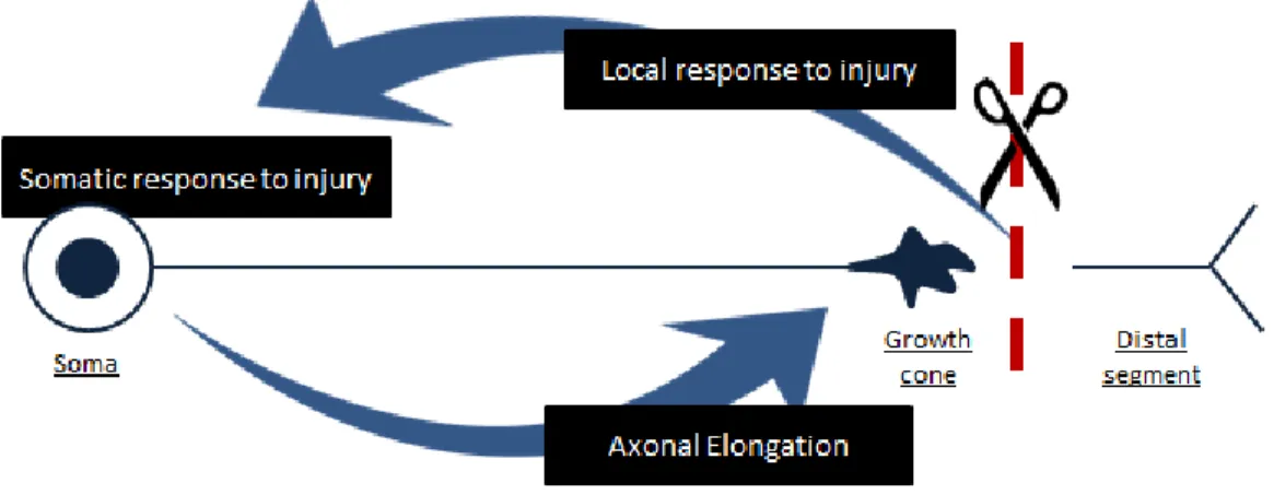
So far, it is known that switching to a growth-competent state following injury requires in first place a local response, which involves the rearrangement of the cytoskeleton at the cut tip of the axon and the activation of several injury signals (Bradke et al 2012, Mar et al 2014a); followed by a somatic response where nuclear changes occur as a consequence of the signals received from the injury site (Kiryu-Seo & Kiyama 2011); and finally the transport of newly synthesized components to the cut tip (Mar et al 2014a). Altogether, these changes are capable of eliciting axonal elongation and guidance to the proper targets (Fig. 10).
Figure 10 |Activation of the regeneration program following injury
Axonal injury triggers intrinsic changes which in some cases lead to the activation of a regeneration program capable of overcoming the inhibitory environment. Successful axonal regeneration is achieved when local changes elicit the rearrangement of the cytoskeleton at the cut tip and triggers a variety of intrinsic signals that will induce a somatic response. This somatic response to injury involves the synthesis of new components that, once transported to the cut tip, will allow axonal elongation and guidance to the correct targets.
22
3.3.1 Local response to injury
Following injury, axolemma is disrupted and the intracellular milieu is exposed to the extracellular content, leading to changes in homeostatic balance. Within 30 minutes, calcium influx activates calpain proteases that will induce a rapid disintegration of the cytoskeleton for a few hundreds of micrometers - AAD - and consequently membrane sealing (Eddleman et al 2000, Kerschensteiner et al 2005, Knoferle et al 2010).
After membrane sealing, some injured neurons are able to rearrange their cut end to form a competent growth cone in a process dependent on calcium influx and local protein synthesis (Kamber et al 2009, Verma et al 2005). The formation of this structure is crucial for axonal regeneration since it prepares the neuron for the next steps of axonal growth (Bradke et al 2012). In addition, activation of a variety of signaling mechanisms upon injury allows the cell body to receive precise information about the local changes caused by the lesion (Perlson et al 2004).
3.3.1.1 Growth cone assembling
The growth cone is a highly dynamic region found in the tip of growing axons and it is responsible for the integration of extracellular cues with intracellular signaling to guide axonal elongation (Bradke et al 2012, Kaplan et al 2014). Indeed, following injury, calcium influx allows the formation of a growth cone. Moreover, in a calcium free environment, axotomized neurons do not assemble a new growth cone and regeneration fails (Chierzi et al 2005).
Depending on their cytoskeleton distribution, it is possible to identify three different regions, namely the peripheral domain (P), the transition domain (T) and the central domain (C) (Fig. 11). The P domain is the outermost region of the growth cone and is mainly composed by actin filaments (F-actin) that are rearranged to form long bundles of actin (filopodia) separated by mesh-like networks of F-actin (lamellipodia). Due its long extension, the filopodia is the main responsible for exploring the surrounding environment. The C domain, which is the nearest region to the axon, is formed by dynamic microtubules that can protrude into P domain, and houses a variety of cytoplasmic vesicles and membranous organelles. The third and last region, the T domain, is the transition zone between P and C domains and is highly enriched in actin arcs (actomyosin contractile structures) that form a hemicircumferential curve (Lowery & Van Vactor 2009). Together, these domains determine the movement of the growth cone and, as a consequence, the elongation pathway of the growing axon.
23
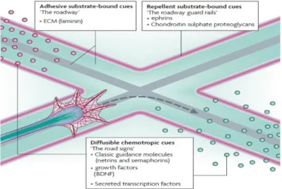
Figure 11 | Growth cone structure and components The growth cone is a complex found in the tip of growing axons and is capable of changing its shape and direction in response to extracellular cues. In the periphery of the growth cone, it is possible to find two types of structures, the filopodia and lamellipodium, which mediates their movements. (Lowery & Van Vactor 2009).Of equal importance are the extracellular cues, since they exhibit repulsive and attractive interactions over the growth cone, which then guide the axon through a pathway that in optimal conditions lead to the right target (Fig. 12). Indeed, some external cues, such as neurotrophins, are required to stimulate axonal growth and cell survival by triggering intracellular signals (Paves & Saarma 1997). When depleted of neurotrophic factors, growing neurons fail to innervate their target or die (Polleux & Snider 2010). Extracellular cues include diffusible chemotropic cues, such as guidance cues (netrins and semaphorins) and neurotropic factors (nerve growth factor (NGF)); adhesive substrate-bound cues, such as the extracellular matrix (ECM) protein laminin; and repellent substrate-substrate-bound cues that include the repulsive cue ephrin
and the ECM inhibitors, CSPG (Lowery & Van Vactor 2009).
Figure 12 | Extracellular cues determine the
elongation pathway of the axon
By exerting over the growth cone repulsive and attractive interactions, extracellular cues determine the elongation pathway of the growing axon. Extracellular cues include adhesive and repellent substrate-bound cues, and diffusible chemotrophic factors. Adapted from (Lowery & Van Vactor 2009)
As referred before, it is the interaction with external cues that allows the growth cone to change its shape and direction. But how the growth cone is capable of such changes is a question that has puzzled investigators for years. It is currently known that these interactions are mediated by cell-surface
24
receptors that trigger a variety of intracellular signaling pathways, thus leading to cytoskeleton rearrangement (Bradke et al 2012). One of the major players in regulation of growth cone dynamics is the Rho family of small GTP-binding proteins (RhoGTPases), being the Cdc42, Rac, and RhoA, the best studied ones (Huber et al 2003). By rearranging actin cytoskeleton they are capable of mediating growth cone guidance and axonal elongation. Indeed, activation of Cdc42 and Rac induce the formation of filopodia and lamellipodia, respectively, by promoting actin polymerization and consequently axon guidance and elongation. On the other hand, by activating ROCKII (Rho-associated protein kinase II) which phosphorylates myosin light chain (MLC), RhoA stimulates the constraining of the actin arcs and promotes the assembling of stress fibers (myosin filaments), thus leading to growth cone contraction and reduced motility (Fig. 13).
Figure 13 | RhoA/ROCKII pathway in growth cone
Increased activity of RhoA leads to the activation of ROCKII which then phosphorylates myosin light chain (MLC). This will lead to growth cone retraction, loss of motility and consequent elongation failure. Adapted from (Lowery & Van Vactor 2009, Luo 2000).
Typically, attractive cues activate Cdc42 and Rac, and inhibit RhoA; while repellent cues have the opposite effect (Guan & Rao 2003). In the presence of growth inhibitors the growing tip of the axon becomes “trapped” and the disregulation of intracellular signaling leads to growth cone retraction or even growth cone collapse in a process dependent on ROCKII activity (Mueller et al 2005). In addition, inhibition of Rho kinase activity has also been shown to enhance axonal regeneration either in injured central or peripheral nervous systems (Fournier et al 2003, Hiraga et al 2006), supporting the inhibitory role of RhoA/ROCKII and its effectors in axonal regeneration.
25
3.3.1.2 Injury signaling mechanisms
Regeneration of injured axons is a process that requires the synthesis of new components in soma, including cytoskeleton proteins, such as tubulin and actin, transmembrane proteins, like membrane receptors and voltage gated channels, membrane lipids, signaling molecules and metabolic enzymes (Goldberg 2003). Since in most cases the cell body stands several centimeters from the lesion site, retrograde signaling is required to “inform” the cell body that an injury has taken place (Ambron & Walters 1996, Rishal & Fainzilber 2010). Below some of the injury signals that elicit a proper somatic response (Fig. 14) are summarized.
Figure 14 | Injury signaling mechanisms
Injury triggers a variety of signals that will inform the soma that an injury occurred. a. The first signal that arrives the cell body is the electrophysiological response caused by membrane depolarization. This is followed by motor-dependent signals either the loss of suppressors of axonal growth (Negative injury signals) b., or the activation of injury signals at lesion site (Positive injury signals) that will be retrogradely transported to the cell body c. Adapted from (Rishal & Fainzilber 2010)
3.3.1.2.1 Depolarization
Membrane depolarization due to calcium and sodium entry through the axonal cut ends is one of the first changes that occur upon injury. These local alterations in ionic concentrations lead to activation of voltage-gated Ca2+ channels and reversal of the Na/Ca exchanger, thus increasing free-intracellular Ca2+ concentration and causing the propagation of a calcium wave from the lesion site to the cell body (Mandolesi et al 2004). In the soma, calcium elicits the activation of a variety of signals associated with axonal regeneration. In fact, increased intracellular calcium stimulates Ca2+-dependent
26
enzymes, such as adenyl cyclase (AC), which will then convert ATP to cyclic adenosine monophosphate, cAMP (Fig. 15). cAMP is a signaling molecule that is increased following peripheral nerve injury, and it has been linked to a robust axonal regeneration (Lau et al 2013). Experiments that include the injection of the membrane-permeable analog of cAMP, dibutyryl-cAMP, in lumbar dorsal ganglia (Neumann et al 2002); or administration of rolipram, a phosphodiesterase inhibitor that impairs the conversion of cAMP into AMP (Nikulina et al 2004), have supported the importance of this signal to a successful axonal regeneration.
Figure 15 | Activation of cAMP in regeneration-competent neurons
Following lesion there is backpropagation of a calcium wave from the injury site to the soma leading to a general increase in free intracellular calcium concentration. Calcium will then activate adenyl cyclase (AC) responsible for converting adenosine triphosphate (ATP) into cAMP (cyclic-AMP). Increased levels of cAMP in the soma are associated with a robust regenerative response. Phosphodiesterases (PDE) are responsible for diminuishing cAMP levels by converting cAMP into AMP. This last conversion can be abolished by administrating Rolipram, a phosphodiesterase inhibitor, promoting axonal regeneration. Adapted from (Hannila & Filbin 2008, Hofer 2012, Nikulina et al 2004)
Recently, it has also been shown that the back-propagation of a calcium wave results in nuclear export of HDAC5 that leads to the activation of a proregenerative gene-expression program and successful axonal regeneration (Cho et al 2013).
Additionally to the calcium wave, it has been reported that in some neurons local depolarization may reach a threshold that triggers a burst of action potentials, thus contributing to the increased calcium levels found in the cell body upon injury and axonal regeneration (Mandolesi et al 2004, Ziv & Spira 1995). Therefore, it would be expected that electric stimulation would improve axonal regeneration. However, it is not clear whether this type of stimulation is capable of eliciting a regenerative response, since reports about this field are not consistent. For example, while electrical
27
stimulation caused axonal growth in sensory neurons (Udina et al 2008), transected rubrospinal tract was not able to regenerate when stimulated (Harvey et al 2005). A good explanation for these differences may rely on the paradigm used to stimulate the injured neurons, i.e, the pulse duration, numbered of pulses applied, the chosen system, among other aspects. In fact, Harvey and colleagues (2005) refer that the system used by them was not able to replicate other studies where electric stimulation had elicited axonal regeneration. Moreover, in the study performed in Fouad’s lab, although electric stimulation was capable of promoting axonal outgrowth, regeneration was not sustained over long distances (Udina et al 2008).
Together these results suggest that electric stimulation may be required in the early stages of axonal regeneration, but it is not sufficient to trigger a capable regenerative response.
3.3.1.2.2 Negative Injury signals
Although little is known about this type of signals, they are referred in literature as being suppressors of axonal growth so that synaptic formation could take place. In the same line of thoughts, it is hypothesized that, as a consequence of injury, neuronal disconnection from its targets leads to down-regulation of these signals, thus allowing axonal regeneration. As for my knowledge the only signal that completes these requisites is SMAD2 since is down-regulated following peripheral nerve injury (Abe & Cavalli 2008).
3.3.1.2.3 Positive injury signals
In the 90s, studies in Aplysia suggested that axotomy was able to produce a variety of axoplasmic molecules responsible for the regenerative response (Ambron et al 1995). Today, it is known that the activation of these molecules – the positive injury signals - is dependent on local phosphorylation of proteins as well as local translation, and that retrograde transport machinery ensures that these signals are properly carried to the nucleus (Mar et al 2014a, Rishal & Fainzilber 2014). Moreover, the improvement of new techniques and several studies had allowed so far the recognition of three positive injury signals. Those are the signal transducer and activator of transcription factor 3 (STAT3) (Ben-Yaakov et al 2012, Sheu et al 2000), the extracellular signal regulated kinase (ERK) (Perlson et al 2005, Sheu et al 2000) and the c-Jun N-terminal kinases (JNK) (Cavalli et al 2005). In the following paragraphs the mechanisms that trigger the activation of these signals and their retrograde transport will be discussed, as well as their importance in axonal regeneration.
STAT3 is a transcription factor associated with JAK-STAT pathway and belongs to the STAT family. Extracellular signals, such as cytokines and growth factors, lead to the activation of this pathway and subsequent phosphorylation of STAT proteins. In PNS, after injury, specifically STAT3 is locally translated, subsequent phosphorylated and retrogradely transported to the nucleus (Ben-Yaakov et al 2012, Sheu et