Transcriptomic differential lncRNA expression is
involved in neuropathic pain in rat dorsal root
ganglion after spared sciatic nerve injury
P. Mao
1, C.R. Li
1, S.Z. Zhang
2, Y. Zhang
1, B.T. Liu
1and B.F. Fan
1 1Department of Pain Medicine, China-Japan Friendship Hospital, Beijing, China 2
State Key Laboratory of Toxicology and Medical Countermeasures, Department of Biochemical Pharmacology, Beijing Institute of Pharmacology and Toxicology, Beijing, China
Abstract
Dorsal root ganglia (DRG) neurons regenerate spontaneously after traumatic or surgical injury. Long noncoding RNAs (lncRNAs) are involved in various biological regulation processes. Conditions of lncRNAs in DRG neuron injury deserve to be further investigated. Transcriptomic analysis was performed by high-throughput Illumina HiSeq2500 sequencing to profile the differen-tial genes in L4–L6 DRGs following rat sciatic nerve tying. A total of 1,228 genes were up-regulated and 1,415 down-regulated.
By comparing to rat lncRNA database, 86 known and 26 novel lncRNA genes were found to be differential. The 86 known lncRNA genes modulated 866 target genes subject to gene ontology (GO) and KEGG enrichment analysis. The genes involved in the neurotransmitter status of neurons were downregulated and those involved in a neuronal regeneration were upregulated. Known lncRNA generno-Cntnap2was downregulated. There were 13 credible GO terms for therno-Cntnap2gene, which had a putative function in cell component of voltage-gated potassium channel complex on the cell surface for neurites. In 26 novel lncRNA genes, 4 were related to 21 mRNA genes. A novel lncRNA geneAC111653.1improvedrno-Hypmsynthesizing hunt-ingtin during sciatic nerve regeneration. Real time qPCR results attested the down-regulation ofrno-CntnaplncRNA gene and the upregulation ofAC111653.1lncRNA gene. A total of 26 novel lncRNAs were found. Known lncRNA generno-Cntnap2and novel lncRNAAC111653.1were involved in neuropathic pain of DRGs after spared sciatic nerve injury. They contributed to peripheral nerve regeneration via the putative mechanisms.
Key words: Spared nerve injury; Neuropathic pain; Long noncoding RNA; Transcriptome;Hypmgene
Introduction
After traumatic damage, the peripheral nervous system can regenerate spontaneously by activating the inherent growth ability of neurons, while the central nervous sys-tem cannot do so (1,2). The sciatic nerve is commonly used as a model to study peripheral nerve regeneration. It includes a complex bunch of motor and sensory axons, in which the sensory neurons are situated in the L4–L6 dorsal root ganglion (DRG) (3,4). After sciatic nerve damage, the damaged neurons initiate a regeneration process but cease having the neurotransmitter status (3,5). Axon regeneration and pathfinding after damage involves a complex mechanism involving axon cross-talk with neuro-gliocytes, nerve growth factors, neurotrophic factors, and receptors (6,7).
Neuropathic pain after traumatic or surgical nerve injury challenges doctors and patients and regulatory noncoding RNAs (ncRNAs) are key molecules for understanding and treating this pain (8,9). Regulatory ncRNAs are transcribed
from protein noncoding genes to interfere in gene expression and they include, but are not limited to, miRNAs (21–24 nt), siRNAs (21–25 bp), piRNAs (26–31 nt), and long non-coding RNAs (lncRNAs, 200 bp to more than 100,000 bp) (8,10,11). The role of miRNAs, lncRNAs, and piRNAs in neuropathic pain after nerve injury has been reviewed by Bali and Kuner (8). The role and regulatory mechanisms of lncRNAs in vertebrate central nervous system and human nervous system diseases have been reported in the literature (8,12–15).
In peripheral nerve injury, lncRNAs play an important role in stress responses, plasticity, and axonal outgrowth of DRG neurons (8,16–21). Yu et al. (16) investigated the lncRNA transcriptome of DRG neurons after sciatic nerve injury in rat models and found that lncRNAs modulate DRG neurons responses to ligation stimuli. Zhao et al. (17)
identi-fied the modulating effect of the lncRNA (Kcna2 antisense RNA) on a voltage-dependent potassium channel mRNA
Correspondence: B.F. Fan:<[email protected]>
Received September 10, 2017 | Accepted June 11, 2018
Kcna2 in primary afferent neurons. Yao et al. (18) reported that the lncRNA uc.217 modulates neurite outgrowth of DRG neurons after sciatic nerve injury. lncRNA NONRATT 021972 and lncRNA uc.48 modulate neuropathic pain mediated by the P2X(3/7) purinergic receptors (the cation-permeable ATP-binding ligand-gated ion channels) in the DRG neurons in diabetic rat models, and siRNA therapy alleviate the pain significantly (19–23). However, all these studies are based on microarray analysis (24). A sequencing study to elucidate vital roles of lncRNAs in peripheral nerve pathology will help to understand neuropathic pain.
In this study, we described the lncRNAs expression and mRNA expression in DRGs during nerve regeneration by a transcriptome-level deep sequencing. The results reveal a novel layer of regulation of the inherent growth ability of neurons by lncRNAs.
Material and Methods
Animal surgery and sample preparation
Six male Sprague-Dawley rats (180–220 g) were housed in large cages with sawdust bedding at 25°C in 12 h/12 h dark/light cycle and allowed free access to food and water in the Animal Center of Beijing China-Japan Friendship Hospital. Rats were randomly divided into test group and sham-operation group, three in each group. Surgery was performed as described in the literature with modifications (16). Briefly, rats were anesthetized by intra-peritoneal injection of 10 wt.% chloral hydrate (3 mL/kg, Tianjin Fuchen Chemical Reagent Factory, China). The sciatic nerves were exposed and lifted through an incision on the right lateral thigh. Sciatic nerve segments were tied at the site proximal to the bifurcation of tibial and common peroneal nerves. Rats in the sham-operation group only had the sciatic nerves exposed without tying. L4–L6 DRGs were harvested from each animal a week later. All animal experiments were performed in accordance with institu-tional animal welfare and care guidelines and approved by the Animal Ethics Committee of Beijing China-Japan Friendship Hospital.
RNA isolation, cDNA library preparation, and sequencing
Total RNAs were extracted from the L4–L6 DRG tissues using Trizol reagent according to the instructions of the manufacturer (Invitrogen, USA). RNAs were cleaned, including a DNase I digestion step, using RNeasy spin columns (Qiagen, USA). RNA integrity was detected by agarose gel electrophoresis and RNA was quantified using Nanodrop2000 (Bio-Rad, USA). After rRNA was removed, RNA was interrupted into short fragments by adding fragmentation buffer. These short RNA frag-ments were used as templates to synthesize the fi rst-strand cDNA using FastQuant RT Kit (with gDNAase) (Tiangen, China). Then, double-strand cDNA was obtained.
The cDNA products were purified with QiaQuick PCR extraction kit (Qiagen, Germany) and the purified cDNA were dissolved in EB buffer, followed by end reparation and poly(A) addition. The cDNA fragments were con-nected to sequencing adaptors. The cDNA fragments at 150–200 bp in size were separated on gel-electrophor-esis and were used as the templates for PCR amplifi ca-tion. Two cDNA libraries for the test and sham-operation groups were sequenced using the Illumina HiSeq2500 at Beijing Ori-Gene Science and Technology Co., LTD (China).
Data processing
Raw images generated by sequencers were converted by Illumina software (USA) to nucleotide sequences, called raw reads. FastQC software package (USA) was used to generate clean reads by removing adaptor reads, low quality reads (QC30), sequences containing fuzzy N bases and sequences less than 60 bp. All the following analyses were based on clean reads. The clean reads were mapped to the genome using Tophat2 software package (USA). RSeQC software package with all default parameters (USA) was used to detect the splice junction sites for evaluating the saturation of sequencing.
Differential expression and enrichment
The RPKM method (reads per kilobase per million mapped reads) was used to calculate the differential expression level using the Cufflinks software package (USA). Cufflinks Cuffdiff was used to screen the dif-ferential expression genes (DEGs). The criterion to identify the DEGs between two groups was as follows: sum of mapping reads of two samplesX10; |log2RPKM fold change| 41; P value was corrected by false dis-covery rate (FDR) to get a Q-value; both Pp0.05 and Qp0.05 were required to determine the significance of differential expression. In order to get biological functions, DEGs were subjected to gene ontology (GO) and Kyoto Encyclopedia of Genes and Genomes (KEGG) enrich-ment analyses. R package was used for the analysis. GO terms of DEGs were compared with the genome background and the corrected P value (FDR correction)o
0.05 was set to judge the significantly enriched GO terms. For the KEGG pathway enrichment analysis, P value o
0.05 was the threshold.
Known and novel lncRNA
Coexpression network and target gene enrichment analysis
By comparing the differential gene lists, we obtained gene pairs of novel lncRNA and differential mRNA. FPKM values for each pair were used to calculate the Pearson correlation, where we chose significantly correlated gene pairs (correlation coefficient40.995 oro–0.995, Po0.05
as threshold) to build a coexpression network using Cyto-scape software package (USA). GO and KEGG enrich-ment analyses were performed as described above but the corrected P value threshold was set too0.1. The cor-related mRNA genes that coexpressed with lncRNAs served as the candidate target genes for lncRNAs.
Real-time qPCR quantification
To quantify lncRNA and target mRNAs, real-time qPCR were performed on representative rno-Cntnap2-201lncRNA gene,rno-Fam171bmRNA gene,rno-Hebp2
mRNA gene, rno-Gde1 mRNA gene, rno-AC111653.1
lncRNA gene, and rno-Hypm mRNA gene in the sham-operation and test DRG groups. Briefly, the first-strand cDNA was synthesized using FastQuant RT Kit (with gDNAase) (Tiangen). Then, double-strand cDNA was syn-thesized. Real time qPCR was performed on a Roche LightCyclers96
fluorescence quantifying PCR machine. Primer sequences are listed in Table 1.
The reaction system included 1mL of cDNA, 10mL of RealStar Green Mixture (2), 0.6mL of primers, andfilled up to 20 mL with PCR-grade water. The PCR program included a pre-denaturation at 95°C for 5 min, 40 cycles (95°C for 15 s, 60°C for 20 s, 72°C for 15 s) for amplifi ca-tion, and a default condition for dissociation. The cycle threshold (Ct) values were obtained. Relative interest/ reference mRNA expression was calculated by the formula: 2-DCt (interest-reference). rno-GAPDH was used as inner reference.
Cell assays
Wistar rat DRG cells were isolated and cultured as reported in the literature (25). Cell assays were performed as reported in the literature with a minor modification (26). Briefly, one-day-old Wistar rat DRG cells were isolated and cultured in 95% Eagle’s DMEM feeding medium with 600 mg/mL glucose, 10% fetal bovine serum, 5% horse serum, 20 ng/mL nerve growth factors, and 1mg/100 mL neurotrophins (25). For RNA silencing, siRNA sequences targeting lnc-AC111653.1were designed and synthetized (GenePharma, China), and afinal concentration of 50 nM was used for transient transfection. For overexpression of lnc-AC111653.1, full-length rat lnc-AC111653.1 cDNA was cloned into the pcDNA3.1 expression vector (Gene-Pharma). Lipofectamine 3000 (Invitrogen) was used for transfection according to manufacturer’s instructions (26).
Western blotting
Total proteins were extracted from cell lysates and separated by 10% SDS-polyacrylamide gel. Then, they were electroblotted to PVDF membranes (Beyotime, China). Membranes were incubated in rabbit polyclonal HYPM antibody (Novus Biologicals, China), followed by horse-radish peroxidase-labeled goat rabbit secondary anti-body (Boster, China). Signals were revealed using ECL detection reagent (Beyotime). GAPDH served as control. Images were analyzed by Image-Pro Plus 6 software (Media Cybernetics, USA). The intensity of test protein bands was normalized to the GAPDH bands.
Results
Gene mapping and differential expression genes
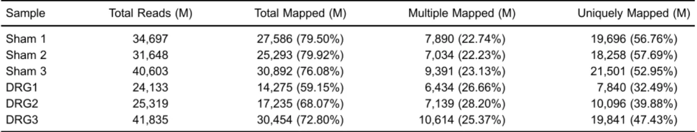
The RNA quality and sequencing quality were guar-anteed. Clean reads were obtained. The results of gene mapping to the rat genome is shown in Table 2. Known gene expression is shown in Tables 3 and 4.
On differential expression analysis, a total of 18,824 genes were included, of which there were 2643 differential genes between DRG test group and sham-operation group. By comparison of the DRG group with the sham-operation group, 1228 were up-regulated and 1415 down-regulated. On enrichment analysis of DEGs, up-regulated differential genes were attributed to 624 GO terms and 50 KEGG pathways; down-regulated differential genes were attrib-uted to 424 GO terms and 30 KEGG pathways. DEGs were clustered into a heatmap (Figure 1).
Known lncRNA, co-expression network, and target gene enrichment
We found 69 neurite-associated known lncRNA genes linking to 866 target mRNA genes (Table 5). After the GO and KEGG enrichment information was presented at a P value threshold o0.1, the 866 targets were enriched to 737 GO terms and 40 KEGG pathways. They were involved either in the downregulation of neurotransmitter
Table 1.Primer sequences.
Gene name Sequence (50-30)
status of neurons or in the upregulation of peripheral neuronal regeneration. The GO terms and KEGG path-ways involved in the downregulation effects included, but were not limited to, synaptic vesicle exocytosis, neuro-transmitter secretion, voltage-gated potassium channel activity, regulation of synaptic transmission, GABAergic synapse, response to pain, endocytosis, neuronal action potential, detection of mechanical stimulus involved in sensory perception of pain, neurotransmitter transport, the GABAergic synapse pathway, the cholinergic synapse pathway, the neuroactive ligand-receptor interaction path-way, the dopaminergic synapse pathpath-way, and the synaptic vesicle cycle pathway. The GO terms and KEGG path-ways involved in the upregulation effects included, but were not limited to, response to mechanical stim-ulus, regulation of cell growth, positive regulation of cell migration, positive regulation of ERK1 and ERK2 cas-cade, positive regulation of PI3K signaling, activation of MAPKK activity, cell differentiation, regulation of neuron projection regeneration, regulation of nerve growth factor receptor activity, peripheral nervous system axon regen-eration, glial cell differentiation, the AMPK signaling pathway, the calcium signaling pathway, the PI3K-Akt signaling pathway, the glucose metabolism pathway, the MAPK signaling pathway, and the cGMP-PKG signaling pathway.
Table 2.Mapping results of transcriptome to referenced genome.
Sample Total Reads (M) Total Mapped (M) Multiple Mapped (M) Uniquely Mapped (M)
Sham 1 34,697 27,586 (79.50%) 7,890 (22.74%) 19,696 (56.76%)
Sham 2 31,648 25,293 (79.92%) 7,034 (22.23%) 18,258 (57.69%)
Sham 3 40,603 30,892 (76.08%) 9,391 (23.13%) 21,501 (52.95%)
DRG1 24,133 14,275 (59.15%) 6,434 (26.66%) 7,840 (32.49%)
DRG2 25,319 17,235 (68.07%) 7,139 (28.20%) 10,096 (39.88%)
DRG3 41,835 30,454 (72.80%) 10,614 (25.37%) 19,841 (47.43%)
DRG: dorsal root ganglia.
Table 3.Number and distribution of known gene expression.
Sample Genes Min. 1st Qu. Median Mean 3rd Qu. Max. Sd. Sum.
Sham 17856 0 0.83 4.75 151.65 14.99 482618 5987.02 2707906
DRG 17296 0 1.15 5.15 400.95 14.28 1828940 19976.57 6934761
DRG: dorsal root ganglia.
Table 4.Number and percentage of known gene expression.
Sample 0–0.5 40.5–1 41–5 45–10 410–50 450
Sham 3634 (20.35%) 1184 (6.63%) 4322 (24%) 2764 (15.48%) 4488 (25.13%) 1464 (0.08%) DRG 2859 (16.53%) 1175 (6.79%) 4504 (26%) 2996 (17.32%) 4416 (25.53%) 1346 (0.08%)
DRG: dorsal root ganglia.
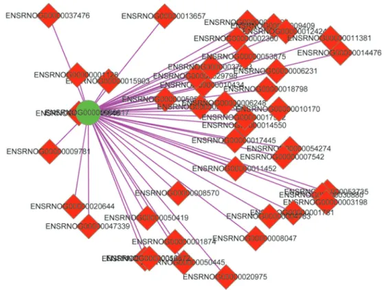
The target gene from GO enrichment of known lncRNA apparently pointed to ENSRNOG00000006617
(Po0.05), thus we singled out the known lncRNA gene
ENSRNOG00000006617 named rno-Cntnap2 (contactin
associated protein-like 2). Through serial analyses of
molecular network (Figure 2) and GO enrichment on gene
rno-Cntnap2,we found 13 credible GO terms at Po0.05
(Table 6).
According to the 13 GO terms, rno-Cntnap2 had a putative gene function that is involved in the cell com-ponent of voltage-gated potassium channel complex on cell surface of brain neurites where it has an enzyme binding activity. Considering the condition of the current study, it was assumed thatrno-Cntnap2is involved in the cell component of voltage-gated potassium channel com-plex on cell surface of sciatic nerve neurites.
We reviewed the differential expression and co-expression network analysis ofrno-Cntnap2 (ENSRNOG
00000006617)gene, which was down-regulated (20.34 and
3.94 for sham group vs DRG group, fold change –2.37, Po0.001, Q=0.0003).
Novel lncRNA, co-expression network, and target gene features
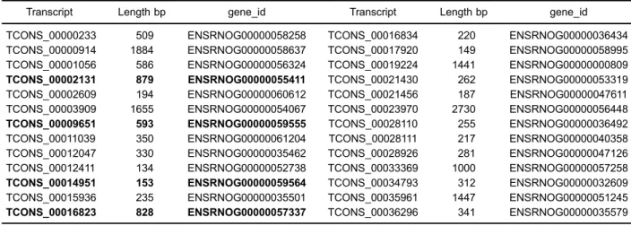
We found 525 novel transcripts containing 26 novel lncRNAs referenced to rat lncRNA database (Table 7). We constructed the co-network of novel lncRNAs with
mRNAs, and only 4 lncRNAs were related to 21 mRNAs under the conditions of thresholds ofo0.05 or 0.1 (Figure 3). The 4 lncRNAs were ENSRNOG00000055411, 000000 59555, 00000059564 and 00000057337 (Table 7).
We noticed that the transcript TCONS_00016823 included only one novel lncRNA gene, AC111653.1
(ENSRNOG00000057337), with a sense strand length
of 828 nt. GeneAC111653.1was null expressed in the sham-operation group and upregulated to 0.527889 in the DRG group. GeneAC111653.1was correlated to the target gene ENSRNOG00000021452 named huntingtin
interacting protein M (rno-Hypm). The rno-Hypm gene
GO annotations included the molecular function of DNA binding and protein heterodimerization activity, the bio-logical process of chromatin silencing, and the cellular component of nuclear chromatin and nucleosome. Huntingtin is essential for neuron survival, and the lack of hunting-tin synthesis may lead to Hunhunting-tington’s disease (27). Up-regulation of bothAC111653.1andrno-Hypmgenes after sciatic nerve injury implies a rescue course that triggers the regeneration of injured neurons. However, the func-tion ofHypmgene is not completely understood.
Quantification of several genes
For known lncRNAs detection, we selectedrno-Cntnap2
lncRNA gene and three down-regulated gene represen-tatives, Fam171b (ENSRNOG00000004783), Hebp2 Table 5.Known long non-coding RNA genes and their node degrees in Cytoscape co-expression network.
nodes_label nodes_degree nodes_label nodes_degree nodes_label nodes_degree
ENSRNOG00000002734 47 ENSRNOG00000052027 3 ENSRNOG00000056599 4
ENSRNOG00000003025 47 ENSRNOG00000052373 1 ENSRNOG00000056608 2
ENSRNOG00000005811 1 ENSRNOG00000052439 3 ENSRNOG00000056656 1
ENSRNOG00000006617 43 ENSRNOG00000052563 37 ENSRNOG00000056824 3
ENSRNOG00000009373 1 ENSRNOG00000052573 2 ENSRNOG00000057161 12
ENSRNOG00000011160 45 ENSRNOG00000053160 2 ENSRNOG00000057278 13
ENSRNOG00000017974 4 ENSRNOG00000053367 6 ENSRNOG00000057291 1
ENSRNOG00000019648 122 ENSRNOG00000053827 1 ENSRNOG00000057463 1
ENSRNOG00000024799 55 ENSRNOG00000054418 2 ENSRNOG00000057991 1
ENSRNOG00000031706 4 ENSRNOG00000054489 1 ENSRNOG00000058263 1
ENSRNOG00000033581 88 ENSRNOG00000054529 2 ENSRNOG00000058571 12
ENSRNOG00000043199 13 ENSRNOG00000054533 5 ENSRNOG00000058935 2
ENSRNOG00000043866 5 ENSRNOG00000054867 3 ENSRNOG00000058944 3
ENSRNOG00000046171 21 ENSRNOG00000054897 3 ENSRNOG00000059449 3
ENSRNOG00000046774 3 ENSRNOG00000054935 1 ENSRNOG00000059660 1
ENSRNOG00000047117 1 ENSRNOG00000054984 1 ENSRNOG00000060090 6
ENSRNOG00000048929 31 ENSRNOG00000055021 2 ENSRNOG00000060430 2
ENSRNOG00000049537 12 ENSRNOG00000055067 1 ENSRNOG00000060483 1
ENSRNOG00000051356 2 ENSRNOG00000055278 2 ENSRNOG00000060700 1
ENSRNOG00000051492 13 ENSRNOG00000055939 42 ENSRNOG00000060863 64
ENSRNOG00000051664 29 ENSRNOG00000056040 7 ENSRNOG00000061151 2
ENSRNOG00000051722 3 ENSRNOG00000056054 5 ENSRNOG00000061536 3
(ENSRNOG00000053735), andGde1(ENSRNOG00000
050445) from the rno-Cntnap2 gene coexpression
net-work (Figure 2). Real time qPCR was performed to quantify expression levels of the four genes. The quan-tification results are shown in Table 8. Expression levels of the four genes were down-regulated. This result was consistent with the sequencing outcomes.
For novel lncRNAs identification, we selectedAC111653.1
gene (ENSRNOG00000057337) and rno-Hypm gene
(ENSRNOG00000021452) from the AC111653.1 gene
coexpression network (Figure 3). They had a correlation coefficient of 1 (significance P=0). Up-regulation of both AC111653.1 andrno-Hypmgenes after sciatic nerve injury may imply a rescue course that triggers the regeneration
Figure 2.Co-expression network of generno-Cntnap2(ENSRNOG00000006617).
Table 6.Gene Ontology (GO) terms ofrno-Cntnap2long non-coding RNA gene.
Category Term Class Gene_id
GO:0071205 protein localization to juxtaparanode region of axon biological_process ENSRNOG00000006617 GO:0044224 juxtaparanode region of axon cellular_component ENSRNOG00000006617
GO:0030673 axolemma cellular_component ENSRNOG00000006617
GO:0008076 voltage-gated potassium channel complex cellular_component ENSRNOG00000006617
GO:0019899 enzyme binding molecular_function ENSRNOG00000006617
GO:0043204 perikaryon cellular_component ENSRNOG00000006617
GO:0031175 neuron projection development biological_process ENSRNOG00000006617
GO:0005769 early endosome cellular_component ENSRNOG00000006617
GO:0007420 brain development biological_process ENSRNOG00000006617
GO:0030424 axon cellular_component ENSRNOG00000006617
GO:0030425 dendrite cellular_component ENSRNOG00000006617
GO:0043025 neuronal cell body cellular_component ENSRNOG00000006617
of injured neurons, thus the function of Hypm gene deserved to be studied. Real time qPCR was performed to quantify expression levels of the two genes (Table 8), which were up-regulated. This result was consistent with the sequencing outcomes.
Cell assays
To test the biological function of novel lncRNA
AC111653.1gene, we detectedAC111653.1and its target
Hypm in primarily cultured Norway rat DRG cellsin vitro. QPCR results are shown in Figure 4, and western blots in
Table 7.Transcripts of 26 novel long non-coding RNA genes, of which 4 (in bold) are involved in co-expression network with target mRNA genes.
Transcript Length bp gene_id Transcript Length bp gene_id
TCONS_00000233 509 ENSRNOG00000058258 TCONS_00016834 220 ENSRNOG00000036434 TCONS_00000914 1884 ENSRNOG00000058637 TCONS_00017920 149 ENSRNOG00000058995 TCONS_00001056 586 ENSRNOG00000056324 TCONS_00019224 1441 ENSRNOG00000000809 TCONS_00002131 879 ENSRNOG00000055411 TCONS_00021430 262 ENSRNOG00000053319 TCONS_00002609 194 ENSRNOG00000060612 TCONS_00021456 187 ENSRNOG00000047611 TCONS_00003909 1655 ENSRNOG00000054067 TCONS_00023970 2730 ENSRNOG00000056448 TCONS_00009651 593 ENSRNOG00000059555 TCONS_00028110 255 ENSRNOG00000036492 TCONS_00011039 350 ENSRNOG00000061204 TCONS_00028111 217 ENSRNOG00000040358 TCONS_00012047 330 ENSRNOG00000035462 TCONS_00028926 281 ENSRNOG00000047126 TCONS_00012411 134 ENSRNOG00000052738 TCONS_00033369 1000 ENSRNOG00000057258 TCONS_00014951 153 ENSRNOG00000059564 TCONS_00034793 312 ENSRNOG00000032609 TCONS_00015936 235 ENSRNOG00000035501 TCONS_00035961 1447 ENSRNOG00000051245 TCONS_00016823 828 ENSRNOG00000057337 TCONS_00036296 341 ENSRNOG00000035579
Figure 5. Novel lncRNAAC111653.1was overexpressed after pcDNA3.1-lnc-AC111653.1 transfection. At the same time, its target hypm was also upregulated. Expression of AC111653.1 was reduced after siRNA transfection, and at the same time, its target hypm was downregulated. This suggested that novel lncRNA AC111653.1 was positively associated with hypm regulation in rats.
Discussion
In this study, we used a common sciatic nerve injury model to investigate gene expression conditions in rat
DRGs using a high-throughput Illumina HiSeq2500 se-quencing. In total, 86 known lncRNAs and 26 novel lncRNAs were altered during nerve regeneration. To understand the functions of the 86 known lncRNAs, we analyzed the molecular network including 866 co-expressed target genes. After sciatic nerve damage, the nerve systems switched from a neurotransmitter status to a neuronal regeneration status (1,3,5).
Based on the GO and KEGG enrichment results, we found that the neurotransmitter status of neurons are deregulated by the molecular mechanisms linking to the deregulation of the neuroactive neurotransmitter secretion, transmission, and ligand-receptor interaction pathway, while the neuronal regeneration was activated through the molecular mechanisms linking to the positive regulation of cell migration, cell differentiation, cell growth, PI3K signaling, MAPK cascade activity, nerve growth factor receptor activity, and peripheral nerve regeneration.
Table 8.Real time qPCR quantification of interest/GAPDH (interest DRG/Sham) gene expression levels.
Sham DRG1 DRG2 DRG3 Sequencing (Sham/DRG)
rno-Cntnap2-20 lncRNA 0.0530 (1.0000) 0.0074 (0.1411) 0.0069 (0.1310) 0.0039 (0.0736) 20.3371/3.9422 rno-Fam171b 0.0265 (1.0000) 0.0053 (0.1993) 0.0012 (0.0446) 0.0026 (0.0981) 19.7562/6.0137 rno-Hebp2 0.0417 (1.0000) 0.0112 (0.2673) 0.0145 (0.3475) 0.0082 (0.1966) 97.8422/22.9642 rno-Gde1 0.0128 (1.0000) 0.0097 (0.7526) 0.0042 (0.3231) 0.0065 (0.5078) 93.1178/36.7535
rno-AC111653.1 lncRNA 0 0.08652 0.04228 0.03684 0/0.52789
rno-Hypm 0 0.29365 0.1263 0.1046 0/2.6653
DRG: dorsal root ganglia. The numbers outside parenthesis indicate the gene expression ratio of interest to GAPDH and those inside indicate the gene expression ratio of interest DRG groups to the Sham group.
Figure 4. QPCR relative mRNA quantification. Upper panel shows rno-lncRNA AC111653.1 levels. Lower panel shows rno-Hypm mRNA levels in each group. Negative ctrl: pcDNA3.1 vector transfection; overexpress: pcDNA3.1-lnc-AC111653.1 transfection; siRNA interfere: siRNA-lnc-AC111653.1 transfec-tion; normal ctrl: normal cells without treatment.
Glial cells migration, dedifferentiation, differentiation, pro-liferation, and growth play important roles in peripheral nerve regeneration (1–4). The results in this study showed the promotion of glial cell migration and growth by mul-tiple signaling pathways. After sciatic nerve damage, local Schwann cells can shed off the myelin sheaths and trans-form to a neuroblast status, where their proliferation and migration capacities can help to sweep away myelin remnants and generate a conduit for the axonal pathfi nd-ing, and consequently form the beneficial conditions for neurite outgrowth (1–3). The same lncRNA-linked nerve regeneration mechanism is identified by Yu et al. (16) and Yao et al. (18).
We singled out the knownrno-Cntnap2lncRNA gene, thought to be involved in the cell component of voltage-gated potassium channel complex on cell surface for the neurites of the sciatic nerve system. We speculated that sciatic injury might trigger a switch from a neurotransmitter status to a regeneration status of neurons. The gene
rno-Cntnap2may be involved in a neurotransmitter delivery process linking to the function of voltage-gated potassium channel complex. Thus,rno-Cntnap2gene expression was down-regulated because a neurotransmitter status was ceased. The modulation of voltage-gated potassium chan-nels by a lncRNA has been identified in DRG first-order sensory neurons in a spinal nerve ligation rat model (17). In this previous study, peripheral nerve injury increased a conserved lncRNA (Kcna2 antisense RNA) expression in injured DRG through activation of the transcription factor myeloid zincfinger protein 1. This increase of lncRNA
downregulates the voltage-dependent potassium channel Kcna2 mRNA, consequently reducing total potassium current. The decrease of potassium current increases the neural excitability, namely neuropathy-induced sensitivity to mechanical stimuli in DRG neurons, resulting in neuropathic pain symptoms. The modulation of rno-Cntnap2mRNA may also follow this molecular mechanism, though identification is required.
We further selected the transcript TCONS_00016823 containing only one novel lncRNA gene AC111653.1
(ENSRNOG0000005733). This lncRNA’s upregulation
improved therno-Hypmgene expression, which promoted huntingtin synthesis regenerating sciatic nerves. We tested the biological function of novel lncRNAAC111653.1in rat dorsal root ganglion cells. The overexpression of lncRNA
AC111653.1 upregulated rno-Hypm gene substantially,
indicating that this novel lncRNA is accurately associated with the huntingtin protein regulation.
The time-course factor should be considered a limita-tion because transcript levels vary depending on the time between the mechanic stimuli of nerve tying until the detection starts (16). Thus, time-dependent gene expres-sion change and more testing on lncRNA functions should be done in the future. In addition, more annotations on genes should be investigated.
In conclusion, a total of 26 novel lncRNAs were found. Both down-regulatedrno-Cntnap2gene and up-regulated
rno-Hypm gene were involved in neuropathic pain of
DRGs after spared sciatic nerve injury, thus contributing to peripheral nerve regeneration via putative mechanisms.
References
1. Reier PJ, Lane MA. Degeneration, regeneration, and plas-ticity in the nervous system. In: Conn PM (Editors), Neuro-science in Medicine, 3rd Edition. Totowa, NJ: Humana Press, 2008. p 691-727, doi: 10.1007/978-1-60327-455-5. 2. DeFrancesco-Lisowitz A, Lindborg JA, Niemi JP, Zigmond RE. The neuroimmunology of degeneration and regenera-tion in the peripheral nervous system.Neuroscience2015; 302: 174–203, doi: 10.1016/j.neuroscience.2014.09.027. 3. Rodríguez FJ, Valero-Cabré A, Navarro X. Regeneration
and functional recovery following peripheral nerve injury. Drug Discovery Today Disease Models2004; 1: 177–185, doi: 10.1016/j.ddmod.2004.09.008.
4. Angius D, Wang H, Spinner RJ, Gutierrez-Cotto Y, Yaszemski MJ, Windebank AJ. A systematic review of animal models used to study nerve regeneration in tissue-engineered scaffolds.Biomaterials2012; 33: 8034–8039, doi: 10.1016/ j.biomaterials.2012.07.056.
5. Vogelaar CF, Hoekman MF, Gispen WH, Burbach JP. Homeobox gene expression in adult dorsal root ganglia during sciatic nerve regeneration: is regeneration a recapi-tulation of development?Eur J Pharmacol2003; 480: 233– 250, doi: 10.1016/j.ejphar.2003.08.110.
6. Moore DL, Goldberg JL. Multiple transcription factor families regulate axon growth and regeneration.Dev Neurobiol2011; 71: 1186–1211, doi: 10.1002/dneu.20934.
7. Allodi I, Udina E, Navarro X. Specificity of peripheral nerve regeneration: interactions at the axon level.Prog Neurobiol 2012; 98: 16–37, doi: 10.1016/j.pneurobio.2012.05.005. 8. Bali KK, Kuner R. Noncoding RNAs: key molecules in
understanding and treating pain.Trends Mol Med2014; 20: 437–448, doi: 10.1016/j.molmed.2014.05.006.
9. Jiang BC, Sun WX, He LN, Cao DL, Zhang ZJ, Gao YJ. Identification of lncRNA expression profile in the spinal cord of mice following spinal nerve ligation-induced neuro-pathic pain.Mol Pain2015; 11: 43, doi: 10.1186/s12990-015-0047-9.
10. Ponting CP, Oliver PL, Reik W. Evolution and functions of long noncoding RNAs.Cell2009; 136: 629–641, doi: 10.1016/ j.cell.2009.02.006.
12. Qureshi IA, Mehler MF. Long non-coding RNAs: novel targets for nervous system disease diagnosis and therapy. Neurotherapeutics2013; 10: 632–646, doi: 10.1007/s13311-013-0199-0.
13. Knauss JL, Sun T. Regulatory mechanisms of long noncod-ing RNAs in vertebrate central nervous system development and function.Neuroscience2013; 235: 200–214, doi: 10.1016/ j.neuroscience.2013.01.022.
14. Wu P, Zuo X, Deng H, Liu X, Liu L, Ji A. Roles of long noncoding RNAs in brain development, functional diversifi -cation and neurodegenerative diseases. Brain Res Bull 2013; 97: 69–80, doi: 10.1016/j.brainresbull.2013.06.001. 15. Zuo L, Tan Y, Wang Z, Wang KS, Zhang X, Chen X, et al.
Long noncoding RNAs in psychiatric disorders. Psychiatr Genet 2016; 26: 109–116, doi: 10.1097/YPG.000000000 0000129.
16. Yu B, Zhou S, Hu W, Qian T, Gao R, Ding G, et al. Altered long noncoding RNA expressions in dorsal root ganglion after rat sciatic nerve injury.Neurosci Lett2013; 534: 117–122, doi: 10.1016/j.neulet.2012.12.014.
17. Zhao X, Tang Z, Zhang H, Atianjoh FE, Zhao JY, Liang L, et al. A long noncoding RNA contributes to neuropathic pain by silencing Kcna2 in primary afferent neurons. Nat Neurosci2013; 16: 1024–1031, doi: 10.1038/nn.3438. 18. Yao C, Wang J, Zhang H, Zhou S, Qian T, Ding F, et al.
Long non-coding RNA uc.217 regulates neurite outgrowth in dorsal root ganglion neurons following peripheral nerve injury.Eur J Neurosci2015; 42: 1718–1725, doi: 10.1111/ ejn.12966.
19. Liu S, Zou L, Xie J, Xie W, Wen S, Xie Q, et al. LncRNA NONRATT021972 siRNA regulates neuropathic pain beha-viors in type 2 diabetic rats through the P2X7receptor in dorsal root ganglia.Mol Brain 2016; 9: 44, doi: 10.1186/ s13041-016-0226-2.
20. Peng H, Zou L, Xie J, Wu H, Wu B, Zhu G, et al. LncRNA NONRATT021972 siRNA decreases diabetic neuropathic pain mediated by the P2X3 receptor in dorsal root ganglia. Mol Neurobiol2017; 54: 511–523, doi: 10.1007/s12035-015-9632-1.
21. Wang S, Xu H, Zou L, Xie J, Wu H, Wu B, et al. LncRNA uc.48+is involved in diabetic neuropathic pain mediated by the P2X3 receptor in the dorsal root ganglia. Purinergic Signal2016; 12: 139–148, doi: 10.1007/s11302-015-9488-x. 22. North RA. Molecular physiology of P2X receptors.Physiol Rev2002; 82: 1013–1067, doi: 10.1152/physrev.00015.2002. 23. Burnstock G. P2X receptors in sensory neurones. Br J Anaesth 2000; 84: 476–488, doi: 10.1093/oxfordjournals. bja.a013473.
24. Smith SM, Murray DW. An overview of microRNA methods: expression profiling and target identification. In: Espina V (Editor). Molecular Profiling: Methods and Protocols, Meth-ods in Molecular Biology. Germany, Berlin: Springer Press, 2012. p 119-138, doi: 10.1007/978-1-60327-216-2. 25. Haller I, Lirk P, Keller C, Wang GK, Gerner P, Klimaschewski
L. Differential neurotoxicity of tricyclic antidepressants and novel derivatives in vitro in a dorsal root ganglion cell culture model.Eur J Anaesthesiol2007; 24: 702–708, doi: 10.1017/ S0265021507000154.
26. Ye C, Shen Z, Wang B, Li Y, Li T, Yang Y, et al. A novel long non-coding RNA lnc-GNAT1-1 is low expressed in colorectal cancer and acts as a tumor suppressor through regulating RKIP-NF-kB-Snail circuit.J Exp Clin Cancer Res2016; 35: 187, doi: 10.1186/s13046-016-0467-z.