Ano 2013/2014
Sandra Catarina
Oliveira Braz
Alternative Polyadenylation of Rho GTPases: a
gene/cell specific process
Poliadenilação alternativa das Rho GTPases: um
processo específico de cada gene e de cada tipo
celular
Ano
Sandra Catarina
Oliveira Braz
Alternative Polyadenylation
gene
Poliadenilação alternativa das Rho GTPases: um
processo específico de cada gene e de cada tipo
celular
Dissertação apresentada à Universidade de Aveiro para cumprimento dos requisitos necessários à obtenção do grau de Mestre em Biologia Molecular e Celular, realizada sob a orientação científica da Doutora
Ribeiro da Cruz Celular da Universidade do Moreira Molecular e da Silva Universidade de Aveiro
This work was supported by “NORTE 0124
Tissue Organization and Organism Biology” co
Norte, under the FEDER and by FCT.
Ano 2013/2014
Alternative Polyadenylation of Rho GTPases: a
gene/cell specific process
Poliadenilação alternativa das Rho GTPases: um
processo específico de cada gene e de cada tipo
celular
Dissertação apresentada à Universidade de Aveiro para cumprimento dos requisitos necessários à obtenção do grau de Mestre em Biologia Molecular e Celular, realizada sob a orientação científica da Doutora
Ribeiro da Cruz, Investigadora Pós-Doc no Instituto de Celular da Universidade do Porto, da Doutora Maria Moreira Mourão do Carmo, Investigadora Principal
olecular e Celular da Universidade do Porto, e do Doutor Manuel
da Silva Santos, Professor Associado do Departamento de Biologia da Universidade de Aveiro
This work was supported by “NORTE-07-0124-FEDER-000003-Cell Homeostasis Tissue Organization and Organism Biology” co-funded by ON.2—O Novo Norte, under the QREN, through the FEDER and by FCT.
The work was supported by FEDER through the Operational Competitiveness Programme
National Funds through FCT
para a Ciência e a Tecnologia under the projects FCOMP
028252 (PTDC/BEX and FCOMP
(PTDC/SAU-of Rho GTPases: a
Poliadenilação alternativa das Rho GTPases: um
processo específico de cada gene e de cada tipo
Dissertação apresentada à Universidade de Aveiro para cumprimento dos requisitos necessários à obtenção do grau de Mestre em Biologia Molecular e Celular, realizada sob a orientação científica da Doutora Andrea Patrícia Doc no Instituto de Biologia Molecular e Maria Alexandra Marques , Investigadora Principal no Instituto de Biologia , e do Doutor Manuel António Santos, Professor Associado do Departamento de Biologia daThe work was supported by FEDER through the Operational Competitiveness Programme – COMPETE and by National Funds through FCT– Fundação para a Ciência e a Tecnologia under the projects
FCOMP-01-0124-FEDER-(PTDC/BEX-BCM/0468/2012) and FCOMP-01-0124-FEDER-021201
“Choose a job you love, and you will never have to work a day in your life.”
O júri
Presidente Professora Doutora Maria de Lourdes Gomes Pereira
Professora Associada com Agregação do Departamento de Biologia da Universidade de Aveiro
Professora Doutora Maria Teresa Burnay Summavielle
Investigadora Principal no Instituto de Biologia Molecular e Celular da Universidade do Porto
Doutora Andrea Patrícia Ribeiro da Cruz
Durante a realização deste trabalho foram efectuadas as
seguintes publicações científicas e comunicações:
Publicações científicas:
Curinha A, Braz SO, Pereira-Castro I, Cruz A, Moreira A (2014) Implications of polyadenylation in health in disease, Nucleus. Sep 5;5(6)
Andrea Cruz, Diogo Teixeira, Sandra O. Braz, Rui Camacho, João Relvas, Alexandra Moreira PBS Finder, a bioinformatic tool for clustering analysis of protein binding to mRNA UTRs Under revision in RNA
Comunicações em formato de Poster:
Braz SO, Cruz A, Relvas JB, Moreira A (2014) Expression regulation of Rho GTPases family members during olygodendrocytes differentiation and myelination, 7º Encontro Investigação Jovem da Universidade do Porto, Porto, Portugal, 12 -14 Fevereiro
Braz SO, Relvas JB, Moreira A, Cruz A (2014) Alternative Polyadenylation of Rho GTPases: a gene/cell specific process, The Complex Life of mRNA, EMBL Heidelberg, Alemanha, 5 – 8 Outubro
Cruz A, Domingues S, Braz SO, Moreira A, Relvas JB (2014) Role of PABPC1 and YBX1 in oligodendrocyte differentiation, The Complex Life of mRNA, EMBL Heidelberg, Alemanha, 5 – 8 Outubro
S.O. Braz, J.B. Relvas, A. Moreira, A. Cruz (2014) Alternative polyadenylation of Rho GTPases in central nervous system cells, I3S 4thAnnual Meeting, Póvoa de Varzim, Portugal, 30 – 31 Outubro
Andrea Cruz, Sofia Domingues, Sandra O. Braz, Alexandra Moreira, João B.Relvas (2014) Role of RNA-binding proteins in oligodendrocyte differentiation, I3S 4thAnnual Meeting, Póvoa de Varzim, Portugal, 30 – 31 Outubro
Agradecimentos Agradeço em primeiro lugar à Alexandra por me ter cativado para este fabuloso mundo, e por me ter proporcionado a oportunidade de ‘viver a ciência’ de uma forma tão próxima e intensa e, também a oportunidade de pertencer a um grupo tão fantástico como o GR. Agradeço também o apoio e entusiasmo que sempre me dedicou.
O maior dos obrigados é necessariamente para a minha Andrea, que tão bem me acolheu e sem a qual certamente este trabalho não teria sido realizado, ou pelo menos não com o mesmo sucesso. Obrigada por toda a ajuda, por todos os ensinamentos, por toda a partilha e paciência… Obrigada por me apoiares sempre, principalmente quando as coisas foram menos fáceis, por me dares a liberdade de pensar e fazer escolhas. De certo modo fizeste-me perceber que é isto que eu quero fazer de agora em diante. Agradeço-te as discussões científicas que me fizeram evoluir e, principalmente aquelas menos científicas que temos nos nossos momentos solitários de manhã no laboratório ☺ Agora que está prestes a acabar a orientação fica o mais importante, a amizade. OBRIGADA POR TUDO… Em segundo lugar tenho que agradecer a este GR fantástico que me acolheu, fazendo-me sentir desde o primeiro dia parte integrante. Obrigada a todos os elementos que co-habitaram comigo, contribuindo para o meu crescimento científico e pessoal.
À Dra. Isabelocas um obrigado do tamanho do mundo por toda a ajuda em todo o meu percurso, mas principalmente na minha luta com as clonagens… Mafis, Boal & Baldi obrigada por todos os momentos de relax e perfeita parvalheira, tornaram a minha estadia aqui muito mais divertida, vou-me lembrar sempre de vocês e dos nossos momentos.
Necessariamente tenho também que agradecer ao Dr. João por todos os ‘laivos de sapiência’ que me dispensou e por toda a dedicação a este projecto, mas principalmente pelo constante incentivo à prática do ‘thinking out of the box’. Obrigado também ao GCB, em particular à Sofia pela incansável ajuda e disponibilidade.
Aos meus preferidos: Ana, obrigada por teres sido a melhor parceira que eu podia ter, sempre comigo em todos os momentos. Obrigada por toda a ajuda, por todas as conversas até às tantas (excepto aquela sobre qPCR até à meia noite…) e por toda a amizade; Rita, obrigado por todas as vezes que ouviste os meus desabafos, e por teres sempre uma palavra certa no momento certo; Marcos, obrigado pelos momentos de pausa, tão preciosos muitas vezes. Aos três agradeço por me acolherem como membro mais recente do vosso grupo, pelos momentos de descontracção e companhia nas idas ao Mac. Sem vocês esta jornada teria sido bem mais difícil, foram (talvez) a melhor coisa que este mestrado me deu.
Um eterno obrigado aos meus Pais e Irmão e Avó, pela paciência para os meus horários loucos e pelo apoio mas sobretudo pelo exemplo, devo-vos o que sou.
Finalmente, a ti Zé agradeço-te o teres acreditado sempre mais em mim que eu mesma, sem ti dificilmente teria chegado até aqui, és a minha inspiração. Obrigado pelo apoio incondicional, pela cumplicidade e por desdramatizares a minha vida.
Palavras-chave Poliadenilação alternativa, Rho GTPases, diferenciação, RNA mensageiro, região 3’ não traduzida, regulação transcripcional, RNA-binding proteins
Resumo A poliadenilação alternativa (APA) é um mecanismo importante de regulação genética que ocorre em 70% dos organismos eucariotas. Este mecanismo compreende a formação de extremidades 3’ alternativas por poliadenilação em diferentes locais do mRNA, de acordo com os sinais de poliadenilação (pAs). Na APA, a escolha dos pAs é um mecanismo co-transcripcional que depende de factores auxiliares cis e trans necessários para os processos de clivagem e poliadenilação de todos os pré-mRNAs. Além disso, o uso dos pAs proximais ou distais está relacionado com eventos fisiológicos gerais. Consensualmente assume-se que em estados de proliferação ocorre o encurtamento, enquanto em estados de desenvolvimento e diferenciação ocorre o alongamento das extremidades 3’ não traduzidas (3’UTRs). Este padrão de APA é confirmado em tecidos cerebrais, onde a maior parte das células são diferenciadas, no entanto não existe uma alteração completa para a isoforma de mRNA longa uma vez que a isoforma curta continua a ser expressa. As Rho GTPases são ‘interruptores’ moleculares essenciais a vários processos celulares, incluindo a diferenciação, no entanto nada é conhecido sobre a sua regulação transcripcional. Assim, começamos a explorar se estes genes são regulados por APA. Descobrimos por análise de 3´RACE que, as Rho GTPases clássicas, expressam duas formas alternativas de mRNA. Contudo durante a diferenciação dos oligodendrócitos (OLs), eles expressam preferencialmente a isoforma mRNA mais curta, e não se observou uma alteração para a escolha da isoforma mais longa, em contraste com os dados de estudos globais do genoma em tecido cerebral. Uma vez que estas proteínas são altamente reguladas por GEFs e por GAPs, provavelmente não necessitam de regulação a nível transcripcional. As Rho GTPases atípicas, que estão constitutivamente activas, apresentam um indução global dos pAs distais, distintas das Rho GTPases clássicas. Curiosamente, este padrão sugere que APA é um mecanismo específico do gene. Como 3’UTRs mais longas providenciam mais locais de ligação para microRNA ou proteínas de ligação ao RNA (RBPs), isto sugere que as Rho GTPases atípicas requerem uma regulação mais fina ao nível co-transcriptional, por APA. Adicionalmente, mostramos que a APA é também específica de cada tipo celular, pela análise da expressão do mRNA em outras células da glia (microglia, astrócitos), e em diferentes tipos de neurónios (corticais, estriatais e hipocampais). Nós observamos o mesmo padrão de APA para as Rho GTPases selecionadas em todas as células da glia. No entanto, em neurónios corticais e do estriado, observámos a existência do alongamento do 3’UTR no mRNA da Rac1 durante o crescimento axonal, o que resulta num aumento da quantidade total de proteína. Em resumo, estes resultados indicam, pela primeira vez, que a APA é um mecanismo específico de cada gene e de cada tipo celular. Para além disso, descobrimos uma expressão diferencial de ambas as isoformas da Cdc42 durante a diferenciação dos OLs e do nervo ciático. Durante a diferenciação in vitro de OLs e in vivo do nervo ciático, observámos um aumento do rácio da expressão entre Cdc42 Iso1/Cdc42 Iso2. Mais ainda, a expressão constitutiva de Cdc42 Iso2 em OLs induz um atraso na diferenciação, enquanto a expressão constitutiva da Cdc42 Iso1 induz um aumento das ramificações, sugerindo uma exacerbação do fenótipo de diferenciação. Assim, estas observações sugerem um papel distinto para as diferentes isoformas de Cdc42 durante a diferenciação de OLs. Globalmente, esta tese abre novos caminhos para explorar no futuro, que podem ter um impacto no nosso conhecimento, na regulação do processo de mielinização/remielinização.
Keywords Alternative polyadenylation, Rho GTPases, messenger RNA, 3’ untranslated region, transcriptional regulation, differentiation, RNA-binding proteins
Abstract Alternative polyadenylation (APA) is an important mechanism of gene regulation that occurs in 70% of eukaryotic organisms. This process comprises the formation of alternative 3’ ends of an mRNA by cleavage of the pre-mRNA and polyadenylation at different sites according to the polyadenylation signals (pAs). The choice of pAs in APA is a co-transcriptional mechanism that depends on auxiliary cis- and trans-acting factors. The usage of the proximal or the distal pAs has been related to global physiologic events. It is consensually assumed that in proliferative conditions there is preferential usage of proximal pAs, while during development and in differentiated cellular states occurs lengthening of the 3’UTRs by selection of the distal pAs. This pattern is also confirmed in brain tissues, where most of the cells are differentiated, and where it was observed a lengthening of the 3’ UTRs. However, there is not a complete switch for the distal pA, since the shortest mRNA is still expressed. Rho GTPases are key molecular switchers essential for several cellular processes, including differentiation, however nothing is known about transcriptional regulation in these genes. Therefore, we started to explore if Rho GTPases genes undergo APA. We found by 3’RACE analyses, that classical Rho GTPAses express two alternative mRNA isoforms. However during oligodendrocytes differentiation, they preferentially express the shortest mRNA isoform, and we did not observe a switch towards the distal pA usage, in contrast with the published genome-wide data obtained from brain tissues. Since Rho GTPases are tightly regulated at the protein level by GEFs and GAPs, they may not require this mode of co-transcriptional regulation. The atypical RhoBTB2, which is constitutively active, present a global induction of distal pA sites, distinct from the classical Rho GTPases. Interestingly, this pattern suggests that APA is a gene specific mechanism. As longer 3'UTRs contain more binding sites for miRNAs and RNA binding proteins (RBPs) this suggests that atypical Rho GTPases require a fine-tune regulation at the co-transcriptional level, by APA. Additionally, we showed that APA is also cell-specific, by analyzing the expression of the different mRNA isoforms of Rho GTPases in other glial cells (microglia, astrocytes) and different types of neurons (cortical, striatal and hippocampal). We observed the same APA profile for the selected Rho GTPases in all glial cells types. However, in cortical and striatal neurons we observed a lengthening in the 3’UTR Rac1 mRNA during axonal growth, which results in the increase of the total protein levels. Taken together, our results indicate for the first time that APA is a gene- and cell- specific mechanism. In addition, we have found a differential expression of both Cdc42 isoforms during OL and sciatic nerve differentiation. During in vitro OL and in vivo sciatic nerve differentiation we observed an increase in the expression ratio between Cdc42 Iso1/Cdc42 Iso2. Further, constitutive expression of Cdc42 Iso2 in OLs induces a delay in differentiation, whereas constitutive expression of Cdc42 Iso1 induces an increase in OL branching, suggesting an exacerbation of the differentiated phenotype. Thus, these observations suggest a distinct role for the different Cdc42 isoforms during OL differentiation. Overall, this thesis opens new avenues to explore in the future that can impact our understanding on the regulation of the myelination/remyelination processes.
Index
INTRODUCTION ... 1
1. Central Nervous System Cells ... 3
1.1. Neurons ... 3
1.2. Glial Cells ... 3
2. Rho GTPases ... 5
2.1. ‘Classically activated’ Rho GTPases (Rac1, Cdc42 and RhoA) ... 6
2.2. Atypical Rho GTPase RhoBTB2 ... 10
3. Gene expression regulation ... 10
3.1. Co-transcriptional regulation ... 11
3.2. Polyadenylation ... 11
3.3. Alternative polyadenylation ... 13
3.4. RNA Binding Proteins ... 14
3.5. MicroRNAs ... 15
Aims ... 19
MATERIAL & METHODS ... 21
In silico analysis... 23
Cell Culture ... 23
RNA extraction ... 24
Reverse transcription ... 25
RACE (Rapid Amplification of cDNA Ends) ... 25
Cloning into TOPO® vector ... 26
Transformation of competent bacteria ... 27
Colony PCR ... 27
Real-Time qPCR ... 27
MicroRNA expression quantification ... 28
Actinomycin D treatment ... 29 RNA immunoprecipitation ... 29 Cell transfection ... 30 Immunofluorescence (IF) ... 31 Western-blot ... 31 Antibodies ... 32
RESULTS & DISCUSSION ... 35
1. Alternative Polyadenylation in Rho GTPases ... 37
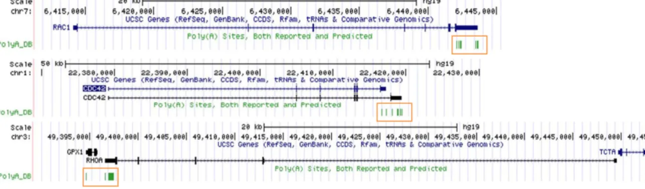
1.1. Rho GTPases produce two mRNA isoforms with different 3’UTR lengths due to the usage of two conserved pA sites ... 37
1.2. Classical Rho GTPases preferentially use the proximal pAs during OL differentiation 44 1.3. Atypical RhoBTB2 mRNA expression during OLs differentiation ... 53
1.4. In glial cells, Rho GTPases show the same pattern of APA, using preferentially the
proximal pAs. ... 56
1.5. Rac1 mRNA undergoes 3’UTR lengthening during axonal growth of the cortical neurons, concomitant with an increase in Rac1 protein levels ... 58
1.6. Candidate RBPs to regulate Rac1 3’UTR. ... 64
2. Cdc42 isoforms functions in CNS and PNS myelination ... 70
2.1. Potential different roles of Cdc42 isoforms in OL and SC differentiation. ... 70
2.2. Role of miRNAs in regulation of the Cdc42 isoforms expression. ... 74
CONCLUSION... 76
FUTURE PERSPECTIVES ... 76
Figures
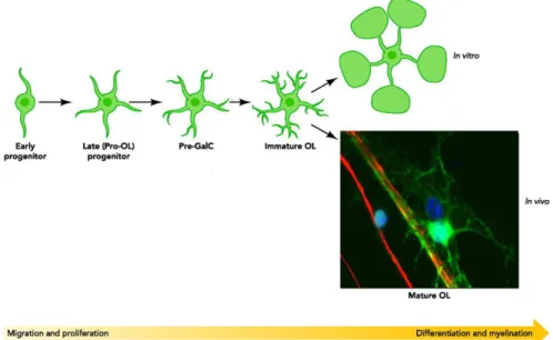
Figure 1. Oligodendrocyte development and differentiation. ... 4
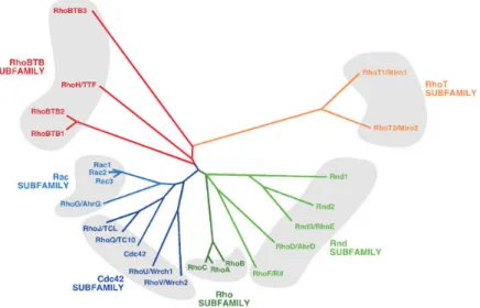
Figure 2. Dendrogram showing the classification of Rho/Rac subfamily members according to structural similarity criteria. ... 5
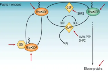
Figure 3. GEFs, GAPs and GDIs regulating Rho GTPases. ... 6
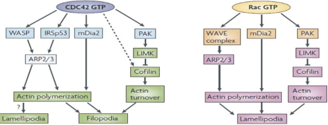
Figure 4. Cdc42 and Rac1 effectors in lamelipodia and filopodia pathways. ... 7
Figure 5. RhoA effectors linked to actin reorganization. ... 8
Figure 6. Regulation of SC and OL development by Rho GTPases. ... 9
Figure 7. Core pre-mRNA 3’end processing machinery involved in cleavage and polyadenylation... 12
Figure 8. Alternative cleavage and polyadenylation sites (PAS). ... 13
Figure 9. Regulation by cis- and trans-acting elements in the 3’ UTR originated by APA. ... 17
Figure 10. Reported and predicted PAS for human Rac1, Cdc42 Iso1/2 and RhoA. ... 37
Figure 11. 3’RACE products using rat OLs and schematic representation of mRNA isoforms of Rho GTPases. ... 39
Figure 12. Mapping of the mRNA 3’ ends of Rac1, Cdc42 Iso1 and Iso2 and RhoA isoforms. . 41
Figure 13. Genomic alignment of Rho GTPases 3’UTR sequences (rat, mouse and human). . 43
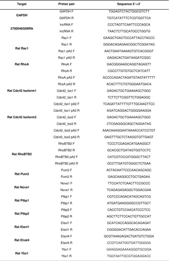
Figure 14. Schematic representation of the primer pairs used in RT-qPCR analyses. ... 44
Figure 15. Relative mRNA expression levels quantified by RT-qPCR, of the different mRNA isoforms during OL differentiation. ... 45
Figure 16. Relative mRNA expression levels of Rho GTPases mRNA isoforms during ON development. ... 48
Figure 17. Stability of Rho GTPases mRNA isoforms. ... 50
Figure 18. Quantification of the relative mRNAs expression levels by RT-qPCR, of the different RhoBTB2 isoforms during in vitro OL differentiation. ... 53
Figure 19. Quantification of the relative mRNAs expression levels by RT-qPCR, of the different RhoBTB2 isoforms during ON development. ... 54
Figure 20. Stability of RhoBTB2 mRNA isoforms. ... 54
Figure 21. Rho GTPases mRNA isoforms quantification in different cell types. ... 57
Figure 22. Rho GTPases mRNA isoforms quantification during axonal growth of cortical neurons. ... 59
Figure 23. Quantification of Rac1 protein levels during the axonal growth of cortical neurons. 60 Figure 24. Rac1 mRNA isoforms quantification during axonal growth of hippocampal and striatal neurons. ... 61
Figure 25. Comparison of the mRNA isoform expression ratio (pA2/CDS) in different types of neurons during axonal growth. ... 62
Figure 26. mRNA sequence of the Rac1 3´UTR. The putative binding sites for several RBPs are highlighted. ... 64
Figure27. Quantification of mRNA expression levels of RBPs during OLs differentiation and axonal growth of neurons. ... 66
Figure 28. Quantification of mRNA expression levels of RBPs during OLs axonal growth of cortical neurons. ... 68
Figure 29. YBX1 expression and Rac1 mRNA binding during OL differentiation. ... 69
Figure 30. Schematic representation of the Cdc42 gene splicing variants. ... 70
Figure 31. mRNA expression ratios of the Cdc42 isoforms during OL, ON and SC differentiation. ... 71
Figure 32. Constitutive expression of Cdc42 Isoforms in CG4 cells. ... 73
Figure 33. Representation of the Cdc42 Iso1 and Cdc42 Iso2 shortest 3’UTR highlighting the putative target sites for miRNAs. ... 74
Abbreviations
APA Alternative polyadenylation
mRNA Messenger RNA
pA polyA signal
Pre-mRNA Precursor mRNA
3’UTR 3’ Untranslated Region
Rho GTPases Ras homologue family of small guanosine triphosphatases
GEFs GTPase exchange factor
GAPs GTPase-activating proteins
miRNAs microRNAs
RBPs RNA-binding proteins
Rac1 Ras-related C3 botulinum toxin substrate 1 Cdc42 Cell division control protein 42 homolog
CNS Central nervous system
E Embryonic day
div Days in vitro
OL Oligodendrocytes
PNS Peripheral Nervous System
SC Schwann cells
OPC Oligodendrocyte precursor cell
P Post-natal day
PDGF-A Platelet-derived growth factor alpha FGF-2 Fibroblast growth factor
GDIs guanine nucleotide dissociation inhibitors RhoA Ras homolog gene family, member A WAVE WASP-family verprolin-homologous protein
mDia Mammalian Diaphanous
PAK p21-activated kinase
LIMK LIM domain kinase
WASP Wiskot-Aldrich syndrome protein IRSp53 Insulin-receptor insulin substrate p53
a.a. aminoacid
ROCKI/II Rho-associated, coiled-coil containing protein kinase 1
MLC myosin light chain
Par partitioning-defective proteins PI3 phosphatidylinositol 3–kinase
IQGAP IQ motif containing GTPase activating protein N-WASP Neural- Wiskot-Aldrich syndrome protein
NRG1 Neuregulin 1
CA Constitutive active
DN Dominant negative
RhoBTB2 Rho-related BTB domain containing 2
PA Polyadenylation
snRNA Small nuclear RNA
polyA tail Poly adenosine tail
PAP polyA polymerase
nt nucleotide
PAS Polyadenylation site
DSE Downstream sequence elements
USE Upstream sequence elements
CPSF cleavage and polyadenylation specificity factor CstF cleavage stimulation factor
CFI/II cleavage factor I/II
PABP PolyA binding protein
mRNP messenger ribonucleoprotein particles
RBD RNA-binding domains
PABPN1 PolyA binding protein nuclear 1 Elavl ELAV Like RNA Binding Protein
Abbreviations (continuation) Pre-miRNA Precursor microRNA
miRISC miRNA-induced silencing complex
AGO Argonaut
GW182 Glycine-trytophan protein of 182kDa
CDS Coding sequence
3’RACE 3’ Rapid amplification of cDNA ends
ON Optic nerve
BDNF Brain-derived neurotrophic factor GFAP Glial Fibrillary Acidic Protein
Pum2 Pumilio RNA-binding family member 2 Ptbp1/2 Polypyrimidine tract binding protein 1/2
YBX1 Y box binding protein1
AREs Au-rich elements
SN Sciatic nerve
QK Quanking
MBP Myelin basic protein
hnRNP Heterogeneous nuclear ribonucleoprotein
bp Base pairs
1. Central Nervous System Cells
1.1. Neurons
The nervous system of higher vertebrates, besides its role in coordination the vital functions, enables its members to perform fast and coordinated movements, respond to pain stimuli, and exert complex cognitive functions such as learning, memory and social behavior. The cellular basis of the vertebrate nervous system is built upon two major cell types: neurons and glia cells. Neurons are the main signal relaying cells and differ from other cell types present in the nervous system in their organization of fibrillar or tubular proteins that constitute the cytoskeleton.1 During maturation, neurons undergo a transition
trough several stages of differentiation until becoming fully mature, i.e. acquiring axon-dendrite polarity.2 During this process, neurons develop lamellipodial and filopodial
protrusions (at embryonic day 18 (E18)), followed by extension of multiple immature neuritis (at E18+1-2 days of in vitro culture (div)) which undergo axonal specification (E18+2-4div) and branching (E18+4-15div), and harbour dendritic spines and axon initiation at the fully differentiated stage (at E18+15-25div).3 The molecular mechanisms
underlying neuronal development comprise: local protein translation and degradation; a wide range of signalling pathways; transcription regulation by microRNAs4 (miRNAs) or
RNA-binding proteins (RBPs)5; and extracellular cues3. Although sharing the mechanisms
involved in neuronal growth, different neurons have particular gene expression6 and gene
regulation7 programs according to their sub type.
1.2. Glial Cells
Glial cells, derived from the Greek word “γλία”, meaning “glue”, provide important support to the neurons. In the CNS, there are three types of glial cells: astrocytes that provide trophic and structural support to the neurons and act in several other aspects of the neuronal homeostasis and metabolism; oligodendrocytes (OL) that produce myelin sheaths around axons in order to increase nervous conduction speed and microglia, the resident immune cells. In the peripheral nervous system (PNS) the glial population is constituted by Schwann cells (SC).8 Glial cells, are essential for the correct development,
maintenance and function of the brain network. They are the most abundant cells in the mammalian brain, and exist in a ratio of 3 glial cells for 1 neuron.1 Although glia cells also
extend complex processes, these are less prominent and have completely different functions than those of neurons. During OL maturation, bipolar and proliferative progenitor cells (OPC) will give rise to more differentiated myelinating-competent OL containing long radial processes and branching (Figure1). Between these two developmental stages OPC undergo a complete cellular reprogramming in which they stop proliferating and initiate a differentiation program.9, 10 This developmental program is regulated in response to
extracellular cues, which are sensed by the OL. Such cues could be integrin, growth factor or chemokine receptor activation. Since OPC have a strong tendency to differentiate in absence of proliferative stimulus, this proliferation-differentiation transition needs to have fine-tune regulation. Similar to neurons, OL differentiation is regulated trough several mechanisms, such as epigenetic control11, 12; gene expression programs
(whereas during morphological alterations, cells express a well defined panel of markers that define each stage)10, 13; and transcriptional and post-transcriptional regulation 14. As
OL-neuron interactions are fundamental for neurons to function, the other way around is equally vital for OL development and myelination. The OL depends on neuronal signals, such as platelet-derived growth factor alpha (PDGF-A) or fibroblast growth factor (FGF-2), to decide the time and place for differentiation15.
Figure 1. Oligodendrocyte development and differentiation. Representative scheme of the morphology alteration of oligodendrocytes during development and differentiation and an in vivo image of a mature oligodendrocyte (in green) myelinating an axon (in red). (Adapted from Jackman N. et al. 2009, Physiology)
2. Rho GTPases
Rho (Ras homologue) family of small guanosine triphosphatases (Rho GTPases) includes a large subgroup of the Ras superfamily of 20-30kDa GTP-binding proteins.16 They act as
molecular switches integrating signals from the environment to intracellular signal transduction pathways, thereby controlling a wide range of essential biochemical responses in eukaryotic cells.17 The network involving Rho GTPases is thought to be very
complex since approximately one percent of the human genome encodes proteins that either regulate or are regulated by direct interaction with members of the Rho family.18
Rho GTPases are ubiquitously expressed from yeast to mammalians, and within these they share over 50% of sequence identity.19 They are divided in 6 subfamilies, according
to their sequence similarity and/or functionality: the classical RhoA (Ras homolog gene family, member A)-, Rac1 (Ras-related C3 botulinum toxin substrate 1)- and Cdc42 (Cell division control protein 42 homolog)-related subfamilies; and the atypical Rnd and RhoBTB (Rho-related BTB (Broad Complex/Tramtrack/Bric-a-brac) domain) subfamilies (Figure 2).20, 21 As molecular switches, typical Rho GTPases cycle between two
conformatial states: inactive, in GDP-bound state or active, when binding to GTP. This alternation is controlated by three classes of regulatores: GTPase exchange factors (GEFs), which promotes the dissociation of GDP to allow the binding of GTP; GTPase-activating proteins (GAPs), which increase the activity of Rho GTPases by promoting the
Figure 2. Dendrogram showing the classification of Rho/Rac subfamily members according to structural similarity criteria. Members of each subfamily are highlighted using the same color code and grouped by shaded areas. The first symbol used for each GTPase corresponds to that approved by the Human Genome Organization Gene Nomenclature Committee. (Bustelo X. et al. 2007, Bioessays)
return to the inactive state; and guanine nucleotide dissociation inhibitors (GDIs), that stabilize the GDP-bound form and inhibit the association of GTPases to the membrane (Figure 3).22 In addition to this specfic regulation, Rho GTPases are also tightly regulated
at expression level.21 When activated, these GTPases interact with their downstream
effectors, which in turn regulate a wide range of mechanisms such as microtubles dynamics, transcription activation or membrane trafficking. These interactions, as well the resulting functions, are likely to be specific for each cell-type and physiologic condition. Rho GTPases are therefore fundamental for diverse cellular processes including cell growth, cytokinesis, cell motility, cell adhesion, cell transformation, invasion and neuronal development. Our knowledge about Rho GTPases arises mostly from the best studied proteins Rac1, Cdc42 and RhoA.17, 23-26
Figure 3. GEFs, GAPs and GDIs regulating Rho GTPases. The activity of the Rho GTPases is modulated by Rho regulatory proteins of the following classes: GEFs, which activate Rho; GAPs, that facilitate inactivation of GTP-bound Rho by increasing GTPase activity; and GDIs that prevent dissociation of GDP and inhibit activation. Phosphatases could potentially regulate the Rho GTPases at many levels, as shown by the red arrows. (Adapted from Larsen M. et al. 2003 Nature)
2.1. ‘Classically activated’ Rho GTPases (Rac1, Cdc42 and RhoA)
Rac1 is known to regulate signaling pathways for cell proliferation, apoptosis and activation of immune cells. However, its major function is the organization of the actin cytoskeleton (adhesion and differentiation) and the lamellipodium.18, 27, 28 Activated Rac1
triggers a broad list of downstream effectors including proteins from the Wiskot-Aldrich syndrome protein (WASP)-family verprolin-homologous protein (WAVE), mammalian Diaphanous (mDia) and p21-activated kinase (PAK), which are implicated in the major
functions described below (Figure 4). The fact that the Rac1-knockout is embryonic lethal and the Rac2-, Rac3- and RhoG-knockout are viable and do not have developmental defects demonstrates the importance of Rac1 relatively to the other members of the Rac subfamily.28
Cdc42 function (as the other Rho GTPases) is related with receptor-mediated signal transduction leading to induction of gene transcription, cell cycle progression, apoptosis, migration, chemotaxis, and cell fate determination. 18, 28, 29 However, the primordial
function of the Cdc42 is the regulation of cell polarity and actin cytoskeleton resulting in the filipodium formation. When activated, Cdc42 signals through WASP, mDia, the insulin-receptor substrate p53 (IRSp53) and PAK to perform its functions in actin polymerization (Figure 4).28, 29 Its function is thought to be essential during embryonic development as
Cdc42-knockout mice that die at embryonic day 7.5.30 Recently, two spliced isoforms were
described for Cdc42: the canonical isoform that is ubiquitously expressed and the brain specific isoform (further referred as Cdc42 Isoform 1 and Isoform 2 respectively).31 These
isoforms differ only in their C-terminal exon, a region where they have a few amino acidic (a.a.) changes resulting in different lipidations of the proteins.32 Thus, both Cdc42
Isoforms have a prenylation in that region, however Isoform 2 has an additional palmitoylation.31, 32 The palmitoylation of the Cdc42 Isoform 2 is essential for its
membrane localization and in neurons plays a role in dendritic protrusion and dendritic spine formation.32 Additionally, the dual lipidation increases the affinity of Isoform 2 for the
Figure 4. Cdc42 and Rac1 effectors in lamelipodia and filopodia pathways. Cdc42 induces actin polymerization by binding to WASP, or through IRSp53 Tyr kinase to induce branched actin filaments using the ARP2/3 complex. Rac activates the ARP2/3 complex through the WAVE complex. Cdc42 and Rac also induce actin polymerization by activation of mDia2. Rac- or Cdc42- mediated activation of the Ser/Thr kinase PAK phosphorylates, LIM kinase (LIMK), which phosphorylates and inhibits cofilin, thereby regulating actin-filament turnover. In the neuronal growth cone, Cdc42 might result in reduced cofilin phosphorylation by an unknown mechanism (dotted line), thereby stimulating actin polymerization and filopodium formation. (Adapted from Heasman and Ridley 2008, Nature)
plasma membrane.33 Although its biologic function is poorly known, this specific feature of
Cdc42 represents an additional mechanism of regulation, highlighting the fine-tuned activities of Rho GTPases.
RhoA was the first identified member of Rho GTPases. Similar to Rac1 and Cdc42, RhoA transduces extracellular signals and, through it effectors, regulate cell migration, adhesion, survival, cell division, gene expression and vesicle trafficking in a cell-type dependent manner.18, 34 Rho has been postulated to be a major regulator of actomyosin
and focal adhesion dynamics, but in several cell types, these effects are only seen in cooperation either with the other members of the Rho subfamily (RhoB and RhoC) or with its effectors (e.g. ROCKI/II (Rho-associated, coiled-coil containing protein kinase 1/2) or mDia) or with other Rho GTPases (Figure 5).34 In the past few years, several papers
highlighted the important functions of Rho GTPases in regulating both the actin and microtubule cytoskeletons and the machinery involved in establishing polarity, during CNS and PNS development.
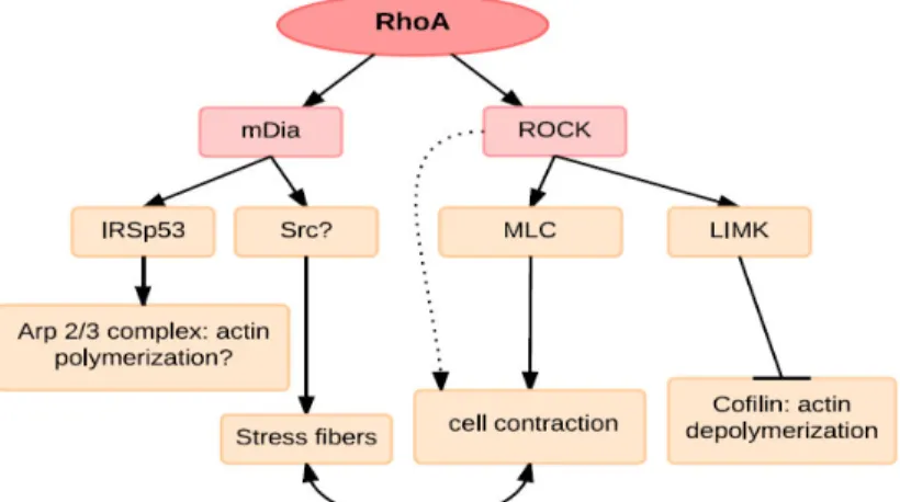
Figure 5. RhoA effectors linked to actin reorganization. Rho via ROCK can stimulate myosin light chain (MLC) phosphorylation and together with mDia induces stress fibre formation. mDia is dependent on Src for its contribution to stress fibres and has also been reported to interact with IRSp53, which can mediate Arp2/3-complex-induced actin polymerization. ROCK can also phosphorylate a number of other target proteins that may contribute to actin reorganization, including LIMK, which inhibits cofilin-mediated actin depolymerization. (Adapted from Ridley A.J. 2001, Journal of Cell Science)
In the CNS neuronal development requires Rac1 and Cdc42 activity for neurite formation and outgrowth, as well as for axonal guidance.23, 26, 35 During axon polarization Cdc42
activates Rac1 via the Par3/Par6 complex and Rac1 GEFs (Tiam1 or Tiam2/STEF), which are known to be essential to neuronal polarization and extension. This mechanism is a positive feedback loop between Rac1 and Cdc42, whereas the former could activate the latter through activation of PI3-kinase (phosphatidylinositol 3–kinase). In addition, Rac1 is
activated by GEF DOCK7 to allow the microtubule growth in the nascent axon. Neuronal differentiation progresses with axon extension and, for this is required an additional effector of Rac1 and Cdc42, the IQGAP3 (IQ motif containing GTPase activating protein) and, N-WASP (Neuronal- WASP) that is targeted only by Cdc42.23, 36 Nevertheless, Rac1
functions are not restricted to the axon, and they also affect branching of the dendrites.35, 37 Regarding axon guidance, Rac1 and Cdc42 are associated with attractive cues and
forward protrusion of growth-cone, while RhoA is associated with repulsive cues and growth-cone collapse. The RhoA and Rac1/Cdc42 are also thought to have antagonistic roles during axon formation and outgrowth.23, 34, 36 The functions of these Rho GTPases
are not restricted to neurons, and it are equally important for the appropriate differentiation of OL and SC, even though through different mechanisms.24 Rac1-null SC lead to
hypomyelination of the sciatic nerves and defects in radial sorting (similar to those seen on laminin or β1 integrin mutants). Rac1 activity, which is regulated by β1 integrin, is necessary for process extension and stabilization, and consequently for efficient radial sorting of axon bundles in a β1 integrin-dependent way.38 Recently two downstream
effectors of Rac1 were described that directly influence myelination: NF2/merlin and cAMP.39 In the other hand, Cdc42, which activity is regulated by NRG1 affects SC
proliferation and consequently radial sorting of axons (Figure 6).38In OL, differentiation
and the proper axon ensheatment and myelination depend on Rac1 and Cdc42 activity, and on RhoA inactivity.
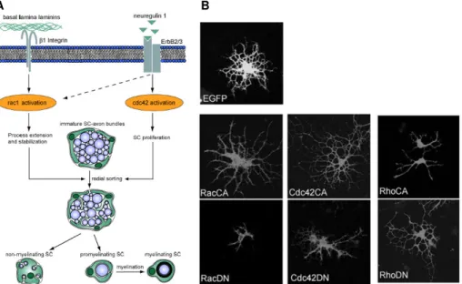
Figure 6. Regulation of SC and OL development by Rho GTPases. A) Proposed mechanism for regulation of SC by Cdc42 and Rac1, which plays different but essential roles during SC development. Both Rac1 and Cdc42 are required for efficient radial sorting of axon bundles, a prerequisite for subsequent axon
B A
myelination. B) Effects on OL differentiation upon expression of constitutive active (CA) or dominant negative (DN) Rho GTPases. Constitutively active Cdc42 and Rac1 and dominant negative RhoA induce outgrowth of OL, whereas dominant-negative Cdc42 and Rac1 or constitutive active RhoA inhibit OPC differentiation, when compared with the empty vector (EGFP). (Adapted from Benninger Y. et al. 2007, The Journal of Cell Biology and Liang X. et al. 2004, The Journal of Neuroscience)
Similar to neurons, Rac1 and Cdc42 act synergistically in process extension and branching during the OL differentiation and myelination.38, 40 These two proteins are both
necessary for proper axon ensheathing and their loss leads to the accumulation of a large amount of cytoplasm in the inner tongue of the process resulting in the formation of myelin outfoldings.40, 41 The Rho GTPase effectors participating in myelination are poorly known,
however a few studies have shown that the Rac1 effector WAVE142 or the Fyn
phosphatase, which affects RhoGAP p190 activity 41. This is in contrast with RhoA, which
has a negative role in the OL differentiation inhibiting process extension. Although the molecular effectors of this regulation are not completely known, ROCK is thought to be one of them41.
2.2. Atypical Rho GTPase RhoBTB2
Rho BTBs are a group of atypical Rho GTPases that differ from the classical in their size; in their regulation by gene expression or protein stability and in the constitutive GTP-bound state. They are much larger than classical Rho GTPases and have additional BTB domains, which are important for protein:protein interactions. Although the inactivation of
RhoBTB genes did not reveal any associated phenotype, they are reported, in particular RhoBTB2, as tumour suppressor genes.21 As classical Rho GTPases, RhoBTBs are
ubiquitously expressed in human, mouse and rat tissues. RhoBTB1 and 3 have higher expression levels then RhoBTB2. Albeit having lower mRNA levels, RhoBTB2 expression is significantly increased in the brain tissues, suggesting a tissue specific role.43
3. Gene expression regulation
The control of gene expression is a biological process essential to all organisms. Thus, gene expression requires an accurate regulation of several events during the development, division and differentiation of the cell. This regulation can be done at the transcriptional and co-transcriptional level or at the post-transcriptional levels. Transcriptional regulation occurs through the binding of regulatory proteins with specific DNA motifs, chromatin remodelling or by the interaction with the transcription machinery.
Regulation at the co-transcriptional level includes pre-mRNA processing: capping, splicing and polyadenylation. Post-transcriptional regulation comprises mRNA stability, transport, and translation mechanisms. Some of the key events that are regulated at the co-transcriptional level are alternative splicing, polyadenylation (PA) and alternative polyadenylation (APA) and include several players cis- and trans-regulatory elements, RBP and small RNAs such as miRNAs. All of these mechanisms are emerging as key components in this regulatory process.44-46
3.1. Co-transcriptional regulation
Pre-mRNA (precursor mRNA) processing is a key step in mRNA maturation and in the specificity of several cellular programs such as tissue-specific gene expression, apoptosis, sex determination or development, implying a tight regulation of the mechanisms involved. The production of a mature and functional mRNA requires three well defined processing steps: (1) capping, that consists in the addition of a cap structure at the 5’end of the nascent transcript and prevents it from degradation; (2) splicing, involving the assemblage of small nuclear RNAs (snRNAs) with proteins in the small nuclear ribonucleic particles (snRNPs) to form the spliceossome, in order to remove the introns from the (pre-mRNA); (3) 3’end formation, that comprises the cleavage of pre-mRNA followed by the polymerization of an adenosine tail (polyA tail).47, 48 Through alternative splicing or PA,
one single transcriptional unit can produce several mRNA molecules that could lead to different proteins or mRNAs with different 3’ UTR lengths. These fundamental processes occur co-transcriptionaly and, although being independent mechanisms, are in close crosstalk and interact with the transcription machinery.48, 49
3.2. Polyadenylation
The maturation of eukaryotic mRNAs requires a precise 3’end formation. As referred above this process comprises two steps, one specific endonucleolytic cleavage and the polymerization of the polyA tail by the polyA polymerase (PAP) (histone genes are the exception). This reaction is called polyadenylation (PA) and is driven by cis-elements present in pre-mRNA and trans-acting factors. The length of the polyA tail was recently described as organism-, development stage-, and tissue-specific and could vary between
30 to 100 nucleotides50, 51, in contrast with what was consensually accepted before (polyA
tails with approximately 200 adenosines)44-46. The PA occurs in almost fully processed
eukaryotic mRNAs48, and influence the stability, translation and transport of mRNA to the
cytoplasm44, 46. However recent studies have, in part, challenged this view showing that
the effect of the polyA tail on mRNA stability is restricted to the early developmental stages.50, 51 The consensus and stronger signal for PA (pAs) is the cis sequence AAUAAA
(though the signal might be a weaker variant) that is recognized by the 3’end processing machinery, and normally is localized 10-35 nucleotides (nt) upstream of the cleavage and polyadenylation site (PAS).52 The cleavage and polyA tail addition occurs between this
hexamer and an U/GU-rich downstream sequence element (DSE), which affects PA efficiency. Upstream U-rich sequence elements (USE) that present before the pAs could also affect the efficiency, providing binding sites for several protein factors.44-46 The core
machinery responsible for cleavage and polyadenylation comprises four main protein complexes: cleavage and polyadenylation specificity factor (CPSF), that identify pAs and catalyse the cleavage reaction; cleavage stimulation factor (CstF), that recognize the DSE; and cleavage factor I (CF Im) and II (CF IIm), which are important for the assembly of
the other members of the protein complex. For correct 3’end processing the core machinery have to engage 25 protein factors and around 60 individual proteins such as symplekin, PAP or polyA binding protein (PABP) (Figure 7).53, 54 This assemblage requires
a fine-tune regulation to allow the accurate mRNA maturation.48, 53 The impairment of one
of these factors or functions leads to a PA defect, related with a wide range of diseases or cellular conditions.55
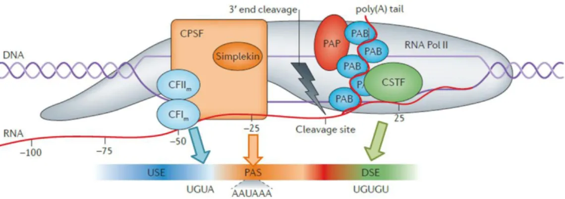
Figure 7. Core pre-mRNA 3’end processing machinery involved in cleavage and polyadenylation. The cleavage factors CF Im, CF IIm bind to their cis regulatory elements, USE, and CPSF binds to the pAs, whereas CstF binds to the U/GU-rich region, DSE. PAP polymerizes the polyA tail and PABP binds along the polyA tail. (Adapted from Elkon R. et al. 2013, Nature)
3.3. Alternative polyadenylation
In the human genome 70% of the genes have multiple pAs in their 3’ untranslated regions (3’UTRs), and in mammalian, 80% of the genes undergo APA.56, 57 APA controls gene
expression by production of alternative mRNA isoforms due to the usage of different pAs present in one gene. Usage of one PAS over another is often attributed to the relative strength of the cis and trans-acting elements and to a close relation with transcription as mentioned above. The different types of APA differ in the localization of pA signal (intronic or exonic), which generates either different mRNA coding sequences (resulting in different proteins produced), or mRNAs with 3’UTRs of different lengths (Figure 8).44, 46, 58
In the past few years, with the increase usage of genome wide methodologies, APA has been related with global physiologic events. Recent studies associate the usage of the shortest 3’UTRs with proliferation and transformation of the cells. The usage of proximal pAs is observed during the activation of T lymphocytes, cell proliferation and in cancer cell lines. Usage of proximal pAs has been related to the production of higher amounts of protein, due to the loss of regulatory elements, present in the longest 3’UTRs, such as target sites for miRNAs.59, 60 On the other hand the longest 3’UTRs are present
preferentially in differentiation and development programs.
Several studies have shown that different cell types were reprogrammed to generate induced pluripotent stem cells through APA modulation61, additionally 3’UTR lengthening
was seen in the mouse embryonic development studies62. In addition, various works using
RNA sequencing (RNA-seq) have shown that in brain tissues, both from Drosophila and
Figure 8. Alternative cleavage and polyadenylation sites (PAS). A) Alternative polyadenylation (APA) in the 3’ most exon. A hypothetical gene is shown, with two pAs located in the 3’UTR originating two mRNAs with different 3’UTR lengths. B) APA in upstream regions producing mRNAs containing different coding regions, and consequently, different proteins (Adapted from Tian B. and Manley J.L. 2013, Trends in Biochemical Sciences )
A
mammalian models, there is a diffuse and extensive lengthening of the 3’UTRs. Curiously was found that brain 3’UTR extensions are significantly longer than previously annotated.63, 64 Alternative mRNA isoforms (resulting from APA) with longer 3’UTRs have
more cis-regulatory elements in pre-mRNA such as binding sites for RBPs (involved in mRNA stability, localization and translation), miRNAs (involved in mRNA translation inhibition, cleavage/degradation), and regions with AU-rich content (important for PA site definition). Consequently, the differential signals usage implies that these RNA sequence elements will be differential used, affecting the transcript produced and ultimately modulating gene expression, both in a negative or positive manner. Therefore the differential selection of pAs in APA is a co-transcriptional mechanism that allows the mature mRNA present alternative places for regulation (Figure 9). 44, 46, 5865
3.4. RNA Binding Proteins
The RBPs are key regulators of gene expression both at co- and post-transcriptional levels. The pre-mRNA processing is intrinsically related with the binding of RBPs to the specific sequences present in the mRNA resulting in the formation of messenger ribonucleoprotein particles (mRNP) complex. This interaction occurs trough the specific RNA-binding domains (RBD) in the RBP, which could be repeats of the same type or a conjugation of different domains. Together, the high variability of these RBD, the alternative splicing of RBPs pre-mRNA, the auxiliary functional domains, and the high diversity of RNA-binding sequences leads to a variety of mechanisms of regulation by RBPs in one cell.65 The importance of the RBPs is denoted by the high evolutionary
conservation of their binding sequences.66 In short, cells are able to generate diverse
mRNPs specific for each mRNA. Several aspects of the RNA biology such as transcription, pre-mRNA splicing, PA/APA, modification, export, localization, translation and turnover can be affected by RBPs. An efficient PA is dependent on specific RBPs, while one of the major players, the CPSF, binds to the pre-mRNA by its RNA-binding subunits. Poly(A) binding protein, nuclear 1 (PABPN1) is another important RBP for PA, that additional to CPSF, its binding promote the activity of PAP. 65 The impairment of
RBPs biology, as mutations or alterations in their relative concentrations in the cell can lead to severe disorders.55 In other way, APA per se is able to escape or induce the
regulation through RBPs according to the production of the shortest or longest mRNA isoforms respectively. Therefore, cells can use this mechanism, in addition to the
expression of cell-specific RBPs to achieve their cellular fate.62, 67 The usage of this type
of regulation is most evident and known in neuronal development and specification, where the couple between the 3’ lengthening64, 68 and the expression of brain-specific RBPs such
as ELAVl/Hu (ELAV Like RNA Binding Protein) proteins or NOVA (neuro-oncological ventral antigen)69 occurs. Another important feature of the neuronal cells involving RNPs
is the mRNA transport and the local translation. As described above neurons extend long axons that need the transport of mRNAs to the apical zone for local translation of proteins in a rapid response manner.69, 70 The deregulation of these processes is one of the major
causes of neuronal diseases.55 Although less is known, some studies have demonstrated
the fundamental role of RBPs in OL differentiation and myelination. The quaking (QK) family of RBPs has identified in glial progenitors and OLs, where the differential expression of spliced isoforms coincides with the development of OLs and the onset of myelination. Mutation on qk gene leads to a failure in the development of mature myelinating OLs.71 Another important family of RBPs is the Musashi (Msi) family, which is
preferential expressed in CNS stem cells, and its downregulation is required to the differentiation of OLs.72 Similar to neurons, in OLs the transport and local translation of
specific mRNAs is also observed. The best studied is the myelin basic protein (Mbp) mRNA, which requires the formation of a RNP complex, where different family members of the RBPs hnRNP (heterogeneous nuclear ribonucleoprotein) play different functions through binding to the 3’UTR.73-75 This co-transcriptional regulation by RBPs is tightly
associated with those of miRNAs, indeed recent studies have confirmed either competition or collaboration between them.76, 77
3.5. MicroRNAs
miRNAs are a class of nonconding RNAs with approximately 21 nucleotides in length that are able to post-transcriptionally silence mRNAs by binding to them through complementary or semi-complementary sequences. In mammals, the activity of approximately 50% of protein-coding genes is controlled by miRNAs. Although a single miRNA is often predicted to target several individual transcripts, their expression can impact the definition of the cell-specific protein profile, since miRNAs are expressed in a tissue-specific and/or developmental-stage-specific manner. The mRNA target sequences for miRNAs could be also binding sites for other miRNAs as well for RBPs, which give a multiple combinations for regulatory effects.76 In short, the biogenesis of miRNAs, initiate
with the formation of the pre-miRNA (precursor miRNA), which start by the transcription of an independent gene or an intron of one protein-coding gene by RNA polymerase II. This pre-miRNA is processed first by Drosha (in the nucleus) and by Dicer (in the cytoplasm) enzymes resulting in a ~20bp miRNA/miRNA duplex. This duplex is then separated and one of the strands becomes the mature miRNA that is incorporated into miRNA-induced silencing complex (miRISC). In miRISC, miRNAs will induce the translational repression or degradation, through the action of Argonaute (AGO) proteins and glycine-tryptophan protein of 182kDa (GW182), of the target mRNA.78, 79 The major determinant of the AGO
action is a 6-8 nucleotidic domain at the 5’ end of the miRNA (this domain is called seed region). The seed region binds to the complementary region (‘seed matches’) on target mRNA and causes a decrease in its expression. The repressive effect is highest when the seed matches occur in the 3’UTR.80
Regulation by miRNA activity has some of the most interesting examples in neurons, where miRNAs have a role in the regulation of localized protein expression. Thereby, the mRNAs can be inactivated by miRNAs to reach the dendritic spines, where it becomes accessible to the translation machinery, and also they can be downregulated in the dendritic spines by the local expression of specific miRNAs.78 In addition, neuronal
determination and development are also dependent on miRNA activity, either through promoting the differentiation of the neural stem cells and progenitors (e.g. miR-124 and miR-9) or inducing their proliferation (e.g. miR-134, miR-25 or miR-137), according to the cluster of genes that are targeted.4 Another important role of the miRNA function in the
CNS is the control of the production, differentiation and health of myelinating OLs. Specific miRNAs act in OPCs and in OLs through: (1) promoting OPC expansion; (2) suppressing OPC-expressed genes to promote differentiation; (3) the overall suppressing of inappropriate non-OL lineage gene expression in OPCs and OLs; and (4) by suppressing transiently genes required at high levels during myelin sheath formation. Several members of the miR-17-92 cluster have been described as necessary and sufficient for OPC proliferation.81 The two most highly induced miRNAs in differentiating OLs are
miR-219 and miR-338; they promote normal OPC differentiation into OLs.82 Recently, in one
study it was shown that myelination of the CNS is promoted by the expression of miR-23a.83 Thus several steps of development and differentiation of CNS can be regulated
either through inducing or repressing miRNAs. The impairment of these functions by the loss of miRNA target sequences can lead to several disorders or cellular conditions55.
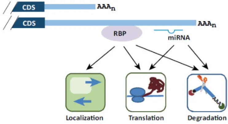
Figure 9. Regulation by cis- and trans-acting elements in the 3’ UTR originated by APA. Two mRNA isoforms are shown containing different RBPs binding sites and miRNA target sites. Their impact on mRNA localization, translation and degradation are indicated. CDS, coding sequence. (Tian B. and Manley J.L. 2013, Trends in Biochemical Sciences)
Aims
In this thesis we addressed three main aims: Part I
• Characterization of Rho GTPases APA during OL differentiation.
• Understand if the mechanism of Rho GTPases APA is cell type specific. Part II
• Study the function of Cdc42 Iso1 and Cdc42 Iso2 in OL differentiation.
In silico
analysis
The UCSC genome browser was accessed to check for APA sites of mRNAs. The nucleotide sequences were obtained from NCBI database. Conservation of nucleotide sequences among different species was performed using the Geneious v4.8 software84.
To predict putative binding sites of RBPs, the PBSFinder85 (tool developed by us),
RBPDB86 and SFmap databases87 were used; while for microRNAs predition, the
microRNA.org88, mirWalk 2.0 database89 and Targetscan.org were used.
Cell Culture
Primary mixed glial cultures. Primary mixed glial cultures (composed of OPC, microglia
and astrocytes) were harvested from post-natal day (P) 1 to P2 neonatal Wistar rat cortex following a standard protocol90 with minor modifications91. In brief, dissociated rat neonatal
cortices were cultured at 37 °C in 7.5 % CO2 in DMEM with 10 % FCS and penicillin/streptomycin for 10 days in vitro.
Microglia. To remove the microglia on the top of the mixed glial cultures, the flasks were
shaken for 1 h at 200 rpm on an orbital shaker. Microglia were plated at 1 x 10⁶ cells per
ml into 10 cm plate in DMEM/F12 supplemented with 1 ng/ml GM-CSF and 10% FBS media. The cells were left 24 h for recover and adhesion, and were used directly for total RNA extraction.
Oligodendrocytes. After shaker to remove microglia, mixed glial cultures were shaken at
240 rpm overnight to dislodge the loosely attached OPC. These OPC were further purified from contaminating microglia by a differential adhesion step. Purified OPC were plated at a density of 20,000 cells per 0.8 cm2 well in proliferation media (SATO media [L-glutamine
(4 mM), putrescine (16 µg/ml), T4 (400 µg/ml), T3 (400 µg/ml), progesterone (6.2 ng/ml), sodium selenite (5 ng/ml), BSA V (100 µg/ml), insulin (5 µg/ml), holo-transferrin (50 µg/ml)] supplemented with PDGF-A (10 ng/ml), FGF-2 (10 ng/ml), 1 % penicillin/ streptomycin). After 2 days the cells are harvested, for day 0 time point, or maintained for induction of differentiation. The proliferation media was replaced with differentiation media (SATO media supplemented with1 % penicillin/ streptomycin and 0.5 % FCS). Cells are collected at 3, 5 or 7 days of differentiation. All cells were cultured in Poly-D-Lysin and Laminin coated plates. All mixed glial cultures were maintained until a total of three shakes were performed.
Astrocytes. Upon the three shakers describe above, the mixed glial cultures were subject
to trypsinization and plated in new flask for further astrocyte purification. A total of three steps of purification were performed. After obtaining a pure astrocytes culture, the cells were used directly for total RNA extraction.
Neurons. High density cultures of rat cortex, hippocampal and striatal neurons were
prepared from E18–E19 Wistar rat embryos as previously described92, and were obtained
from Teresa Summavielle’s laboratory. Briefly, neuronal cultures were maintained in serum-free Neurobasal medium (Gibco Invitrogen), supplemented with B27 (GibcoInvitro- gen), glutamate (25 µM), glutamine (0.5 mM) and gentamycin (0.12 mg/ml). Cells were cultured at a density of 9 × 10⁴ cells/cm2 on poly-D-lysine coated 6-well microplates, and
kept at 37 °C in a humidified incubator with 5% CO2/95% air, for 24 h, 7 or 14 days in vitro (DIV), and were used directly for total RNA extraction.
Optic and sciatic nerves. Optic and sciatic nerves were harvested from P2, P14 and P30
Wistar rats followed by a rapid freeze at -80ºC. The tissues were homogenized in TRIzol (Invitrogen) reagent before proceeding to the total RNA extraction.
RNA extraction
Total RNA was extracted with TRIzol (Invitrogen) reagent following the standard protocol.
The cells were incubated 5min at RT with 1 mL of TRIzol and then overnight at -80ºC. To separate the RNA fraction chloroform was added to the lysates, mixed and centrifuge for 15 min at 11400 rpm at 4ºC. The aqueous phase was transferred to a fresh tube, where Glicoblue (15 mg/mL, Ambion Life Technologies) and the same volume as the aqueous phase of Isopropanol was added and mixed. The samples were frozen overnight at -80ºC and after, centrifuged twice, 20 min and 10 min at 11400 rpm at 4ºC, with a washing step with 75% cold ethanol between centrifugations. The pellet was air dried and resuspended in Nuclease-free water (Thermo Scientific). RNA quantification was performed in a NanoDropTM 1000 Spectrophotometer (Thermo Scientific) and the RNA stored at -80ºC.
The extraction of RNA enriched with small RNAs was performed using mirVanaTM
isolation kit (Ambion, Life Technologies) and following the manufacturer’s protocol. Briefly, the cells were counted and 102 to 107 cells were pelleted and washed in cold PBS (1x).
Cells were lysed in the lysis/binding buffer and incubated with miRNA Homogenate Additive provided in the kit. For RNA fraction extraction were added one volume of Acid-Phenol:Chloroform and centrifuged. The aqueous phase was recovered into a fresh tube and 1.25 volumes of 100% ethanol were added and mixed. This mixture was passed
through a filter cartridge before a wash step with miRNA Wash Solution 1. At this point was preformed the DNAse (DNase I recombinant, RNase free, 10 U/µL, Roche) treatment on column for 15 min. Samples were washed again with miRNA Wash Solution 1 and twice with mRNA Wash Solution 2/3. After discard the flow-through, RNA was eluted in 95ºC pre-heated Elution Solution. RNA quantification and storage was performed as described previously.
Reverse transcription
DNase treatment. To avoid DNA contamination, the total RNA was treated with DNase
(DNase I recombinant, RNase free, 10 U/µL, Roche) in DNase I Incubation Buffer and Nuclease-free water (Thermo Scientific), for 1h-1,5h at 37ºC. The incubation was followed for 10 min at 75ºCfor enzyme denaturation.
Reverse transcription reaction. cDNA was prepared using SuperScript IIITM Reverse
Transcriptase enzyme (Invitrogen, Life Technologies) and 500ng of total RNA following the manufacture’s guidelines. Briefly, the RNA was denatured for 5 min at 65ºC with dNTPs (10 mM), Random Hexamers (50 mM) and Nuclease-free water (Thermo Scientific), followed for 5 min at 4ºC. The mixture for reverse transcription was prepared with the cDNA synthesis buffer (5X), DTT (0,1 M), RiboLock RNase Inhibitor (Thermo scientific) and SuperScript III RT (200 units/µL, Invitrogen) reverse transcriptase enzyme, added to the denaturated RNA samples and gently mixed. The samples were then incubated for 10 min at 25ºC, 60 min at 50ºC and 10 min at 70ºC to inactivate the enzyme. For discard genomic DNA contaminations, negative reactions were performed without SuperScript III.
RACE (Rapid Amplification of cDNA Ends)
Reverse transcription reaction. The cDNA synthesis was performed using the
SMARTerTM RACE cDNA Amplification kit (Clontech) following the manufacturer’s
protocol, shortly described here. Initially, the RNA and 3’-CDS Primer A were added to sterile water. The samples were incubated at 72ºC for 3 min, cooled at 42ºC for 2 min and then briefly centrifuged. During the incubation step were prepared the Master Mix with Buffer Mix for 3’RACE-Ready cDNA synthesis (5x First-Strand Buffer, DTT (20mM) and dNTP Mix (10 mM)), RNases Inhibitor (40 U/µL) and SMARTSribeTM Reverse