UNIVERSIDADE TÉCNICA DE LISBOA
Faculdade de Medicina Veterinária
Structural and functional insights into the role of
Carbohydrate Esterases and Carbohydrate-Binding
Modules in plant cell wall hydrolysis
Márcia Alexandra da Silva Correia
CONSTITUIÇÃO DO JÚRI PRESIDENTE
Reitor da Universidade Técnica de Lisboa
VOGAIS
Doutora Maria dos Anjos Lopez de Macedo
Doutora Maria Joao Lobo de Reis Madeira Crispim Romao Doutor Shabir Najmudim
Doutor José António Mestre Prates
Doutor Carlos Mendes Godinho de Andrade Fontes
2009 LISBOA
ORIENTADOR
Doutor José António Mestre Prates
CO-ORIENTADOR
UNIVERSIDADE TÉCNICA DE LISBOA
Faculdade de Medicina Veterinária
Structural and functional insights into the role of
Carbohydrate Esterases and Carbohydrate-Binding
Modules in plant cell wall hydrolysis
Márcia Alexandra da Silva Correia
Tese de Doutoramento em Ciências e Tecnologia AnimalCONSTITUIÇÃO DO JÚRI PRESIDENTE
Reitor da Universidade Técnica de Lisboa
VOGAIS
Doutora Maria dos Anjos Lopez de Macedo
Doutora Maria Joao Lobo de Reis Madeira Crispim Romao Doutor Shabir Najmudim
Doutor José António Mestre Prates
Doutor Carlos Mendes Godinho de Andrade Fontes
2009 LISBOA
ORIENTADOR
Doutor José António Mestre Prates
CO-ORIENTADOR
V
Ao Henrique e à minha pequena Inês Aos meus pais, Maria Helena e José, e à minha irmã Joana
VII
À Faculdade de Medicina Veterinária da Universidade Técnica de Lisboa por me ter proporcionado meios humanos e materiais para a execução da maior parte do trabalho que originou esta tese.
Ao CIISA – Centro de Investigação Interdisciplinar em Sanidade Animal (FMV-UTL), na pessoa do seu coordenador, Professor Doutor Luís Tavares, pelos meios técnicos disponibilizados para a realização deste projectos, bem como no apoio e na divulgação em vários congressos dos resultados obtidos neste trabalho.
To the Institute for Cell and Molecular Biosciences from the Newcastle University, for have given me the human and material means for accomplishing part of this thesis and for the wellcoming way they have received me.
Ao Professor Doutor José Mestre Prates, meu orientador ciêntifico, pela forma como me integrou no Laboratório de Bioquímica, pela total disponibilidade, pelo esclarecimento de todas as duvidas e pela sua extrema organização.
Ao Professor Doutor Carlos Fontes, meu co-orientador cientifico, por ter estado sempre presente, pela sua imensa capacidade de trabalho, por todo o seu empenho, esforço e conselhos preciosos ao longo de todo este projecto, por me ensinar o caminho a percorrer, e finalmente, não menos importante, pela boa disposição presente ao longo deste anos. Um muito obrigada.
To Professor Harry Gilbert, from Newcastle University Upon Tyne, UK, for received me so warmly in his laboratory and for all is knowledge and assistance given when i was working in Newcastle.
To Cedric Montaneir for all the help during my stay in Newcastle and for his contribution to the Rhe-CBM35 and CtCBM55 study.
VIII
Ao prof. Doutor Luis Ferreira pelos conselhos, bom humor e pela forma hospitaleira como me recebeu;
À Professora Doutora Maria João Romão, e à Doutora Ana Luisa Carvalho, da Faculdade de Ciência e Tecnologia da Universidade Nova de Lisboa, pela partilha de meios técnico e de conhecimento ao longo deste Doutoramento.
Aos meus amigos e colegas de laboratório, à Benedita, à Catarina, à Cristina, ao Fernando, à Helena, à Joana, à Marija, à Marisa, à Patricia, à Paula, à Susana, à Teresa, à virginia, à Vânia e à Zé, pela boa disposição, pelo incentivo, por toda a ajuda e pela amizade demonstradra em momentos únicos da minha vida. À Benedita, um obrigada especial por me acompanhar juntamente com a sua amizade desde os inicios da faculdade. À Helena, para além da amizade e simpatia, pela grande técnica que ela é, sempre presente com a sua ajuda.
Aos meus amigos extra-trabalho, que apesar de nem sempre estarmos juntos a quantidade de vezes desejada, estiveram sempre presentes, com alegria, divertimento e apoio, nos momentos mais marcantes da minha vida.
A toda a minha familia, que esteve sempre presente em todos os momentos felizes, calmos e stressantes deste percuso, por cada um deles ser como é.
Aos meus pais, pelos seus valores, pelo seu amor, por terem contribuido para eu ser a pessoa que sou hoje e por me acompanharem em todas a fases desta grande caminhada que é a Vida. Adoro-vos.
À minha irmã Joana, pelo seu afecto, pelo seu orgulho e confiança que sempre depositou em mim.
Ao Henrique, à minha luz, por ser um marido incansável, que me apoiou em todos os momentos cheio de orgulho e de Amor. Amo-te muito.
À minha Croquetita, à minha Estrela, à minha Inês, que esteve inevitavelmente comigo em todas as ocasiões e que me deu inspiração e força para finalizar a escrita deste trabalho.
Por último gostaria de estender os meus agradecimentos a todos aqueles que de uma forma ou de outra (fornecendo ideias e/ou criticando), foram ajudando nas inúmeras etapas ao longo desta caminhada.
XI
This work was funded by Fundação para a Ciência e Tecnologia through individual fellowship SFRH/BD/23784/2005.
Co-funded by POCTI/CVT/2004/61162, POCTI/BIA-PRO/2004/59118 and FSE from Ministério da Ciência, Tecnologia e Ensino Superior.
XIII
Structural and functional insights into the role of Carbohydrate Esterases and Carbohydrate-Binding Modules in plant cell wall hydrolysis
Plant cell wall polysaccharides offer an extraordinary source of carbon and energy that can be used by various microorganisms, thus constituting a central component of the carbon cycle. The anaerobic thermophilic bacterium Clostridium thermocellum is one of the most prolific degraders of plant cell wall polysaccharides. It produces a multi-enzyme extra-cellular complex of cellulases and hemicellulases, the cellulosome, and these enzymes were shown to have a remarkable biotechnological potential. Based on the recently determined genome sequence of Clostridium thermocellum, we aimed to address several unresolved questions concerning the mechanism of plant cell wall hydrolysis by microbial multi-enzyme complexes.The crystal structure and biochemical properties of the N-terminal carbohydrate esterase domain of Clostridium thermocellum CtCes3-1 were determined (chapter 2). The enzyme is a thermostable acetyl-specific esterase that exhibits a strong preference for acetylated xylan. In adition, we report, in Chapter 3, the characterization of four carbohydrate-binding modules (CBMs) of family 35 that display specificity for ∆4,5-anhydrogalacturonic acid (∆4,5-GalA), although two of the proteins also interact with glucuronic acid (GlcA). X-ray crystallographic data revealed that the ligand binding site is highly conserved in the four CBM35s. In chapter 4, biochemical properties of a CBM6 (CBM6-1) from Cellvibrio mixtus CmCel5A are presented. The data revealed that CBM6-1 recognizes specifically β1,3-glucans through a previous unknown ligand binding plataform. These studies reveal the different mechanisms by which a highly conserved protein platform (CBM6) can recognise a common ligand. Finally, we identified a novel CBM within the large C. thermocellum cellulosomal protein Cthe_2193 (CtCBM55), which is the founder member of a new CBM family. CtCBM55, in contrast to the previously characterized cellulosomal CBMs, binds to D-galactose and L-arabinose in either anomeric configuration in complex polysaccharides. Ligand specificity is conferred through numerous interactions with the axial O4 of the target sugars, a feature that distinguishes galactose and arabinose from the other major sugars located into plant cell walls.
Key-words: Clostridium thermocellum, carbohydrate esterases, carbohydrate-binding
XV
Perspectivas Estruturais e funcionais do papel das carbohidrato esterases e dos modulos de ligação a hidratos de carbono na hidrólise da parede cellular vegetal.
Os polissacáridos da parede celular vegetal constituem uma fonte de carbono e energia que pode ser utilizada por diversos microorganismos desempenhando assim um papel de relevo no ciclo do carbono. A bactéria termofílica anaeróbia Clostridium thermocellum é muito eficaz na degradação dos polissacáridos da parede celular das plantas. Esta produz um complexo multi-enzimático extra-celular de celulases e hemicelulases, denominado celulossoma, demonstrando estas enzimas um extraordinário potencial biotecnológico. Neste trabalho foram determinadas as propriedades bioquímicas e a estrutura cristalográfica do domínio catalítico N-terminal da enzima CtCes3 (CtCes3-1) do Clostridium thermocellum (Capítulo 2). Esta enzima é uma esterase termostável específica para grupos acetilo e que demonstra uma forte preferência por xilano acetilado. É também feita, no Capitulo 3, a caracterização de quatro módulos de ligação a hidratos de carbono (CBMs) da familia 35 que demonstram afinidade para o ácido ∆4,5-anidrogalacturónico (∆4,5-GalA), apesar de duas das proteínas também interagirem com o acido glucurónico (GlcA). Os dados cristalográficos demonstram que o local de ligação é altamente conservado nos quatro CBM35s. No Capítulo 4, são reveladas as propriedades bioquímicas de um CBM6 da enzima CmCel5A (CBM6-1) do Cellvibrio mixtus. Os dados revelaram que o CBM6-1 reconhece especificamente β1,3-glucanos através de uma plataforma de ligação previamente desconhecida. Estes estudos demonstram os diferentes mecanismos pelos quais uma plataforma proteíca altamente conservada (CBM6) pode reconhecer o mesmo ligando. Finalmente, identificámos um novo CBM da enzima Cthe_2193 (CtCBM55), pertencente ao celulossoma do C. thermocellum, estabelecendo-se assim uma nova família de CBMs. O CtCBM55, ao contrário de outros CBMs do celulossoma, liga-se à D-galactose e à L-arabinose em qualquer configuração anomérica em polissacáridos complexos. A sua especificidade é conferida por varias interacções com o O4 axial dos açucares alvo, uma característica que distingue a galactose e a arabinose dos outros hidratos de carbono que compoêm a parede celular vegetal.
Palavras-Chave: Clostridium thermocellum, carbohidrato esterases, módulos de
XVII
PAPERS
This thesis is based on the following publications:
Correia, M. A. S., Prates, J. A. M., Brás, J., Fontes, C. M. G. A., Newman, J. A.,
Lewis, R. J., Gilbert, H. J. and Flint, J. E. (2008). Crystal Structure of a Cellulosomal Family 3 Carbohydrate Esterase from Clostridium thermocellum Provides Insights into the Mechanism of Substrate Recognition. Journal of Molecular Biology. 379, 64-72.
Montanier, C., Bueren, A. L. v., Dumon, C., Flint, J. E., Correia, M. A., Prates, J. A., Firbank, S. J., Lewis, R. J., Grondin, G. G., Ghinet, M. G., Gloster, T. M., Cecile Hervef, Knox, J. P., Talbot, B. G., Turkenburg, J. P., Kerovuo, J., Brzezinski, R., Fontes, C. M. G. A., Davies, G. J., Boraston, A. B. and Gilbert, H. J. (2009). Evidence that family 35 carbohydrate-binding modules display conserved specificity but divergent function. PNAS, 106 (9), 3065–3070.
Correia, M. A. S., Pires, V. M. R., Gilbert, H. J., Bolam, D. N., Prates, J. A. M., Alves,
V. D., Ferreira, L. M. A. and Fontes, C. M. G. A. (2009). Family 6 carbohydrate-binding modules display multiple -1,3-linked glucan specific binding interfaces. Submitted for publication.
Correia, M. A. S., Montanier, C., Flint, J. E., Prates, J. A. M., Faribanks, S., Lewis, R.
J., Coutinho, P. M., Fontes, C. M. G. A. and Gilbert, H. J. (2009). A novel non-catalytic carbohydrate-binding modules displays specificity for galactose-containing polysaccharides. Submitted for publication.
XIX
CONTENTS
TABLES ... XXIII
FIGURES ... XXIV
ABBREVIATIONS AND SYMBOLS ... XXVI
INTRODUTION ... 1
CHAPTER 1 Scientific background and objectives ... 5
1.1. The plant cell wall ... 5
1.1.1. Cellulose ... 7
1.1.2. Xyloglucan ... 8
1.1.3. Mannan, glucomannan and galactomannan ... 9
1.1.4. Pectins ... 10
1.2. Hydrolysis of plant cell wall polysaccharides by high molecular mass multi enzyme complexes ... 11
1.2.1. Structure and funtion of Carbohydrate-Active Enzymes ... 15
1.2.1.1. Glycoside hydrolases ... 16
1.2.1.1.1. Nomenclature of glycoside hydrolases ... 16
1.2.1.2. Carbohydrate Esterases ... 18
1.2.1.3. Carbohydrate-Binding Modules... 19
1.2.1.3.1. Nomenclature and types of CBMs ... 21
1.2.1.3.2. Biological role of CBM. ... 26
1.2.1.3.3. Multivalency of CBMs ... 30
1.3. Objectives of this work ... 31
CHAPTER 2 Crystal structure of a cellulosomal family 3 carbohydrate esterase from clostridium thermocellum provides insights into the mechanism of substrate recognition. ... 33
2.1. Introduction ... 34
2.2. Materials and methods ... 35
XX
2.2.2. Protein expression and purification ... 36
2.2.3. Mutagenesis ... 36
2.2.4. Enzyme assays ... 37
2.2.5. Crystallization, data collection, structure solution and refinement ... 37
2.2.6. Protein Data Bank accession code ... 38
2.3. Results and discussion ... 38
2.3.1. Biochemical properties of CtCes3 ... 38
2.3.2. Crystal structure of CtCes3-1 ... 40
2.3.3. Overall structure ... 41
2.3.4. Structural homologues ... 44
2.3.5. CtCes3-1 active site ... 44
2.4. Conclusions ... 48
CHAPTER 3 Evidence that family 35 carbohydrate-binding modules display conserved specificity but divergent function ... 51
3.1. Introdution ... 52
3.2. Materials and methods ... 53
3.2.1. Gene cloning and protein expression ... 53
3.2.2. Protein expression and purification of CBM35s ... 53
3.2.3. Mutagenesis ... 55
3.2.4. Carbohydrate and metal binding studies ... 55
3.2.5. Amycolatopsis orientalis cell-binding studies. ... 56
3.2.6. Immunofluorescence microscopy. ... 56
3.2.7. Crystallisation and structure solution ... 57
3.3. Results ... 60
3.3.1. Uronic Acid Recognition by CBM35. ... 60
3.3.2. Chi-CBM35 Is a Bacterial Adhesion Molecule. ... 64
3.3.3. CBM35 Targeting of Enzymes to Degraded Regions of the Plant Cell Wall. ... 65
3.3.4. Structure of Unliganded CBM35s. ... 66
3.3.5. Structure of CBM35-Ligand Complexes. ... 67
XXI
CHAPTER 4 Family 6 carbohydrate-binding modules display multiple -1,3-linked glucan specific binding interfaces. ... 73 4.1. Introdution ... 74 4.2. Materials and methods ... 75 4.2.1. Protein expression and purification ... 75 4.2.2. Source of sugars used ... 77 4.2.3. Mutagenesis ... 78 4.2.4. Affinity Gel Electrophoresis ... 78 4.2.5. Isothermal Titration Calorimetry ... 78 4.2.6. Enzyme assays ... 79 4.3. Results and discussion ... 79 4.3.1. Ligand specificity of CBM6-1 ... 79 4.3.2. CBM6-1 and CBM6-2 do not act cooperatively to bind polysaccharides 83 4.3.3. Mapping the ligand binding site by mutagenesis ... 84 4.3.4. Role of CBM6 modules in the function of CmCel5B ... 85 4.4. Conclusion: Biological rational for 1,3-glucan recognition by CmCel5B ... 86
CHAPTER 5A novel non-catalytic Carbohydrate-Binding Module (CBM) displays specificity for galactose-containing polysaccharides... 89 5.1. Introduction ... 90 5.2. Experimental procedures ... 92 5.2.1. Cloning, expression and purification of components of Cthe_2193 ... 92 5.2.2. Mutagenesis ... 92 5.2.3. Assays ... 93 5.2.4. Crystallography ... 94 5.3. Results ... 96 5.3.1. CtCBM55 is a component of the C. thermocellum cellulosomal protein Cthe_2193. ... 96 5.3.2. Biochemical properties of CtCBM55 ... 97 5.3.3. The crystal structure of CtCBM55 ... 99 5.3.4. Role of calcium in the function of CtCBM55 ... 106 5.3.5. CtCBM55 defines a new CBM family ... 108
XXII
5.3.6. Description of the biochemistry and structure of the catalytic module appended to the CBM6 of Cthe_2193 ... 109 5.4. Discussion ... 113
CHAPTER 6General Discussion and Future Perspectives ... 115
XXIII
TABLES
Table 1.1 Glycoside hydrolases fold superfamilies. ... 17 Table 1.2 Classification of CBM fold families. ... 22 Table 1.3 The different types of CBMs. ... 23 Table 2.1 Primers used for the cloning and mutagenesis of CBM30 and CBM44. ... 35 Table 2.2 X-ray diffraction collection and structure refinement of CtCE3-1 ... 41 Table 2.3 Catalytic activity of the wild type and active site mutants of CtCes3-1. ... 48 Table 3.1 Primers used for the cloning of Rhe-CBM35. ... 53 Table 3.2 Primers used to mutate Pel-CBM35 ... 55 Table 3.3 Data processing and refinement for Rhe-CBM35 and their complex ... 60 Table 3.4 Thermodynamics of the binding of CBM35s to uronic acids ... 61 Table 3.5 Affinity of wild type and mutants of Pel-CBM35 for metal and carbohydrate ligands ... 62 Table 3.6 Assessment of the binding of CBM35s to sugars ... 63 Table 3.7 Evaluation of the binding of Xyl-CBM35 to Cellvibrio japonicas cells ... 65 Table 4.1 Primers used to obtain the genes encoding CmCel5B derivatives used in this work and for the mutagenesis of CBM6-1. ... 77 Table 4.2 Specific activities of GH5 and GH5-CBM6-1/2 for a range of plant cell wall polysaccharides and targeting effect of CBM6-1/2 ... 86 Table 5.1 Primers used for cloning components of Cthe_2193 and for the mutagenesis of CtCBM55 ... 93 Table 5.2 Crystal parameters and refinement statistics of native CtCBM55 and in complexes with ligands... 95 Table 5.3 Polysaccharide specificity of CtCBM55 determined by affinity polyacrylamide gel electrophoresis ... 98 Table 5.4 Affinity and thermodynamic parameters of CtCBM55 binding to polysaccharides, oligosaccharides and monosaccharides. ... 99 Table 5.5 The influence of calcium on the affinity of CtCBM55 for xyloglucan and galactose ... 106
XXIV
FIGURES
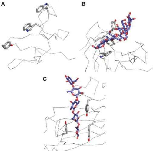
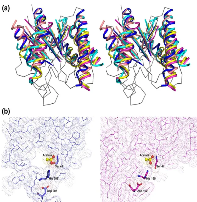
Figure 1.1 Scale model of the Arabidopsis leaf primary cell wall. ... 6 Figure 1.2 Structure of cellulose... 7 Figure 1.3 Xyloglucan architecture and the one letter code to define polysaccharide structure. ... 8 Figure 1.4 Schematic representation of XXXG-type xyloglucans. ... 9 Figure 1.5 A portion (Glucose, Glucose, Manose, Manose) of the glucomannan repeating unit. ... 10 Figure 1.6 Ultrastructure of Clostridium thermocellum cell surface and cellulosomes. 12 Figure 1.7 Schematic representation of Clostridium thermocellum cellulosome and mechanisms of cellulosome cell surface attachment. ... 14 Figure 1.8 Mechanism of action of carbohydrate esterase. ... 18 Figure 1.9 The role of calcium in Xylan recognition by the family 36 CBM from Paenibacillus polymyxa Xyn43A... 22 Figure 1.10 Example of a CBM belonging to the functional Type A. ... 24 Figure 1.11 Example of a CBM belonging to the functional Type B. ... 25 Figure 1.12 Example of a CBM belonging to the functional Type C. ... 26 Figure 1.13 The three types of binding-site „platforms‟ formed by aromatic amino acid residues. ... 28 Figure 1.14 Multivalent CBMs. ... 31 Figure 2.1 Molecular architecture of CtCE3 from Clostridium thermocellum. ... 40 Figure 2.2 Crystals of Selenomethionine-CtCe3-1... 41 Figure 2.3 Structure of the CtCes3-1 monomer coloured as a blue to red gradient from the N terminus to the C terminus with key secondary structural elements labelled. ... 42 Figure 2.4 The 1.4 Å resolution 2Fo–1Fc electron density map contoured at 2ζ covering the final refined coordinates of CtCes3-1 in the vicinity of the calcium ion-binding site. ... 43 Figure 2.5 Comparison of the overall fold and active sites of enzymes displaying structural similarity to CtCes3-1. ... 46 Figure 2.6 The enzymatic breakdown of xylan. ... 49
XXV
Figure 3.1 Purification of recombinant Rhe-CBM35 through affinity chromatography evaluated through SDS-PAGE in a 14% (w/v) polyacrylamide gel. ... 54 Figure 3.2 Crystals of Rhe-CBM35. ... 58 Figure 3.3 Molecular architecture of Rgae12A (Cthe_3141) of Clostridium thermocellum. ... 60 Figure 3.4 Ligands targeted by the CBM35 domains described in this work. ... 62 Figure 3.5 Binding of CBM35s to their target ligands. ... 64 Figure 3.6 structure of family 35 CBMs. ... 67 Figure 3.7 Comparison of the Chi-CBM35 and Xyl-CBM35 binding sites. ... 69 Figure 4.1 Molecular architecture of truncated derivatives of the CmCel5B used in this study. ... 76 Figure 4.2. Structural alignment of CBM6-1 and CBM6-2 and Three-dimensional structure of CmCBM6-2. ... 80 Figure 4.3 Interaction of CmCel5B derivatives with lichenan analysed by affinity gel electrophoresis (AGE). ... 82 Figure 4.4 Isothermal titration calorimetry of wild type (CBM6-1) and mutant derivatives of CBM6-1 with laminarin. ... 83 Figure 4.5 The three-dimensional structure of BhCBM6 (PDB ID: 1W9W). ... 85 Figure 5.1 Crystals of CtCBM55. ... 96 Figure 5.2 Schematic of Cthe_2193 ... 96 Figure 5.3 Representative ITC data of CtCBM55 binding to soluble ligands. ... 98 Figure 5.4 The crystal structure of CtCBM55 and the location of residues that interact with galactose ... 100 Figure 5.5 Structure of CtCBM55 in complex with xyloglucan ... 102 Figure 5.6 Structure of CtCBM55 in complex with galactomannan-derived oligosaccharide (61-α-D-GalMan3). ... 103
Figure 5.7 Affinity gel electrophoresis of wide type and mutants of CtCBM55 against xyloglucan. ... 104 Figure 5.8 Structure of CtCBM55 in complex with arabinopyranose ... 105 Figure 5.9 The interface between the CtCBM55 protomers in the homodimeric protein
... 108 Figure 5.10 Sequence alignment of CtCBM55 with related protein modules... 109 Figure 5.11 The crystal structure of CBM6- Cthe_2193 linked to GH5‟ ... 111
XXVI
ABBREVIATIONS AND SYMBOLS
A Alanine
Å Angstrom
AGE Affinity gel electrophoresis
BhCBM6 CBM6 from Bacillus haludurans
BSA Bovine serum albumine
°C Degree Celsius
Cα Alpha carbon
CAZy Carbohydrate-active enzyme
CBD Cellulose-binding domain
CBM Carbohydrate-binding module
CBM6 Carbohydrate-binding module family 6
CBM22 Carbohydrate-binding module family 22
CBM35 Carbohydrate-binding module family 35
CCP4 Collaborative computational project number 4
CE Carbohydrate esterase
CE3 Carbohydrate esterase family 3
Chi-CBM35 CBM35 of the exo-β-D-glucosaminidase CsxA from
Amycolatopsis orientalis
CMC Carboxymethylcellulose
CsxA exo-β-D-glucosaminidase from Amycolatopsis
orientalis
Csn endo-chitosanase from Amycolatopsis orientalis
Ct Clostridium thermocellum
CtCBM55 Clostridium thermocellum CBM55
CtCes3 Clostridium thermocellum CE3
∆4,5-GalA ∆ 4,5-anhydrogalaturonic acid
Da Dalton
DP Degree of polymerization
DNA Deoxyribonucleic acid
DTT Dithiothreitol
E Glutamic acid / Glutamate
XXVII
EDTA Ethylenediamine tetraacetic acid
ESRF European synchrotron radiation facility
Fe Iron
FPLC Fast Protein Liquid Chromatography
Fuc Fucose
Gal Galactose
Gal2Man5 63, 64-di-α-D-galactosyl mannopentaose
GalMan3 61-α-D-galactosyl mannotriose
GAX Glucuronoarabinoxylan
GH Glycoside hydrolase
GH5 Glycoside hydrolase family 5
GH10 Glycoside hydrolase family 10
GH11 Glycoside hydrolase family 11
GlcA Glucuronic acid
Gln Glutamine
Glu Glutamic acid / Glutamate
HEC Hydroxyethylcellulose
Hepes 4-(2-Hydroxyethyl)-1-piperazine-ethanesulfonic acid
HG Homogalacturonan
HPLC High-performance liquid chromatography
IMAC Immobilized metal ion affinity chromatography
IPTG Isopropyl 1-thio-β-d-galactopyranoside
ITC Isothermal titration calorimetry
IU International units
IUB-MB International Union of Biochemistry and Molecular
Biology
Ka Equilibrium affinity constant
kcat Catalytic efficiency
kDa KiloDalton
Km Michaelis constant
kW Kilowatt
LB Luria-Bertani groth medium
LK Linker
XXVIII
MAD Multi-wavelengh anomalous dispersion
mA Miliampere
MES 4-Morpholine-ethanesulfonic acid
NMR Nuclear Magnetic Resonance spectroscopy
˚ Degree
OD Optical density
PCR Polimerase Chain Reaction
PDB Protein data bank
PEG Polyethyleneglycol
Pel-CBM35 CBM35 of a pectate lyase
pH Potential of hidrogen
PL Polysaccharide Lyases
PNPAc 4-ρ-nitrophenyl acetate
R Arginine
Rhe-CBM35 CBM35 of the rhamnogalauronan acetyl esterase from
Clostridium thermocellum
RGI Rhamnogalacturonan I
RGII rhamnogalacturonan II
rpm Rotation per minute
SAD single-wavelength anomalous dispersion
SDS-PAGE Sodium dodecyl sulfate-polyacrylamide gel electrophoresis
SeMet Seleno-methionine
SLH S-layer homology module
Tris 2-Amino-2-hydroxymethyl-1,3-propanediol
U Units
V Volt
X Glucose decorated α-1,6 with xylose
XG Xyloglucan
XGOs Xylogluco-oligosaccharides
X-gal 5-bromo-4-chloro-3-indolyl β-D-galactoside
Xyl Α-1,6 D-xylosyl
Xyl-CBM35 CBM35 of a xylanase from Cellvibrio japonicus
1
INTRODUTION
Plant cells are encased within a complex polysaccharide rich cell wall, which constitutes the raw material that is used to manufacture textiles, paper, lumber, films, thickeners and other products. The plant cell wall is also the primary source of cellulose, one of the most abundant and useful biopolymers on Earth. The biological degradation of plant cell wall polysaccharides involves the cooperative action of a large range of Glycoside Hydrolases (GHs), Carbohydrtae Esterases (CEs) and Polysaccharide Lyases (PLs), the majority of these being secreted by microorganisms like bacteria and fungi. Enzymes that digest plant cell wall polysaccharides generally contain non-catalytic domains, carbohydrate-binding domains (CBMs), that function by anchoring the enzyme to the substrate, thus potentiating catalytic activity (Tunnicliffe et al., 2005).
A generic feature of enzymes that hydrolyze complex carbohydrates is their modular structure. The most common noncatalytic modules are the carbohydrate-binding modules (CBMs), which are grouped into sequence-based families (Boraston et al., 2004). The general function of CBMs is to promote the interaction of the enzyme with the target substrate, thereby increasing the efficiency of catalysis (Boraston et al., 2003; Hall et al., 1995; Tomme et al.¸1988). CBMs are prevalent in plant cell wall degrading enzymes where they direct the cognate catalytic modules to their target substrate within these complex composite structures (Boraston et al., 2004). CBMs are the perfect candidates for a range of biotechnological applications since they present three basic properties that were described by Shoseyov et al. (2006): (i) CBMs are usually independently folding units and, therefore, can function autonomously in chimeric proteins; (ii) the attachment matrices are abundant and inexpensive and also have excellent chemical and physical properties; and (iii) the binding specificities can be controlled, and therefore the right solution can be adapted to an existing problem (Shoseyov et al. 2006).
Large-scale recovery and purification of biologically active molecules continues to be a challenge for many biotechnology companies. Various purification procedures have been developed, of which biospecific affinity purification (affinity chromatography) has become one of the most rapidly developing divisions of immobilized affinity ligand technology. To date, several affinity tags have been developed that vary in size from several amino acids to a complete protein and each
2
individual affinity-based purification system embodies specific advantages. When compared with most immobilization systems, cellulose is an economical support-matrix for large-scale protein purification (Levy and Shoseyov, 2002). CBMs are high-capacity purification tags that may be used for the isolation of biologically active target peptides and many protein entities at relatively low cost, (Shoseyov et al. 2006). Protein engineering using CBMs is also an emerging field and it is well established that expression of foreign proteins fused to CBMs results, for the most part, in high expression levels, and as a result, expression vectors (pET34 to pET38 from Novagen) incorporating CBMs as fusion tags were developed. Several studies have shown the potential of CBMs for modifying the characteristics of several enzymes and therefore, the basic approach in CBM engineering was to replace or add a CBM in order to improve hydrolytic activity (Shoseyov et al. 2006).
Another area of increasing interest for CBMs is the production of bioethanol from cellulosic material. Lignocellulose is the most abundant renewable natural resource on earth. CBMs are pivotal for the conversion of this organic substrate into fuels since they are able to target the catalytic modules of polysaccharidases to their target bonds. Resulting sugars may then be converted to liquid fuel (Shoseyov et al. 2006). The strong affinity that exists between cellulose and CBMs is also used in many applications associated with the textile industry. Numerous laundry powders contain recombinant enzymes that do not possess a native affinity to the cellulosic fabric (amylases, proteases, lipases, and oxidoreductases). The performance of these enzymes, under conventional washing conditions, can be improved by increasing their affinity to the textile substrate by fusion to CBMs (Levy and Shoseyov, 2002). Additional substances can also be targeted to cellulosic fabrics through the use of CBMs, as for instant fragrance-bearing particles in the case of laundry powder. It was also discovered that applying CBMs to cellulose fibers has a potential for use in paper recycling since it has been demonstrated that the application of CBMs on secondary fibers, such as old paperboard containers, results in increased tensile and burst indexes as well as improvement in pulp drainage (Levy and Shoseyov, 2002).
Additional biotechnological uses for CBMs are in the production of oral care products, since CBMs may contribute to disperse the polysaccharides present in the dental plaque; in the baking industry, as CBMs can retard staling and aging of baked bread; in diagnosis test kits (Levy and Shoseyov, 2002) and as part of a system for parenteral vaccination of fish (Maurice et al., 2003). Since CBMs specificity for
3 carbohydrate ligands is high, these modules have been used as molecular probes for the analysis for plant cell wall polyssacharides (McCartney et al., 2004). Furthermore, Shoseyov et al. (2001) also showed that CBMs could modulate plant growth on transgenic plants. The introduction of family 3 CBM gene from Clostridium cellulovorans in poplars (Populus tremula) made the transgenic plants grow faster than the wild-type, showing an increase in fiber cell length and in the average degree of polymerization of cellulose (Levy and Shoseyov, 2002). Environmental technology has also been exploring the function of CBMs. Several authors fused CBMs with decontamination enzymes and the fusion molecules, attached to cellulose supports successfully degrade toxic compounds (Richins et al., 2000; Wang et al., 2002; Xu et al., 2002).
Although the mechanisms by which these enzymes act have been extensively studied over the last decades, much is still unknown. Whether it is for use in animal nutrition or to develop and/or optimize novel or current biotechnological applications, it is crucial that we improve our knowledge on the processes involved in plant cell wall hydrolysis. This work aims to provide insightfull contributions in this area. This thesis is divided in 6 Chapters. The scientific background (Chapter 1) begins with a general review of plant cell wall structure, with particular focus on cellulose, xyloglucan and galactomannan. Subsequently, attention is focused into the cellulosome, an enzymatic multi-enzyme complex of cellulases and hemicellulases that is usually produced by anaerobic bacteria. Clostridium thermocellum‟s cellulosome is described and the complexity and functionality of this macromolecule is highlighted. In the following subchapters the role of GHs, CEs and Carbohydrate-Binding Modules (CBMs) in plant cell wall degradation is reviewed. Finally, the chapter finishes with the clear identification of the objectives of this project. In Chapter 2, we address several unresolved questions concerning the mechanism of plant cell wall hydrolysis by microbial multi-enzyme complexes.The crystal structure and biochemical properties of the N-terminal carbohydrate esterase domain of Clostridium thermocellum CtCes3-1 were determined. In Chapter 3, four carbohydrate-biding modules (CBMs) of family 35 that display specificity for ∆4,5-anhydrogalacturonic acid (∆4,5-GalA) were characterized, although two of the proteins also interact with glucuronic acid (GlcA). We present the biochemical properties of a CBM6 (CBM6-1) from Cellvibrio mixtus CmCel5A in Chapter 4 and a novel CBM within the large C. thermocellum cellulosomal protein Cthe_2193 (CtCBM55), which is the founder member of a new CBM family in
4
Chapter 5. Finally Chapter 6, contains a general discussion with the most importante conclusions from each chapter.
5
CHAPTER 1
Scientific background and objectives
1.1. THE PLANT CELL WALL
Plants present two cell wall types that differ in function and composition: the primary and secondary cell walls. Primary walls surround growing and dividing plant cells, providing mechanical strength but allowing the cell to expand. In contrast, secondary walls are much thicker and stronger and are deposited only when cells have ceased to grow. The secondary walls are strengthened by the incorporation of lignin, a phenolic polymer, which cements and anchors the cellulose microfibrils and other matrix polysaccharides and thus stiffens the walls, preventing biochemical degradation and physical damage (Knudsen, 1997).
The primary wall is a composite polymeric structure in which crystalline cellulose microfibrils are embedded in a complex, highly hydrated, and less ordered polysaccharide matrix (Figure 1.1). The most important components of this matrix are hemicelluloses and pectin. Cellulose is present as long unbranched fibrils composed of approximately 30 to 36 hydrogen-bonded chains of -1,4-glucose (Somerville et al., 2004). Hemicelluloses are branched polysaccharides containing backbones of neutral sugars, like β-D-hexosyl or β-D-pentosyl residues, that can form hydrogen bonds to the surface of cellulose fibrils. Pectins form a gel phase in which the cellulose-hemicellulose network is embedded and uronic acids are major components of these molecules. The simplest of these is homogalacturonan (HG), an unbranched polymer of α-1,4-D-galacturonic acid. Rhamnogalacturonan I (RGI) has a backbone composed of alternating α-1,2-L-rhamnose-α-1,4-D-galacturonic acid decorated primarily with arabinan and galactan side chains. It has recently been suggested that RGI functions as a scaffold to which other pectins, such as rhamnogalacturonan II (RGII) and HG, are covalently attached as side chains (Somerville et al., 2004).
6
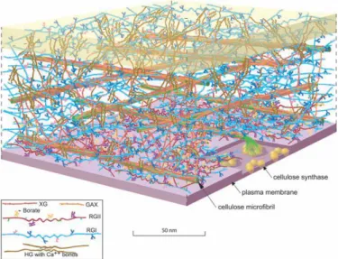
Figure 1.1 Scale model of the Arabidopsis leaf primary cell wall.
The amount of the various polymers is shown based approximately on their ratio to the amount of cellulose. The amount of cellulose, relative to a living cell, was reduced for clarity. Because of the exaggerated distance between microfibrils, the hemicellulose cross-links [shown in dark orange (xyloglucan, XG) or light orange (glucuronoarabinoxylan, GAX)] are abnormally extended. Also, recent solidstate NMR studies have suggested that, in some plants, only about 8% of the surface of the cellulose microfibrils is occluded by XG. The figure is an elaboration of a model originally presented by McCann and Roberts. The figure was rendered by Abbey Ryan.
Several models, which reflect the interactions established between its components, have been proposed to explain the organization of plant cell walls. In the seventies, Keegstra et al. (1973) proposed that polymers from the matrix (xylan, xyloglucan, pectic polysaccharides and structural proteins) were covalently linked and formed a very large macromolecular network. In this model, cellulose fibres are connected through hydrogen-bonding to xyloglucan chains (Cosgrove, 2001). An alternative model was subsequently proposed by Hayashi (1989) and Fry (1989) that suggested that single xyloglucan chains fill the gap between cellulose microfibrils and tether them together; the pectin polysaccharides and structural proteins occupy the space between xyloglucan chains. Although this is presently the most popular model, two other models have, more recently, been proposed: the multicoat model described by Talbott and Ray (1992), in which each cellulose microfibril is coated with successively looser layers of matrix polysaccharides; and the stratified wall model described by Ha et al. (1997), in which cellulose-xyloglucan layers are separated by pectic polysaccharides. All these models have in common the concept that cellulose fibrils are coated with xyloglucan (Cosgrove, 2001). In summary, cellulose microfibrils are linked together by non-covalent interactions with matrix polysaccharides, which determine most of the physical properties of the cell wall. The complexity of the cell-wall network allows for
7 many potential sites where loosening and expansion might be initiated (Cosgrove, 2005).
1.1.1. Cellulose
The primary structure of cellulose is of an unbranched 1,4-linked β-D-glucan the structural repeat in the polymer being the disaccharide cellobiose due to the 180º rotation of the glucose moieties inside the carbohydrate (Figure 1.2). Many parallel glucans interact due to an extensive hydrogen bond network to form a crystalline microfibril that is mechanically strong and highly resistant to enzymatic attack (an almost ideal scaffold material). These long, crystalline ribbons are 3–5 nm wide and, in growing cells, are aligned with each other, giving a structural bias to the cell wall (Cosgrove, 2005).
Figure 1.2 Structure of cellulose.
The cellulose is first sandwiched together in sheets. In these sheets, the cellulose forms hydrogen bonds to
its adjacent neighbours, increasing mechanical strength and enzymatic recalcitrance.Adapted from http://
www.biologie.uni-hamburg.de/b-online/e26/26a.htm.
In nature, cellulose chains have a degree of polymerization of approximately 10,000 glucopyranose units in wood cellulose and 15,000 in native cotton cellulose. There is some evidence for a lower degree of polymerization in primary cell walls as compared with secondary cell walls (O‟Sullivan, 1997). Natural crystalline cellulose is named cellulose I and comprises the two forms Iα and Iβ, in which these chains lie parallel (Jamal et al., 2004), although these cellulose architectures are not the most stable form of the polysaccharide (O‟Sullivan, 1997). Celluloses produced by primitive
8
organisms were said to have the I component dominant, while those produced by the higher plants have the Iβ form dominant (O‟Sullivan, 1997). Many non-natural forms of cellulose and crystalline arrays of cellooligosaccharides form cellulose II in which the chains lie anti-parallel (Jamal et al., 2004). In addition to crystalline regions, many model sources of natural crystalline cellulose appear to contain various proportions of unstructured cellulose, rather loosely termed “amorphous” cellulose. However, amorphous cellulose cannot be considered truly amorphous as, by definition, an amorphous material is one which is formless or lacks a definite shape and areas of amorphous cellulose probably still possess a degree of order (O‟Sullivan, 1997).
1.1.2. Xyloglucan
Xyloglucan is the quantitatively predominant hemicellulosic polysaccharide in the primary walls of dicots. It has a cellulosic semirigid backbone of β-1,4-glucan that is substituted with α-1,6-D-xylosyl units which, in a species-dependent manner, is further derivatized with α-L-arabinose or β-D-galactose. Usually, up to 75% of the residues are substituted at O6 with mono-, di-, or triglycoside side chains. As displayed in Figure 1.3, a single letter nomenclature is used to simplify the naming of xyloglucan side chain structures. For example, a capital G represents an unbranched glucose residue. A capital
F represents a glucose residue with a fucose-containing trisaccharide.
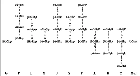
Figure 1.3 Xyloglucan architecture and the one letter code to define polysaccharide structure.
9 Xyloglucans are classified as XXXG-type or XXGG-type based on type of decorations. XXXG have three consecutive backbone residues that are subsituted with xylose and a fourth unbranched backbone sugar (
Figure 1.4). In contrast, XXGG xyloglucans have two consecutive branched backbone residues that are followed by two unbranched backbone residues. It is well established that xyloglucan structures vary between plant species. However, primary walls of several dicotyledons, non-graminaceous monocotyledons and gymnosperms contain fucosylated xyloglucan with a XXFG-type structure.
Figure 1.4 Schematic representation of XXXG-type xyloglucans.
Adapted from http://www.ccrc.uga.edu/~mao/xyloglc/Xtext.htm (Mars 2009).
1.1.3. Mannan, glucomannan and galactomannan
The mannose-containing polysaccharides, mannan and glucomannan, are important components of plant biomass. Mannan consists of a backbone of β-1,4-linked mannose residues, whereas glucomannan comprises a heterogeneous polymer of β-1,4-linked glucose and mannose sugars randomly distributed (Brett et al., 1996). The backbone of both mannan and glucomannan can be decorated with α-1,6-linked galactosyl residues, and thus these polysaccharides are often referred to as galactomannan and galactoglucomannan, respectively (Brett et al., 1996). Glucomannan plays a key structural role in the plant cell walls of Angiosperms, while galactomannan is generally found in the cell walls of seeds such as carob, where it functions as a storage polysaccharide (Hogg et al., 2003). Galactoglucomannan is present mainly in softwoods, whereas inhardwoods glucomannan is the most common form (Vries et al., 2001).
10
Figure 1.5 A portion (Glucose, Glucose, Manose, Manose) of the glucomannan repeating unit. The second glucose has an acetate group.
Galactomannans are present in several vegetable gums that are used to increase the viscosity of food products. The mannose:galactose ratio varies between different gums like Fenugreek gum (1:1), Guar gum (2:1), Tara gum (3:1), Locust bean gum or Carob gum (4:1). Guar is a legume that has been traditionally cultivated as livestock feed. Approximately 85% of guar gum is guaran, a water soluble polysaccharide consisting of linear chains of mannose with β-1,4 linkages to which galactose units are attached with α-1,6 linkages. The ratio of mannose to galactose is 2:1. Guar gum has five to eight times the thickening power of starch and has many uses in the pharmaceutical industry, as a food stabilizer and as a source of dietary fiber.
1.1.4. Pectins
Pectic substances constitute the major matrix polysaccharides in the middle lamella and primary cell walls of dicotyledonous plants, with a backbone composed of alternating homogalacturonan (smooth regions) and rhamnogalacturonan (hairy regions) (Kofod et al., 1994). Rhamnogalacturan I consists of alternating residues of galacturonic acid and rhamnose, and probably has side branches that contain other pectin domains (Cosgrove, 2005). The latter can have acetylations at the C-2 and C-3 positions, and the removal of such acetyl groups facilitates the action of lyases and hydrolases, since the acetylation sterically hinders the cleavage of the glycoside linkages (Navarro-Fernández et al., 2008). Homogalacturonan comprises a linear chain of galacturonic acid residues, whereas xylogalacturonan is modified by the addition of xylose branches. The carboxyl groups of homogalacturonan and xylogalacturonan are often methyl esterified, a modification that „blocks‟ the acidic group and reduces their ability to form gels (Cosgrove, 2005). Rhamnogalacturonan II is a complex pectin domain that contains 11 different sugar residues and forms dimers through borate (B) esters. The neutral arabinans and arabinogalactans are also linked to the acidic pectins and it has been proposed that they promote wall flexibility while they bind to the surface of cellulose.
11
1.2. HYDROLYSIS OF PLANT CELL WALL POLYSACCHARIDES BY HIGH MOLECULAR MASS MULTI ENZYME COMPLEXES
In nature, different mechanisms have evolved for the hydrolysis of plant cell wall structural carbohydrates. Aerobic bacteria and fungi usually secrete a large repertoire of cellulases and hemicellulases to the extracellular space, since structural carbohydrates cannot enter the microbial cells, which individually bind to each specific target polysaccharide through the action of non-catalytic CBMs. In contrast, several anaerobic bacteria and fungi organize cellulases and hemicellulases in high molecular mass multi enzyme complexes that present one of the finest examples of a naturally evolved nono-machine. These multi enzyme complexes were termed as Cellulosomes due to their very high efficiency for cellulose hydrolysis. Anaerobic bacteria producing cellulosomes were identified in the genera Clostridium, Acetivibrio, Bacteroides and Ruminococcus that colonize various environmental niches, including thesoil, wood chip piles, sewage, and the rumen. Cellulosomes may be the largest extracellular multienzyme complexes found in nature,since polycellulosomes have been reported to be as large as100 MDa, although the individual cellulosomes range in massfrom about 650,000 Da to 2.5 MDa (Doi et al., 2003). Generally, enzymatic complexes from anaerobic microorganisms are much more elaborated and complex when compared with their aerobic counterparts where enzymes act individually during cell wall hydrolysis. It is possible that the anaerobic environment presents a greater selective pressure for the evolution of a highly efficient machinery (Bayer et al., 2004). Aerobic cellulolytic fungi, for example, are able to produce and secrete copious amounts of free cellulases and hemicellulases which act individually during plant cell wall hydrolysis. When the first attempt to study bacterial anaerobic cellulases and hemicellulases were made, scientists found it rather odd that those enzymes could not be found in the free state (Bayer et al., 2004). Only later it was recognized that this phenomenon resulted from the association of the hydrolytic enzymes in cellulosomes that are usually attached to the bacterial surface. Studies indicating that C. thermocellum enzymes were probably associated in a multi-enzyme complex date back from the eighties of the last century (Garcia-Martinez et al., 1980; Lamed and Zeikus, 1980). In these initial studies, Bayer et al. (1985; 1986) presented electron microscopy images displaying the C. thermocellum cellulosome, suggesting that this multi-enzyme complex appears to be centralized on protuberant structures primarily located on the bacterial surface (Figure 1.6).
12
Figure 1.6 Ultrastructure of Clostridium thermocellum cell surface and cellulosomes.
(A) Diagrammatic representation of a typical C. thermocelum cell bound to cellulose. The cell is intermittently covered with polycellulosomal protuberance-like organelles, some of which are in the resting state while others have protracted upon binding to the substrate. (B) Transmission electron micrograph of a cell in the free state, prior to contact with cellulose. The cell was stained with cellulosome-specific antibody. (C) A high-resolution magnification of a quiescent cellulosome-specific antibody-labeled polycellulosomal protuberance. Note the label on the outer surface of the protuberances. (D) Schematic interpretation of the cell surface shown in C. (E) A rotary-shadowed, transmission electron micrograph of a cell envelope fragment of C. thermocellum. (F) Transmission electron micrograph of a cellulose-bound cell, stained with cellulosome-specific antibody. Upon binding to the substrate, the polycellulosomal organelle has unfurled. (G) A high-resolution magnification of a protracted, antibody-labeled polycellulosomal protuberance. The cellulosome-specific label is mainly associated with the cellulose surface and connected to the cell via extended fibrous material. Compare with micrograph in C. (H) Schematic interpretation of the cellulose-bound cell surface shown in G. (I) Transmission electron micrograph of negatively stained, purified cellulosomes in the absence of cellulose. Note multicomponent nature of the cellulosomes. (J) Similar micrograph of cellulosome bound to fibers of bacterial microcrystalline cellulose. Bars in B, C, E–G, 100 nm; in I and J, 50 nm. Adapted from Bayer et al. (1998).
13 The initial definition of the cellulosome concept was based on studies of the cellulase system produced by the anaerobic thermophilic cellulolytic bacterium Clostridium thermocellum, where adherence of the microorganism to the cellulose was proven (Bayer et al., 1983). It is now recognized that cellulosomes may actively degrade other plant cell wall components and not only cellulose but also a variety of hemicellulases, by incorporating carbohydrate esterases and pectate lyases in this multi-enzyme complex (Bayer et al., 2004).
Typical bacterial cellulosomes are composed of a scaffolding protein, termed CipA in C. thermocellum, that contains a powerful cellulose-binding module (CBM), usually of family 3 (see below), and a number of cohesin modules which tightly bind to complementary dockerin modules borne by the catalytic cellulosomal subunits (Bayer et al., 2004). Moreover, the scaffoldin contains a C-terminal modular dyad that includes a dockerin that displays a divergent type of specificity to that of the enzyme-borne dockerins. Instead, CipA dockerin, binds selectively to cohesins located on a set of anchoring scaffoldins, each of which contains a C-terminal S-layer homology module that mediates attachment to the bacterial cell surface (Bayer et al., 2008). Due to the different specificities of the cohesin–dockerin pairings, the cohesins of the primary scaffoldin were termed type-I cohesins, whereas those of the anchoring scaffoldins were designated type-II, while dockerin type is set according to that of the cohesin with which it interacts (Bayer et al., 2008). The family-3 CBM (CBM3) located in scaffoldins binds strongly to crystalline cellulose, which accounts for the primary targeting of the cellulosome to its substrate (Tomme et al., 2003). The three-dimensional crystal structure of C. thermocellum CipA CBM3 has been determined, and a general molecular mechanism for binding to the cellulosic surface has been proposed, which is described below (Bayer et al., 2008). A schematic representation of Clostridium thermocellum cellulosome is shown in Figure 1.7. Other bacteria, like Acetivibrio cellulolyticus, Bacteroides cellulosolvens and Ruminococcus flavefaciens also seem to have dockerin-containing scaffoldins and SLH-bearing anchoring proteins (Ding et al. 1999; Ding et al., 2001; Xu et al., 2004).
14
Figure 1.7 Schematic representation of Clostridium thermocellum cellulosome and mechanisms of cellulosome cell surface attachment.
Schematic representation of the supramolecular Lego-like architecture of the Clostridium thermocellum cellulosome paradigm and its disposition on the bacterial cell surface. Dockerin-containing enzymes bind selectively to any of the nine type-I cohesins (enumerated) of the primary CipA scaffoldin. In turn, the terminal X–dockerin dyad of the CipA scaffoldin binds to the type-II cohesins of anchoring scaffoldins SdbA, Orf2p, or OlpB, each of which is connected to the cell surface via an S-layer homology module (SLH). The cellulose-binding module of the primary scaffoldin binds the cellulosome complex and the attached cell to the cellulosic substrate. Adaped from Bayer et al. (2008).
Thus, the scaffoldin subunit is responsible for three critical functions of the C. thermocellum cellulosome: (i) it integrates nine different dockerin-bearing enzyme subunits into the cellulosome complex by virtue of its nine cohesin modules, (ii) it participates in the attachment of the cellulosome to the bacterial cell surface by virtue of its C-terminal dockerin module that binds selectively to cohesins of the anchoring scaffoldins, and (iii) it serves to target the cellulosomal enzymes as well as the entire cell to the cellulosic substrate by virtue of its CBM3 (Bayer et al., 2008).
Cellulosomal enzymes contain minimally a catalytic domain and a 70-amino-acid-long duplicated sequence that binds scaffoldin cohesin domains and which was termed dockerin. The presence of a dockerin in an enzyme usually indicates that it is a
15 cellulosomal enzyme, since the dockerin interacts with the cohesins of the scaffolding protein to form the enzyme complex. Interestingly, most of the known C. thermocellum cellulosomal cellulases and hemicellulases contain additional CBMs, which make the cellulosomal enzymes similar to their free enzyme analogs. Rather than primary binding of the crystalline cellulosic substrate, the latter modules are considered to perform functions of a subtler nature. In some cases, such as the hemicellulases, an enzyme-borne CBM could presumably direct the cellulosome-bound enzyme to the favored site in the hemicellulosic polysaccharide substrate. Finally, in some instances, cellulosomal enzymes from C. thermocellum contain more than one catalytic domain, perhaps indicating a close cooperation between two particular types of catalytic domain, which may promote synergistic action at a given site on the cellulosic or hemicellulosic substrate (Bayer et al., 2004).
There has been a recent interest in constructing designer mini-cellulosomes for several purposes, including studying the functions of cohesins, analyzing synergy between various cellulosomal enzymes and improving the efficiency of cellulosomes (Doi et al., 2003). There is also an increasing interest in developing in vivo systems in various bacteria in order to use relatively inexpensive biomass or agricultural wastes as a substrate to obtain valuable products (Doi et al., 2003). One example of this type of research is to insert cellulosomal genes into organisms that already are able to produce valuable products, such as ethanol, butanol, or amino acids. Cellulosomes are important in that they can degrade crystalline forms of cellulose, which are recalcitrant to many enzymes that can degrade soluble or amorphous forms of cellulose. Furthermore, cellulosomes can degrade hemicelluloses and pectin, two other major components of plant cell wall materials.
1.2.1. Structure and funtion of Carbohydrate-Active Enzymes
In general, enzymes that degrade plant cell wall polysaccharides display a modular architecture, containing one more catalytic domains bound, through flexible linker sequences, to one or more catalytic modules. Most of the characterized non-catalytic binding modules directing the appended non-catalytic regions to their target substrates and are known as Carbohydrate-Binding Modules (CBMs). Below, a detailed description of catalytic and non-catalytic modules found in cellulases and hemicellulases is presented.
16
1.2.1.1. Glycoside hydrolases
The extensive variety of carbohydrates found in plant cell walls requires for their degradation a large multiplicity of enzymes that are thus involved in their metabolism. Glycoside hydrolases (EC 3.2.1.x) are key enzymes in carbohydrate hydrolysis and are found in the three major kingdoms (archaebacteria, eubacteria and eukaryotes) (Henrissat et al., 1991). Glycoside hydrolases cleave the glycosidic bond between two carbohydrates or between a carbohydrate and a non-carbohydrate moiety. Many of these are enzymes produced by saprophytic microorganisms for the degradation of structural polysaccharides (Henrissat et al., 1998).
1.2.1.1.1. Nomenclature of glycoside hydrolases
The International Union of Biochemistry and Molecular Biology enzyme nomenclature (IUB-MB; 1984) is based on the type of reaction that enzymes catalyse and on their substrate-specificity. For GHs (EC 3.2.1.x), the first three digits indicate enzymes hydrolysing O-Glycoside linkages, whereas the last number indicates the substrate and sometimes reflects the molecular mechanism (Henrissat et al., 1991). This classification avoids ambiguities and the proliferation of trivial names, been for that reason very useful. However, in the case of glycoside hydrolases, such classification does not necessarily reflect the structural features of the enzymes and it is also not appropriate for enzymes showing broad substrate specificity (Henrissat et al., 1991). In 1991, Henrissat and colleagues proposed a new classification system for glycoside hydrolases, where enzymes were organized in families based on primary sequence similarities. Since there is a direct relationship between the amino acid sequence and the folding of an enzyme, such classification is expected to: (i) reflect the structural features of these enzymes better than substrate specificity alone; (ii) help to reveal the evolutionary relationships between these enzymes; and (iii) provide a convenient tool to derive mechanistic information from the protein sequence data.
The Henrissat classification does not substitute but rather complements the IUB-MB nomenclature. An advantage of this classification is that a protein, a translated DNA sequence, or even a domain can be classified before even knowing its biochemical properties. In addition, it overcomes the problems associated with the classification of enzymes consisting of several catalytic domains. Significant sequence similarity is a strong indication of folding similarity and, therefore, it was found that members of one family most likely share the same folding characteristics, enabling homology modelling
17 if the three-dimensional structure of one member is known (Henrissat et al., 1991). According to the Henrissat classification, the GHs catalytic modules are currently classified into 114 different families based on amino acid sequence similarities (May 2009). Because the fold of proteins is better conserved than their sequences, some of the families can be grouped in 14 'clans'.
Table 1.1 Glycoside hydrolases fold superfamilies.
Adapted from http://www.cazy.org
Clans of Related Families Protein fold Glycoside Hydrolase Families
GH-A (β/α)8 1 2 5 10 17 26 30 35 39 42 50 51 53 59 72 79 86 113
GH-B β-jelly roll 7 16
GH-C β-jelly roll 11 12
GH-D (β/α)8 27 31 36
GH-E 6-fold β-propeller 33 34 83
GH-F 5-fold β-propeller 43 62 GH-G ( α/α )6 37 63 GH-H (β/α)8 13 70 77 GH-I α+β 24 46 80 GH-J 5-fold β-propeller 32 68 GH-K (β/α)8 18 20 85 GH-L ( α/α )6 15 65 GH-M ( α/α )6 8 48 GH-N β-helix 28 49
Henrissat and colleagues (1998) also proposed, that enzymes could be named according to their target substrate, following the three-letter standard used in bacterial genetics for genes, although this designation would comprehend the family to which the enzyme belongs. Thus, a family 5 GH will be named Cel5 or Man5, depending on its substrate, which could be cellulose or mannan, respectively. If an organism produces multiple enzymes from the same family displaying similar substrate specificities, then they would be designated Cel5A and Cel5B, and so on, where the letters after the family number correspond to the order in which the enzymes were first reported (Henrissat et al., 1998). If an enzyme contains more than two catalytic domains, the designation would include all of them (Henrissat et al., 1998). For example, endoglucanase CelA from Caldocellulosiruptor saccharolyticum is composed of two GHs, one from family 9 and the other from family 48. So, according to this new nomenclature, the enzyme is named CsCel9A-Cel48A, written in the conventional sense from the amino to the carboxyl terminus. The microorganism abbreviation is also included, before the enzyme name, to differentiate similar enzymes of different origins.
18
1.2.1.2. Carbohydrate Esterases
As with the classification of glycoside hydrolases, the Carbohydrate-Active Enzyme Server (www.cazy.org; July 2009) provides a classification of carbohydrate esterases based upon amino acid sequence similarities. Currently, 16 such families have been described (May 2009), ten of which contain enzymes involved in the degradation of plant-cell wall polysaccharides such as xylan, pectin and rhamnogalacturonan. Biologically, these enzymes are involved in the removal of O-(ester) and N-(acetyl) moieties from carbohydrates and indeed, sugar deacetylases display similar catalytic strategies to those employed by more classical esterases and peptidases. In fact, the similarity of different sugar and non-sugar esterases, and the promiscuity of their substrate specificity make the carbohydrate esterase classification in many cases less insightful and predictive than the hydrolase, transferase and lyase families (Davies et al, 2005).
Figure 1.8 mechanism of action of carbohydrate esterase.
Carbohydrate esterases perform the O or N deacetylation of acetylated sugars.
The majority of carbohydrate esterases/deacetylases whose structures have been reported display a classical β/ /β „serine protease‟ fold, as revealed by three-dimensional structures of enzymes from families CE-1 (bacterial ferulate esterases, Prates et al., 2001), CE-5 (acetyl xylan esterases, Molgaard et al., 2002), CE-7 (multifunctional and xylooligosaccharide deacetylases, Vicent et al, 2003), the plethora of enzymes from family CE-10 (www.cazy.org) and the non-classified fungal ferulate esterases (Hermoso et al., 2004). There is also a small deviation from this canonical fold in the CE-12 rhamnogalacturonan acetylesterase (Molgard et al., 2004). In contrast, the pectin methylesterases from family CE-8 use a different mechanism, in which a twin-aspartate catalytic centre is grafted onto a right-handed parallel β-helix and these enzymes may also be considered slightly unusual in that it is the sugar that forms the
19 acid, rather than the „R‟ group (see Figure 1.8) (Jenkins et al.,2001). Some sugar deacetylase structures have also revealed both single and double metal ion catalytic centres. For example, the LpxC zinc-dependent UDP-3-O-acetyl-Nacetylglucosamine deacetylases from family CE-11, which presents a classical zinc hydrolase site on a novel α/β framework (Coggins et al., 2003; Whittington et al., 2003); the single-zinc CE-14 N-acetyl-1-D-myo-inosityl-2-amino-2-deoxy-a-D-glucopyranoside deacetylase (Maynes et al., 2003); the family CE-4 deacetylases (Blair et al., 2005), sometimes refered to as „NodB homologs‟, whose members are involved in the deacetylation, amongst other things, of peptidoglycan, chitin, rhizobial Nod factors and xylan; and the twin-metal, urease-like CE-9 N-acetylglucosamine-6-phosphate deacetylase (Vincent et al., 2004).
1.2.1.3. Carbohydrate-Binding Modules
Enzymes that digest plant cell wall polysaccharides generally contain non-catalytic domains, CBMs, that function by anchoring the enzyme to the substrate, thus potentiating catalytic activity (Tunnicliffe et al., 2005). CBMs were initially classified as cellulose binding domains (CBDs), based on the initial discovery of several modules that bind cellulose (Shoseyov et al., 2006). However, more and more modules in carbohydrate-active enzymes that bind carbohydrates other than cellulose are being found and these domains have assumed the more general terminology of Carbohydrate-Binding Modules (CBMs).
CBMs have three general roles with respect to the function of their cognate catalytic modules: (i) a proximity effect, (ii) a targeting function and (iii) a disruptive function (Boraston et al., 2004). Through their sugar-binding activity, CBMs concentrate enzymes on to the polysaccharide substrates. It is thought that maintaining the enzyme in proximity of the insoluble substrate (i.e. increasing the concentration of the enzyme on the surface of the substrate) leads to a more rapid degradation of the polysaccharide (Bolam et al., 1998), therefore resulting in improved enzyme efficiency. There are numerous examples in the literature where proteolytic excision or genetic truncation of CBMs from the catalytic modules results in significant decreases in the activity of the enzymes on insoluble, but not soluble polysaccharides (Tomme et al., 1988; Gilkes et al., 1988; Bolam et al., 1998; Charnock et al., 2000; Ali et al., 2001; Zverlov et al., 2001; Boraston et al., 2003; Hall et al., 1995). It should be pointed out