Raquel Lopes Costa
Graduated in Biochemistry
Ligand discovery and structural-functional
analysis of proteins involved in plant cell
wall degradation
Dissertation to obtain Master degree in Biochemistry
Supervisor: Maria Angelina de Sá Palma, UCIBIO, FCT/UNL
Co-supervisor: Ana Luísa Carvalho, UCIBIO, FCT/UNL
Jury:
Examiner: Prof. Doutora Filipa Margarida Barradas de Morais Marcelo
Vowel : Prof. Doutora Doutora Maria Angelina de Sá Palma
Raquel Lopes Costa
Graduated in Biochemistry
Ligand discovery and structural-functional
analysis of proteins involved in plant cell
wall degradation
Dissertation to obtain Master degree in Biochemistry
Supervisor: Maria Angelina de Sá Palma, UCIBIO, FCT/UNL
Co-supervisor: Ana Luísa Carvalho, UCIBIO, FCT/UNL
Jury:
Examiner: Prof. Doutora Filipa Margarida Barradas de Morais Marcelo
Vowel: Prof. Doutora Maria Angelina de Sá Palma
I
“Ligand discovery and structural-functional analysis of proteins involved in plant cell wall degradation”
Copyright © em nome de Raquel Lopes Costa, da Faculdade de Ciências e Tecnologia, Universidade Nova de Lisboa.
III À minha orientadora, Doutora Angelina Palma, e co-orientadora, Doutora Ana Luísa Carvalho, por me terem dado esta oportunidade e por acreditarem em mim e no meu trabalho. Obrigada por todo o apoio e ensinamentos que me transmitiram, foram incansáveis. Tive muita sorte em ter-vos como orientadora e co-orientadora, aprendi muito convosco!
À Professora Doutora Maria João Romão não apenas como líder do grupo de investigação XTAL da FCT-UNL (onde amavelmente me recebeu), mas também na qualidade de Presidente do Departamento de Química da FCT-UNL e Directora da Unidade de Investigação UCIBIO@REQUIMTE (Unidade de Ciências Biomoleculares Aplicadas), instituição de acolhimento do trabalho aqui reportado.
À Doutora Márcia Correia e Doutora Benedita Pinheiro pelo acompanhamento e orientação nas técnicas de preparação de células competentes e toda a parte de biologia molecular.
À Diana Ribeiro que esteve comigo em toda a parte laboratorial, que me ajudou e me apoiou bastante durante todo o ano. Sempre disponível quando surgia alguma dúvida. Obrigado pelo teu acompanhamento e apoio durante toda a tese, sei que nem sempre foi fácil principalmente no inicio, não me esquecerei da tua ajuda . Obrigado querida.
À Viviana Correia por suportar as minhas queixas no laboratório e também por tudo o que me transmitiu em termos de conhecimento, gosto muito de ti.
Ao Doutor Filipe Freire e ao Francisco Leisico por todas as dúvidas laboratoriais que esclareceram principalmente em técnicas de expressão e purificação e todo o ensinamento transmitido sobre o thermofluor.
Ao Professor Carlos Fontes e Doutora Joana Brás, da FMV-UL e NZYTech, pela colaboração e discussão científica e pela produção, à escala highthroughput, das proteínas de C. thermocellum e R. flavefaciens FD-1 que foram analisadas nos microarrays de hidratos de carbono.
À Professora Ten Feizi, ao Doutor Wengang Chai e à Doutora Lisete Silva do Glycosciences Laboratory, Imperial College London pela colaboração na construção dos microarrays de hidratos de carbono.
Ao Professor Manuel Coimbra, Doutora Cláudia Nunes e Doutora Cláudia Passos da Universidade de Aveiro. À Professora Berit Smestad Paulsen da Universidade de Oslo. Pela colaboração na obtenção de polissacáridos para a construção dos microarrays.
Aos colegas do grupo XTAL Doutora Catarina Coelho, Doutora Teresa Santos-Silva, Doutor Marino Santos, Rita Otrelo, Cecília Bonifácio por todo o apoio e por tão bem me terem recebido no laboratório. A todos os colegas do XTAL pelo excelente ambiente de trabalho que é proporcionado neste laboratório e pela simpatia e abertura com que recebem quem chega de fora.
IV ao coordenador do mestrado em Bioquímica Professor Ricardo Franco.
Ao BAG Portugal pela oportunidade de realizar trabalho experimental no ESRF (European Synchrotron Radiation Facility, em Grenoble) e assim poder aprender todo o processo de recolher dados de difração de cristais. Um especial obrigado à minha co-orientadora Doutora Ana Luísa Carvalho que me acompanhou nesta viagem.
À Faculdade de Ciências e Tecnologia da Universidade Nova de Lisboa, instituição que me acolheu durante a Licenciatura e Mestrado em Bioquímica onde me foi proporcionada toda a minha formação académica.
O trabalho desenvolvido ao longo deste período e reportado nesta dissertação foi financiado pela
Fundação para a Ciência e Tecnologia (FCT-MCTES) através dos projectos RECI/BBB-BEP/0124/2012 e PTDC-BBB-BEP-0869-2014 e um contrato no âmbito do programa Investigador FCT 2012. A unidade de investigação UCIBIO (Unidade de Ciências Biomoleculares Aplicadas) é financiada por fundos nacionais da FCT-MCTES (UID/Multi/04378/2013) e co-financiada pelo FEDER no âmbito do PT2020 (POCI-01-0145-FEDER-007728).
Obrigado ao meu namorado Mário por todos os dias estar comigo nos bons e maus momentos, por me apoiar e me incentivar mesmo quando queria desistir. AMO-TE. Obrigada também aos meus sogros, que me ajudam SEMPRE!.
Obrigado aos meus pais e maninhas, amo-vos.
Obrigado aos meus avós, que sempre acreditaram em mim, em especial ao meu avô que sempre suportou os custos associados à minha educação. Muito obrigado Avô Carlos.
V
The plant cell wall is constituted by recalcitrant polysaccharides with diverse sequences that comprise an abundant source of terrestrial biomass. To efficiently degrade plant cell wall polysaccharides, some cellulolytic bacterial organisms, such as Clostridium thermocellum and
Ruminococcus flavefaciens, have an extracellular multi-enzyme complex with catalytic and non-catalytic carbohydrate-binding modules (CBMs). CBMs play a crucial role in enhancing the non-catalytic efficiency of the enzymes by proximity effect, cell attachment or targeting and disruptive function. The Carbohydrate Active enZymes database (CAZy) organizes the identified CBMs by sequence similarity into different families. Deposition of CBM sequences in the CAZy database is continually growing for which characterization and structure-function analysis is required.
In this study we aim to characterize the carbohydrate ligand specificities of C. thermocellum ATCC 27405 and R. flavefaciens FD-1 CBMs assigned to different families in the CAZy database. We performed carbohydrate microarray screening analysis for ligand discovery and crystallization screenings aiming to solve the 3D structures of the CBM-ligand complexes by X-ray crystallography. To complement the information provided by these methodologies we also performed ITC (Isothermal Titration Calorimetry), MST (Microscale Thermophoresis) and affinity gel electrophoresis. With the implementation of this approach it was possible to elucidate different carbohydrate binding specificities for biotechnologically relevant CBMs. The results from the initial carbohydrate microarray screening constitute a functional start point to target CBMs for structural-functional analysis of carbohydrate-recognition. C. thermocellum family 50 (CtCBM50) reveals to be a novel chitin binding LysM domain and binding with insoluble chitin and a β-(1-4)-GlcNAc chitin oligosaccharide was identified. R. flavefaciens FD-1 family 62 CBM (RfCBM62) reveals to be highly specific for a pectic polysaccharide for which the structure is being investigated and binding to galacturonan DP4 was observed. In the scope of this thesis, and as the structural characterization was not achieved in due time, the sequence similarity to known structures inspired the attempt to computationally produce similarity models for the two CBMs. The (hypothetical) conservation of the secondary structures revealed some structural features of the proteins under study.
An important outcome from this integrative study is the possibility to understand the versatility of plant and fungal saccharide sequences and their recognition by the different CBM families. The different binding patterns observed could reflect adaptive pressures of the microorganisms to their respective ecological niches, translating in divergent evolution of the proteome.
VII A parede celular vegetal é constituída por polissacáridos com diversas sequências e que constituem uma fonte abundante de biomassa terrestre. Para degradar de forma eficiente os polissacáridos da parede celular vegetal, alguns organismos celulolíticos bacterianos, tais como o Clostridium thermocellum e o
Ruminococcus flavefaciens, desenvolveram um sistema extracellular multienzimático complexo com módulos catalíticos e módulos não catalíticos de ligação a hidratos de carbono (CBMs). Estes CBMs têm um papel crucial em aumentar a eficiência catalítica de algumas enzimas pela sua avidez para o substrato. A base de dados Carbohydrate Active enZymes (CAZy) organiza os CBMs pela similaridade de sequências primárias em diferentes famílias. A deposição de CBMs na CAZy está continuamente a crescer, sendo que a caracterização e análise da estrutura-função destes módulos é necessária.
Este estudo teve como objetivo caracterizar a especificidade para hidratos de carbono de CBMs atribuídos a diferentes famílias na CAZy, pertencentes às bactérias C. thermocellum ATCC 2745 and R. flavefaciens FD-1. Foram realizados ensaios com a tecnologia de microarrays de hidratos de carbono de forma a identificar ligandos para os diferentes CBMs e ensaios de cristalização com o objectivo de resolver a estrutura tridimensional de complexos CBM-ligando por cristalografia de raios-X. Foram aplicados também outros métodos biofísicos para o estudo da interacção CBM-ligando tais como ITC (Isothermal Titration Calorimetry) e MST (Microscale thermophoresis) e ensaios de electroforese de afinidade. Com a implementação desta abordagem foi possível elucidar a especificidade de CBMs para hidratos de carbono que são biologicamente relevantes. A análise nos microarrays constitui uma base funcional para seleccionar CBMs para estudos de estrutura-função de reconhecimento a hidratos de carbono. O CtCBM50 de C. thermocellum revelou ser um um novo dominio LysM que liga a chitina insolúvel e a interacção com o oligossacárido de quitina (β-(1-4)-GlcNAc) foi identificada. O
RfCBM62-de R. flavefaciens FD-1revelou ser altamente específico para um polissacárido de pectina, cuja estrutura está a ser elucidada, e a interacção com um oligossacárido derivado de pectina, galacturonan DP4, foi observada. No âmbito desta tese, e sendo que não foi conseguida a caracterização estrutural, a semelhança com estruturas conhecidas inspirou a produção de modelos de similaridade para os dois CBMs computacionalmente. A conservação (hipotética) da estrutura secundária sugeriu características estruturais e interacção com o ligando para os CBMs em estudo.
Este estudo integrativo permitiu aferir sobre a verstatilidade do reconhecimento de hidratos de carbono de plantas e fungos por diferentes famílias de CBMs, que poderão resultar da adaptação selectiva das bactérias aos seus nichos ecológicos respectivos, resultando na evolução divergente dos seus proteomas.
IX
Acknowledgments ... III
Abstract ... V
Resumo ... VII
Contents ... IX
List of Figures ... XIII
List of Tables ... XVII
Abbreviations and Symbols ... XIX
Chapter 1
–
Introduction and objectives ... 1
1.1 Overview of the plant cell wall ... 3
1.1.1 Plant cell wall morphology ... 3
1.1.2 Plant cell wall composition ... 4
1.1.2.1 Cellulose ... 4
1.1.2.2 Non-cellulosic major components ... 5
1.1.3 Plant cell wall degradation ... 6
1.2 The bacterial Cellulosome: identification and organization ... 6
1.2.1 The highly efficient cellulose-degrading bacterium Clostridium thermocellum ... 7
1.2.2 Ruminococcus flavefaciens ... 8
1.3 The modular organization and recognition mechanisms of Carbohydrate-Active enZymes ... 10
1.3.2 Carbohydrate-binding modules ... 10
1.3.2.1 Classification of CBMs ... 10
1.3.2.2 Functional roles of CBMs ... 13
1.3.2.3 Biotechnological applications of CBMs ... 14
1.4 Overview of the methodologies to study CBM-carbohydrate interactions ... 15
1.4.1 Carbohydrate microarray technology ... 15
1.4.1.1 Carbohydrate probe libraries and immobilization methods ... 16
1.4.1.2 Neoglycolypid-based oligosaccharide technology ... 17
1.4.1.3 Biological Applications ... 18
X
1.4.2.3 Isothermal Titration Calorimetry ... 20
1.4.3 Structural methods ... 21
1.4.3.1 X-ray Crystallography ... 21
1.4.3.2 Saturation-Transfer Difference ... 24
1.5 Objectives of this Thesis ... 24
Chapter 2
–
Carbohydrate microarray screening analysis for ligand discovery ... 27
2.1 Introductory remarks ... 29
2.2 Materials and methods... 29
2.2.1 DNA, bacterial strains and plasmids ... 29
2.2.2 CBMs quantification and SDS-PAGE analysis ... 30
2.2.3 Construction of the carbohydrate microarray ... 30
2.2.4 Carbohydrate microarray binding assay ... 31
2.2.5 Carbohydrate microarray data analysis ... 35
2.3 Results and discussion ... 37
2.3.1 CBMs quantification and SDS-PAGE analysis ... 37
2.3.2 Carbohydrate microarray quality control ... 38
2.3.3 Carbohydrate microarray analysis of CAZy CBMs ... 47
2.4 Conclusions ... 50
Chapter 3
–
Expression, purification and stability analysis of CBMs ... 53
3.1 Introductory remarks ... 55
3.2 Materials and methods... 55
3.2.1 Preparation of competent cells ... 55
3.2.2 Transformation ... 56
3.2.3 DNA amplification, isolation and sequencing ... 56
3.2.4 Small and large-scale expression of CBMs ... 57
3.2.4.1 Protocol for expression with IPTG-induction ... 57
3.2.4.2 Protocol for expression with auto-induction... 57
3.2.5 Cell harvesting and lysis ... 58
3.2.6 Purification of CBMs by affinity chromatography ... 58
3.2.6.1 Purification with ÄKTA START ... 58
3.2.6.2 Affinity purification with His GravitrapTM columns ... 59
3.2.7 Analysis by polyacrylamide gel electrophoresis ... 59
XI
3.3.1 DNA sequencing of CBMs ... 60
3.3.2 Small-scale expression tests... 61
3.3.3 CBMs expression optimization tests ... 62
3.3.4 Large-scale expression and purification ... 65
3.3.5 Protein stability analysis ... 67
3.4 Conclusions ... 71
Chapter 4 - CtCBM50: A novel chitin binding LysM domain ... 73
4.1 Introductiory remarks ... 75
4.2 Materials and methods... 75
4.2.1 Analysis of interaction with insoluble polysaccharides ... 75
4.2.2 Isothermal titration calorimetry ... 75
4.2.3 Crystallization assays ... 76
4.2.4 Phyre2: A bioinformatic tool for 3D structure prediction ... 76
4.3 Results and discussion ... 77
4.3.1 CtCBM50 binding to insoluble chitin ... 77
4.3.2Binding affinity of CtCBM50 for the β-(1-4)-linked GlcNAc chitin pentasaccharide ... 77
4.3.3 Crystallization assays ... 79
4.3.4 CtCBM50 3D structure prediction ... 80
4.4 Conclusions ... 83
Chapter 5 - RfCBM62-1: A highly specific CBM for pectins ... 85
5.1 Introductiory remarks ... 87
5.2 Materials and methods... 87
5.2.1 Analysis of interaction with insoluble polysaccharides ... 87
5.2.2 Affinity gel electrophoresis with soluble polysaccharides ... 87
5.2.3 Microscale thermophoresis ... 88
5.2.4 Crystallization assays ... 88
6.2.5 Phyre2: A bioinformatic tool for 3D structure prediction ... 88
5.3 Results and discussion ... 88
5.3.1 RfCBM62-1 binding with pectin ... 88
5.3.2 Binding affinity of RfCBM62-1 with galacturonan DP4 ... 89
5.3.3 RfCBM62-1 binding with pectin-related polysaccharides ... 90
5.3.4 Crystallization assays ... 92
XII
Chapter 6
–
Integrative conclusions and future work ... 97
References ... 102
XIII
Figure 1.1–Illustration of the different layers of the plant cell wall....…………...4
Figure 1.2–Cellobiose structure.………...4
Figure 1.3–Representation of the plant cell wall and cellulose structure...5
Figure 1.4– Cellulosomes at the surface of Clostridium thermocellumin form of protuberances….7 Figure 1.5 – The Clostridium thermocellum cellulosome. Representation of the plant cell wall degradation by the Clostridium thermocellumcellulosome. ………..8
Figure 1.6– The Ruminococcus flavefaciens FD-1 cellulosome. Representation of the plant cell wall degradation complexity………..………9
Figure 1.7– The crystallographic dimer of CBM63 from Bacillus subtilis shown in complex with cellohexaose ………12
Figure 1.8– The X-ray crystal structure of the family 29 CBM from Piromyces equii in complex with mannohexaose ………...12
Figure 1.9– The X-ray crystal structure of the family 6 CBM from Bacillus halodurans in complex with lamirarihexaose ………...13
Figure 1.10– Schematic representation of the NGL-based technology. ………..18
Figure 1.11–Typical MST binding experiment. ……….20
Figure 1.12 - The ITC binding experiment. ………....21
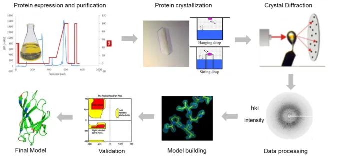
Figure 1.13 - Schematic representation of the crucial steps to obtain a tridimensional structure with X-ray crystallography. From protein expression and purification to the final model………22
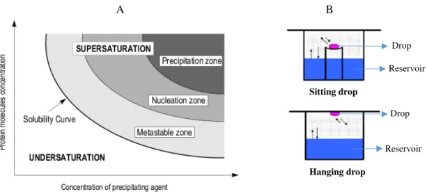
Figure 1.14 - Schematic representation of the vapor diffusion method………...23
Figure 1.15– Scheme of the STD-NMR experiment. ………..24
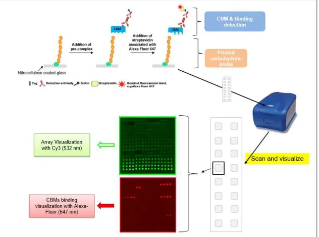
Figure 2.1 - Overview of the carbohydrate microarray binding experiment for CBMs using slides of 16 pads………..35
Figure 2.2 - Schematic representation of an array pad (block) spot visualization and grid construction using the GenePixPro7 Software (Molecular Devices)………36
Figure 2.3 – Schematic representation of the integrated microarray data analysis: database and interactive software. ……….37
Figure 2.4 - Representative SDS-PAGE (13% acrylamide) of the 14 CBMs analyzed………38
Figure 2.5– Carbohydrate microarray data analysis of monoclonal antibodies for quality control of the microarray set……….………41
XIV Low level heat-map of the relative binding intensities represented as a percentage of the
fluorescence signal intensity relative to the probe that binds more strongly to each CBM. ………..43 Figure 2.8– Carbohydrate microarray data analysis of model carbohydrate-binding modules from different families and a plant lectin for quality control of the microarray set………45 Figure 2.9 - Carbohydrate microarray data analysis of CAZy CBMs. ………47 Figure 3.1 - Agarose gel electrophoresis of 14 DNA sequences from CBMs of different families……….60 Figure 3.2- SDS-PAGE (10% acrylamide) analysis showing the expression levels of the 13 CBMs
using two expression protocols……….61
Figure 3.3 - SDS-PAGE (10% acrylamide) results showing expression levels of CBMs from different families………..63 Figure 3.4 - SDS-PAGE (10% acrylamide) and native gel results showing purification of CBMs from different families………..64 Figure 3.5 - Modular organization where the RfCBM62-1 is biologically integrated and respective primary sequence is represented, together with the primary sequence of CtCBM50………65
Figure 3.6 - Results from purification of CtCBM50……….66
Figure 3.7- Results from purification of RfCBM62-1………..67 Figure 3.8– Thermal denaturation assay using Thermofluor for CtCBM50 and for RfCBM62-1…68 Figure 3.9 - Thermal denaturation assay using Thermofluor for RfCBM62-1 in 50mM HEPES, 1M NaCl, 2 mM CaCl2 pH=7 and in H2O with 2 mM CaCl2………...70
Figure 4.1– Qualitative affinity binding analysis of CtCBM50 with insoluble chitin by SDS-PAGE
with 13% acrylamide………77 Figure 4.2–Isothermal titration calorimetry of β-(1-4)-linked GlcNAc pentasaccharide binding to
CtCBM50……….78
Figure 4.3 - CtCBM50 optimization study from 80! screen………...80
Figure 4.4– Ribbon representation of CtCBM50 similarity model and respective alignment with
the template sequence………...81 Figure 4.5 - Representation of the superposition between CtCBM50 similarity model with Mo0v in complex with GlcNAc4 and respective residues involved in ligand binding……….82
Figure 4.6 - Structure-based sequence alignment between CtCBM50 and MoOv………..82
Figure 5.1– Qualitative binding analysis of RfCBM62-1 with pectin from apple by SDS-PAGE with
XV
CtCBM62 Chain A (PDB code 2YFZ) as a template……….…………...93
Figure 5.5 - Representation of the superposition between RfCBM62-1 similarity model and CtCBM62 with 61-α -D-GalMan3 and respectively amino acid residues involved in ligand binding……….94
Figure 5.6 - Structure-based sequence alignment between RfCBM62-1 and CtCBM62.………..95
Supplementary Figure 1 - SDS-PAGE (10% acrylamide) showing IMAC purification of CBMs (expressed with Autoinduction protocol) from different families with His Gravitrap® columns………..120
Supplementary Figure 2 –Thermofluor screen (in house)………...121
Supplementary Figure 3–PEG Ion and PEG Ion2 commercial screens………..122
Supplementary Figure 4– JSCG-plus HT96 commercial screen………..123
Supplementary Figure 5–JBS 1 and JBS 2 commercial screens………..124
Supplementary Figure 6–JBS 3 commercial screen………125
Supplementary Figure 7–JBS 4 commercial screen...126
XVII Table 1.1–Typical ‘fold families’ identified for CBMs and examples of CBM families with these
folds………..11
Table 2.1 -Carbohydrate binding modules (CBMs) investigated for carbohydrate binding using carbohydrate microarrays and their modular organization………29
Table 2.2– List of the carbohydrate-directed antibodies, CBMs and the lectin and their reported specificities used for quality control of the microarray set………32
Table 2.3–Calculated concentration for each CBM………37
Table 2.4 - List of all saccharide probes analyzed in the binding charts and in the matrix (heat-map) ……….………….38
Table 3.1–Primers sequences used to confirm the DNA sequence of the 14 CBMs………57
Table 3.2 – Main differences in the IPTG-induction and auto-induction protocols for protein expression……….57
Table 3.3–Best conditions for large scale expression of the four CBMs………..65
Table 3.4– Buffers used in the IMAC purification of CtCBM50 and RfCBM62-1………..66
Table 3.5 - Summary of the purification yields for CtCBM50 and RfCBM62-1………67
Table 3.6–Results from Thermofluor data analysis………68
Table 3.7– Thermofluor data analysis for RfCBM62-1………...69
Table 4.1 - Data for the ITC analysis of CtCBM50 binding to β-(1-4)-linked GlcNAc pentasaccharide at 25 °C………..78
Table 4.2– Experimental conditions used in crystallization assays for CtCBM50……….79
Table 5.1– Experimental conditions used in crystallization assays for RfCBM62-1………...92
Supplementary Table 1 - Carbohydrate binding modules (CBMs) investigated for carbohydrate binding using carbohydrate microarrays and their modular organization………...111
Supplementary Table 2 - List of all saccharide probes included in the Fungal and Plant polysaccharide set 1………113
Supplementary Table 3–EWI and EWII screen used for preliminary crystallization assays…...128
Supplementary Table 4– PEG Ion 1 and 8k screen used for preliminary crystallization assays...129
XIX % - Percentage
% (w/v) –weight/volume percentage Å – Angstrom
Ala – Alanine Arg - Arginine Asp –Aspartic acid Asn - Aspargine
BSA – Bovine serum albumin CaCl2 – Calcium cloride
CAZymes – Carbohydrate active enzymes
C. termocellum–Clostridium thermocellum
CBM – Carbohydrate-binding module CBMs - Carbohydrate-binding modules D.O.600nm – Optical density at 600 nm DP – Degree of polymerization
E. coli - Escherichia coli
GBPs – Glycan binding proteins Glu – Glutamate
Gly - Glycine
HEPES - 4-(2-hydroxyethyl)-1-piperazineethanesulfonic acid His - Histidine
Ile - Isoleucine
IMAC – Immobilized metal affinity chromatography IPTG - Isopropyl β-D-1-thiogalactopyranoside ITC – Isothermal Titration Calorimetry KDa – Kilodalton
LB –Luria-Bertani Leu – Leucine Lys – Lysine
NGL - Neoglycolipid
NMR - Nuclear Magnetic Ressonance
MAD –Multiple Wavelength Anomalous Dispersion MIR - Multiple Isomorphous Replacement
XX PDB - Protein Data Bank
PEG - Polyethylene glycol Phe - Phenylalanine
R. flavefaciens –Ruminococcus flavefaciens
RMSD - Root-mean-square deviation Rpm – Rotations per minute
SAD - Single Wavelength Anomalous Dispersion Ser – Serine
SPR – Surface Plasmon Ressonance
STD-NMR – Saturation-Tranferer Difference – Nuclear Magnetic Ressonance Thr – Treonine
XXI The symbol notation for monosaccharides is in accord with the proposed in the latest edition of
Essentials of Glycobiology (www.ncbi.nlm.nih.gov/books/NBK310273/).
1
3
1.1 Overview of the plant cell wall
The plant cells are surrounded by a relatively thin but strong and rigid wall. This wall has a functional role in plant growth, development and reproduction by contributing to structural integrity, cell adhesion, and mediation of defense responses (Varner et al. 1989).
In the biotechnology field, plant cell walls are an important source of renewable energy and biomass, as they fix carbon that is then integrated into cell wall polymers. Plant cell wall material is also important for human economics as a natural source of fibers for the production of textile, paper-based products and wood products (Taiz & Zeiger 2002). The organic substances that create humus in the soil and improve its fertility are also derived from the plant cell walls (Taiz & Zeiger 2002).
The architecture of the plant cell wall and its composition is introduced in this section.
1.1.1 Plant cell wall morphology
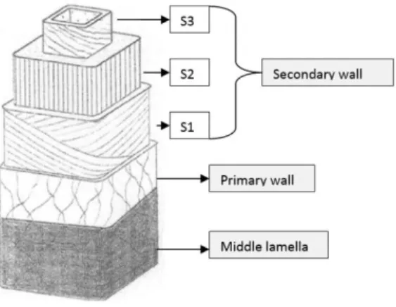
The plant cell wall is a complex extracellular matrix that is present at the surface of the plasma membrane. The cell wall acts as an exoskeleton, conferring resistance to the cell, flexibility to resist to cell disruption and permeability to allow the intercellular transport. The structure and composition of plant cell walls is not uniform; factors such as plant tax, tissue, age, cell type and cell layer have influence on their composition (Taiz & Zeiger 2002). Morphologically, it is possible to differentiate three zones: middle lamella, primary wall and secondary wall (Figure 1.1).
The most external layer is the middle lamella, a thin layer of material that is deposited just after cell division and can usually be seen at the junction between the walls of neighboring cells, acting as a sort of separating panel (Harris & Stone 2008). It is the first zone to be formed and it is constituted almost totally by pectic polysaccharides (Heredia et al. 1995).
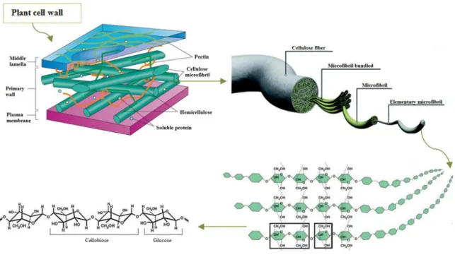
Over the middle lamella is the primary wall that controls the growth of the cell and forms the structural basis of the plant. It is composed of approximately 25% cellulose, 25% hemicelluloses, and 35% pectins, with 1 to 8% structural protein (Taiz & Zeiger 2002). These components form three essential networks: 1) cellulose microfibrils linked to each other by different glucans, 2) gel-forming network of pectin linked by calcium ion bridges and 3) structural proteins that are covalently bound to each other and to other cell wall components. Some cells are formed only by middle lamella and primary wall, but specialized cells have another component, the secondary wall (Heredia et al. 1995). When the secondary wall is formed some changes occur in the middle lamella and primary wall, for example lignification, a complex process in which lignins are deposited on the extracellular polysaccharide matrix and the main function is to strengthen the plant vascular body.
4 1.1.2 Plant cell wall composition
1.1.2.1 Cellulose
Cellulose is the major component of the plant cell wall and represents the most abundant organic polymer on Earth (Overmann 2006). Cellulose is a polysaccharide with approximately 7000 to 15000 D-glucose units that are linked by β-(1-4) glycosidic bonds. The disaccharide cellobiose (Figure 1.2) constitutes the structural repeating unit.
Cellulose molecules are aligned parallel to each other and linked by hydrogen bonds and van der Waals interactions to form elementary fibrils. These elementary fibrils make a highly ordered insoluble (crystalline) ribbon that excludes water and is relatively inaccessible to enzymatic attack.
Elementary fibrils are packed into microfibrils that contribute to the strength and structural basis of the plant cell (Figure 1.3) (Taiz & Zeiger 2002). The cellulose microfibrils include both crystalline and amorphous regions, in ratios dependent on the degree of polymerization (DP), the extent of hydrogen bonding and the source of cellulose (Bayer et al., 2000). The microfibrils are then assembled into cellulose fibers, which are packed together by both intra- and inter-fiber hydrogen bonds (Heredia et al.
1995). Adjacent sheets overlie one another and are held together by weak van der Waals forces. This crystalline structure is tight enough to prevent chemical and biological degradation and the diffusion of small molecules such as water.
Figure 1.1 – Illustration of the different layers of the plant cell wall. Adapted from Plomion et al.
5 1.1.2.2 Non-cellulosic major components
Most important non-cellulosic polysaccharides are divided into two groups: Pectic polysaccharides
and hemicelluloses or “cross-linking glycans”.
Pectic polysaccharides contain galacturonic acid and can be divided into: 1) galacturonans, which are composed of a linear chain of α-(1-4)-linked D-galacturonic acids; 2) rhamnogalacturonan I, which consists of a backbone of alternating α-(1-4)-linked galacturonic acid and α-(1-2)-linked rhamnose units with side branches that contain other pectin domains (primarily α-(1-5)-linked arabinan and β -(1-4)-linked galactan side chains); and 3) rhamnogalacturonan II that is smaller than rhamnogalacturonan I
but has a more complex structure, it comprises an α-(1-4)-homogalacturonan backbone of seven to nine residues substituted by up to five side chains comprising 12 monosaccharides bound by 20 linkages (Gorshkova et al. 2010). Pectins form a hydrated gel phase in which the cellulose-hemicellulose network is surrounded; acting as an hydrophilic filler they prevent aggregation and collapse of the cellulose network (Taiz & Zeiger, 2002).
Hemicelluloses are also designated “cross-linking glycans” because they can be bound to cellulose
microfibrils. Hemicelluloses are polysaccharides that have β-(1-4)-linked backbones with an equatorial configuration in which are included xyloglucans, xylans, mannans, glucomannans, and mixed-linked β -(1-3;1-4)-glucans and that can be decorated with a diverse range of carbohydrate side-chains. The main characteristic of hemicelluloses is their capacity of strengthening the cell wall by interaction with cellulose and, in some cases, with lignin (Scheller & Ulvskov 2010).
6 Besides these two groups of non-cellulosic polysaccharides, the cell wall also contains structural proteins, enzymes and lignin. Lignin is the generic term for cross-linked phenolic polymers. Due to its structure and association with cellulose and hemicellulose, lignin is important to prevent the degradation of these plant polysaccharides (Vanholme et al., 2010).
1.1.3 Plant cell wall degradation
The degradation of the plant cell wall by microorganisms is a fundamental biological process that is of considerable industrial importance, as plant cell wall polysaccharides are a major reservoir of carbon and energy. However, the plant cell wall is very complex and is composed by a variety of carbohydrates that are recalcitrant to the enzymatic attack. Only a restricted number of microorganisms have acquired the ability to deconstruct these structural carbohydrates (Fontes & Gilbert, 2010).
To achieve total or partial degradation of these carbohydrates a consortium of enzymes, free or in complex, is required. The extracellular plant cell wall degrading machinery is different for anaerobic and aerobic microorganisms as these use different strategies to target the polysaccharides (Bayer et al.
2004).
Aerobic microorganisms secrete free extracellular enzymes, such as endoglucanases, exoglucanases and β-glucosidases, which act individually in the degradation of plant cell wall and are considered as non-integrating systems. As example of aerobic microorganisms are bacterium from Bacillus, Micromonospora, Cellvibrio and Pseudomonas (Lynd, 2002) and fungi of genera Aspergillus (de Vries
et al., 2001).
The most anaerobic microorganisms have plant cell wall degrading enzymes associated in a supramolecular complex (molecular weight >3 MDa), termed the ‘cellulosome’ and are considered as integrating systems. As example of anaerobic microorganisms are bacterium of the genera Clostridium,
Ruminococcus, Thermotoga (Bergquist et al., 1999) and fungi of the genera Neocallimastix, Piromyces
and Orpinomyces (Fontes & Gilbert, 2010). Anaerobic bacteria and fungi in the rumen have developed a wide array of multimodular cellulases and hemicellulases that can act individually or as organized cellulosomes for the hydrolysis of plant cell wall polysaccharides to soluble sugars. Many of anaerobic microorganisms are found in the digestive track of invertebrate and vertebrate, as well as other specific ecosystems, such as soils, sediments and water bodies.
1.2 The bacterial Cellulosome: identification and organization
7 In this Section, the two bacterium subject of this Thesis, Clostridium thermocellum and
Ruminococcus flavefaciens, and their respective cellulosomes are described. The C. thermocellum cellulosome was the first to be identified and characterized in the 80’s while R. flavefaciens cellulossome was only recently identified and is not as well characterized.
1.2.1 The highly efficient cellulose-degrading bacterium Clostridium thermocellum
C. thermocellum is an anaerobic, rod shaped and gram-positive thermophile. This bacterium has gained research interest due to its cellulolytic and ethanologenic capabilities as it can convert biomass into energy. The ecological niche of C. thermocellum is abundant in soil worldwide (Freier et al., 1988), however, it can also be found in water bodies and digestive flora of animals (Clostridium thermocellum,
www.microbewiki.kenyon.edu last acessed 16.09.2016)
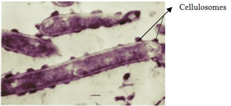
In the 1980, Bayer & Lamed and their colleagues identified and characterized the first cellulosome, on the basis of studies of the cellulolytic system expressed by the anaerobic thermophilic bacterium C. thermocellum (Lamed et al., 1983). Through the 80´s and 90´s there was an effort to elucidate the molecular mechanism for the assembly of C. thermocellum cellulosome, enabling the identification of its different structural components and how it is presented at the surface of the bacterium. In Figure 1.4 the cellulosomes are presented in the form of protuberances (Fontes & Gilbert, 2010).
C. thermocellum has one of the highest rates of cellulose utilization known, and the cellulosome of this bacterium is described as display a specific activity against crystalline cellulose (about 50 times more than Trichoderma) which may have been evolutionary imposed by the anaerobic environment (Fontes & Gilbert, 2010).
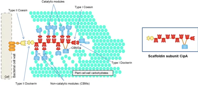
8 the scaffoldin subunit. The type I cohesin – dockerin interactions allow the integration of the hydrolytic enzymes into the complex and potentiates the stability and organization of the cellulossome (Bayer et al. 2004). To lead the scaffoldin subunit to the surface of the bacterium cell, membrane-associated proteins are bound to a type II cohesin. The type II cohesin-dockerin interactions support the anchoring of cellulosomes into cell surfaces. The scaffoldins containing type II cohesins are termed anchoring scaffoldins, while those containing type I are termed primary scaffoldins. Primary scaffoldins usually have a CBM3a that recognizes and binds to the recalcitrant cellulosic substrate and thus play a key role in bringing the cellulosome into close proximity with the plant cell wall.
1.2.2 Ruminococcus flavefaciens
The cellulosome of C. thermocellum is one of the best characterized and one expressing the highest rates of cellulose hydrolysis. Recently, a range of anaerobic bacteria were shown to produce cellulosome systems similar to those of C. thermocellum, such as Ruminococcus flavefaciens.
R. flavefaciens are cellulosic Gram-positive cocci of the order “Clostridiales”, an anaerobic
bacterium that inhabits the rumen community. They are responsible for the digestion of plant cell wall polysaccharides in the large intestine of herbivorous mammals and humans (Berg Miller et al. 2009).
In the past years, the sequencing of the genome of R. flavefaciens FD1 unraveled the complexity and diversity of this rumen bacterial cellulosome, revealing information about enzymatic and structural components of the cellulosome (Berg Miller et al. 2009). Also, the discovery of cellulases and other proteins involved in plant cell wall degradation was important to understand how the host organisms extract energy from their diet. The large number of protein-encoding sequences identified containing dockerin modules dictates that R. flavefaciens FD-1 has the largest collection of cellulosome-associated proteins of any known fiber-degrading bacterium thus far described.
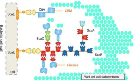
9 This cellulossome is considered as the most intricate and potentially versatile multienzyme complex described (Figure 1.6), with a genome that encodes 275 dockerins (in comparison with the 76 from C. thermocellum) that are likely to integrate the multienzyme complex (Berg Miller et al. 2009). The enzymes with homology for glycosyde hydrolases, carbohydrate esterases and polysaccharide lyases of known function constitute a diverse potential of substrate specificity (Fontes & Gilbert 2010). As the aim of several scientific groups is to elucidate the mechanism for the complete deconstruction of structural polysaccharides, R. flavefaciens cellulosome plays an important role in the discovery of new CAZymes and CBMs and thus to clarify their functions.
Althougth less is known about the R. flavefaciens cellulosome, it is evident that this cellulosome has a different organization from the C. thermocellum cellulosome. Several studies have suggested sequence and structural differences between the cohesins and dockerins of C. thermocellum and R. flavefaciens
(Fontes & Gilbert 2010).
In Figure 1.6, the R. flavefaciens cellulosome, the single cell-anchoring scaffoldin (ScaE) may bind a protein termed CttA, which carries two putative CBMs that mediate the primary anchorage to the plant cell wall. In addition, ScaE cohesin binds to the C-terminal dockerin of the primary scaffoldin (ScaB) which contains nine cohesins with two different specificities: four cohesins (Figure 1.6 in red) recognize the dockerin of the catalytic subunits or ScaC; five cohesins (Figure 1.6 in dark blue) bind to ScaA, which functions as an adaptor and primary scaffoldin. ScaA contains two cohesins that bind the cellulosomal catalytic subunit (presenting a similar specificity to the cohesins of ScaB, Figure 1.6 in red) and thus amplifies the number of enzymes in R. flavefaciens cellulosome. ScaC, an adaptor scaffoldin, can bind to ScaA and ScaB due to the specificity of the catalytic subunits (Fontes & Gilbert 2010).
Cellulosome structural organization varies between different strains of R. flavefaciens, which may reflect the complexity and diversity of substrates found in the rumen.
10
1.3 The modular organization and recognition mechanisms of Carbohydrate-Active
enZymes
Microorganisms that degrade plant-cell carbohydrates produce a high variety of polysaccharide-degrading enzymes, such as glycoside hydrolases, polysaccharide lyases, carbohydrate esterases and polysaccharide oxidases (Gilbert 2010). Many of these enzymes are part of the cellulosome to increase the efficiency for degrading the substrate. These enzymes are multimodular and often contain non-catalytic carbohydrate-binding modules (CBMs), which are connected through highly flexible linker sequences.
CAZymes represent these classes of enzymes that are involved in assembly and modification/cleavage of polysaccharides. CAZymes and CBMs have been classified over 25 years into sequence-based families in the CAZy database (www.CAZy.org) (Davies & Henrissat 2013). Since September 1998 CAZy is dedicated to the display and analysis of genomic, structural and biochemical information on the carbohydrate-active enzymes and associated CBMs involved in the synthesis and degradation of complex carbohydrates (Cantarel et al. 2009). Currently there are numerous different CAZymes and CBM families identified: 135 for glycoside hydrolases, 100 for glycosyl transferases, 24 for polysaccharide lyases, 14 for carbohydrate estereases and 80 for CBMs. In addition, CAZy also displays available PDB codes for those proteins that have been structurally characterized.
1.3.2 Carbohydrate-binding modules
As stated in the previous section, glycoside hydrolases involved in biodegradation of the plant cell wall are normally modular and have appended non-catalytic modules, CBMs. CBMs are defined as a contiguous sequence of 30 to 200 amino acids, that are normally appended to the associated catalytic module by a flexible linker for the recognition of the plant cell wall polysaccharides and to help increase the enzyme activity (Gilbert et al. 2013). CBMs are not always appended to a catalytic module and they can be present in isolated or tandem forms not coupled with an enzyme (Cantarel et al. 2009).
Lectins share some structural similarities with CBMs and they can bind to their target ligand through similar mechanisms, however, CBMs are normally found in enzymes that are degrading complex carbohydrates primarily to provide nutrients. This functionality distinguishes CBMs from lectins into a separated protein group (Gilbert et al. 2013). CBMs were initially classified as cellulose-binding domains (CBDs) based on the initial discovery of several modules that bound cellulose as primary ligand (Boraston et al. 2004). However, the term evolved to carbohydrate-binding modules due to the diverse ligand specificity exhibited.
1.3.2.1 Classification of CBMs
11 protein fold (Boraston et al. 2004). Although there are 7 different-fold families, the majority of CBMs identified to date are included in the β-sandwich fold family (Table 1.1).
Table 2.1 –Typical ‘fold families’ identified for CBMs and examples of CBM families with these folds. Adapted from Boraston et al. 2004 and www.CAZy.org, last acess at 14.09.20016.
Fold Family Fold CBM families
1 β-Sandwich 2,3,4,6,9,15,17,22,27,28,29,32,34,36,,47,51,70
2 β-Trefoil 13,42
3 Cystein knot 1
4 Unique 5,12
5 OB fold 10
6 Hevein fold 18
7 Unique; contains hevein-like fold 14
To provide additional functional relevance to the CBM classification these modules have been grouped into three types: A, B, and C according to its interaction with the carbohydrate. These three types define CBMs that bind crystalline surfaces, oligosaccharide sequences, or short oligosaccharide/monosaccharides, respectively.
CBMs from Type A have a planar hydrophobic surface composed by aromatic residues. This planar configuration in the active site interacts with flat crystalline polysaccharides, such as chitin or cellulose (Boraston et al. 2004). The properties of this CBM-type differ significantly from the other types of carbohydrate-binding proteins.
Recently, the crystal structure of a CBM63-containing Bacillus expansin (proteins that disrupt the cellulose–hemicellulose interface) in complex with cellohexaose was determined. A typical Type A, CBM63 contains a planar surface comprising three aromatic residues that make parallel π/C-H contact with the ligand (Figure1.7) (Georgelis et al. 2012).
Another study showed that a cohort of type A CBMs that belong to families 2 and 3 bind not only to crystalline cellulose but also to xyloglucan (Hernandez-Gomez et al. 2015).
12 CBMs from Type B or endo-type are classified as CBMs that bind to internal oligosaccharide sequences (Gilbert et al. 2013). This type of CBMs display a cleft arrangement in which the binding site accommodates longer glycan chains with four or more monosaccharide units (Figure 1.8). This type of CBMs have clearly evolved a binding site for the interaction with individual glycan chains rather than crystalline surfaces (Boraston et al. 2004).
CBMs from Type C or exo-type are classified as CBMs that bind the termini of an oligosaccharide sequence (Gilbert et al. 2013). This type of CBMs, also described as ‘lectin-like’ CBMs, have the property of binding in an optimal way to mono-, di- or tri-saccharides, due to steric restriction in the binding site (Boraston et al. 2004). Unlike the Type B CBMs, Type C do not contain the extended grooves on binding-sites. One example of this CBM type is the family 6 CBM from Bacillus halodurans in complex with lamirarihexaose (Figure 1.9).
Figure 1.8 – The X-ray crystal structure of the family 29 CBM from Piromyces equii in complex with mannohexaose (PDB code 1GWL). The secondary structures are shown as colored ramped cartoons with relevant amino acid side chains involved in carbohydrate binding shown as sticks. Solvent accessible surfaces are shown in gray with the surfaces contributed by the aromatic residues colored purple. Adapted from Gilbert et al. 2013.
13 1.3.2.2 Functional roles of CBMs
In general, CBMs contribute in the binding of the target substrates to carbohydrate degrading enzymes. According to experiments in the field reviewed by Boraston et al. 2004 and Gilbert et al.
2013, CBMs have four major roles: Proximity effect, targeting function, disruptive function and cell attachment.
Targeting function
As described in the previous section, CBMs are classified into different types according to the architecture of the binding site. Depending on this architecture, CBMs can target the enzyme to distinct regions within a larger macromolecular polysaccharide substrate. In general, there is a tight correlation between the ligands recognized by bacterial CBMs and the substrate specificity of the appended catalytic modules. As an example, CtCBM11 recognizes mixed-linked β-(1-3;1-4) glucans and is associated to the enzymes GH5 and GH6, endoglucanases with catalytic activity for β-(1-3) and β-(1-3;1-4) glucans, respectively (Carvalho et al. 2004).
Another study (Najmudin et al. 2006) shows that the function of CBM44 from C. thermocellum
Cel44A is associated with the targeting of Cel44A to its substrates. Cel44A is a typical endoglucanase that is capable of cleaving a variety of glucan-based plant cell wall polysaccharides such as cellulose,
β-glucans, xyloglucans, and glucomannans. To mediate efficient targeting, ligand recognition by CBM44 needs to mirror the substrate specificity of Cel44A; this study showed that this promiscuity in carbohydrate recognition is intrinsic to CBM44.
Proximity effect
CBMs increase the concentration of enzyme in close proximity to its polysaccharide substrate, which will lead to a rapid and efficient degradation. The proximity between the enzyme and the substrate potentiates the degradation of the polysaccharide (Herve et al. 2010). Herve and colleagues showed that CBMs can potentiate the action of a similar catalytic module toward polysaccharides in intact cell walls through the recognition of non-substrate polysaccharides. The targeting actions of CBMs therefore have strong proximity effects within cell wall structures, explaining why cellulose-directed CBMs are appended to many non-cellulase cell wall hydrolases (Herve et al. 2010).
Disruption of polysaccharide structure
14 This function was first documented for the N-terminal family 2a CBM of Cel6A from Cellulomonas fimi (Din et al. 1994) that appeared to mediate non-catalytic disruption of the crystalline structure of cellulose; furthermore, this disruptive effect enhanced the degradative capacity of the catalytic module.
Cell attachment
CBMs have been shown to adhere enzymes onto the surface of bacterial cell wall components while exhibiting catalytic activity on an external neighboring carbohydrate substrate. A family 35 CBM module has been shown to interact with the surface of glucuronic acid containing sugars in the cell wall of Amycolatopsis orientalis, while the catalytic module is active on external chitosan. This study shows that the biological role of CBM35s is not dictated solely by their carbohydrate specificities but also by the context of their target ligands (Montanier et al. 2009).
A unique family of carbohydrate-binding modules (CBM37) of Ruminococcus albus, located at the C-terminus of different glycoside hydrolases, appears to be responsible both for anchoring these enzymes to the bacterial cell surface and for substrate binding (Ezer et al. 2008).
1.3.2.3 Biotechnological applications of CBMs
The practical uses for CBMs in different fields of biotechnology are constantly on rise. CBMs have three properties that make them perfect candidates for different applications: (i) CBMs are usually independently folded units and therefore can function autonomously in chimeric proteins; (ii) the attachment matrices are abundant and inexpensive and have excellent chemical and physical properties; and (iii) the binding specificities can be controlled (Shoseyov et al. 2006).
CBMs are used to enhance bioprocessing enzymes in biomass degradation for industrial uses in biofuel production. A study demonstrates that the fusion of a CBM to the wild type cellulases Cel9A and Cel5A enhanced their activity as much as three fold on two insoluble lignocellulosic substrates (Reyes-Ortiz et al. 2013).
CBMs are also used for purification of biomolecules in immobilized affinity ligand technology. They act as affinity support for enzyme immobilization with high capacity, while retaining enzymatic activity and in some cases increased enzymatic activity is reported (Shoseyov et al. 2006). Studies demonstrate that CBMs can be utilized as biosensors; an example is a CBM that was used for glucose sensing in bioreactors (Verma et al. 2015).
Cellulose is a major component of numerous commercial products, several of which are capable of being recycled. Therefore, CBMs can also be used for the targeting of functional molecules to materials containing cellulose. The commercial potential of CBMs in this context was first realized for denim stonewashing, where cellulases were used as an alternative to the original abrasive stones (Shoseyov et al. 2006).
15 has been demonstrated for CBMs is that they can modify the characteristics of some enzymes. Replacing or adding a CBM can improve the hydrolytic activity, for example the addition of a CBM derived from cellohydrolase II of Trichoderma reesei to Trichoderma harzianum chitinase resulted in increased hydrolytic activity of insoluble substrates (Shoseyov et al. 2006).
1.4 Overview of the methodologies to study CBM-carbohydrate interactions
Carbohydrates play an intriguing role inside and at the surface of the cell. Carbohydrates occur as mono- and polysaccharides but are also part of glycan structures such as glycoproteins, glycolipids, peptidoglycans. Over the last decade, evidence has accumulated indicating that interactions between carbohydrates and particular proteins that recognize them play critical roles not only in the context of polysaccharide utilization and biotechnological applications but also in many other biological processes in health and disease, such as cell adhesion, signal transduction, host-pathogen interactions and inflammation processes (Park et al. 2008; Liu et al. 2009; De Schutter & Van Damme 2015). In many cases, the protein-carbohydrate interaction is only a first step in a series of events, often leading to a complex recognition process or signaling cascade. To fully understand the protein-carbohydrate interaction, different techniques for its characterization are required.
Biophysical methods such as carbohydrate microarray technology, Microscale Thermophoresis (MST), Isothermal Titration Calorimetry (ITC) and Surface Plasmon Resonance (SPR) and also structural methods such as X-ray crystallography and Saturation Transfer Difference NMR (STD-NMR) are used to study the protein-carbohydrate interactions.
In this Section these methods are briefly described giving more emphasis to the carbohydrate microarray technology and X-ray crystallography.
1.4.1 Carbohydrate microarray technology
The development of the carbohydrate (or glycan) microarrays in the beginning of the twenty-first century addressed the need for high-throughput methods that systematically array glycan libraries and identify glycan binding proteins (GBPs) to enable the investigation of their biological roles. Since their introduction in 2002, applications of the glycan microarrays have grown exponentially (Rillahan & Paulson 2011; Palma et al. 2014; Palma et al. 2015)
16 1.4.1.1 Carbohydrate probe libraries and immobilization methods
The first step in the construction of a carbohydrate microarray is to obtain pure and characterised carbohydrates, either from isolation from natural sources or from chemical or chemoenzymatic synthesis, and prepare these as suitable carbohydrate probes for the microarrays.
The oligosaccharides obtained from natural sources can include those derived from glycoproteins and glycolipids of different sources (mammalian, non-mammalian), bacterial-, fungal- and plant polysaccharides obtained through acid/alkaline hydrolysis and enzymatic degradation or depolymerization, as well as free-oligosaccharides isolated from mammalian milk and urine (Feizi 2003). The isolation of pure compounds, assignment of their structures and their immobilization on the array surface are challenges of using glycans from natural sources.
The polysaccharides or glycoconjugates, such as glycoproteins or glycolipids, can be directly immobilized on solid matrices by hydrophobic physical adsorption (Pedersen et al. 2012; Palma et al.
2015b) or charge-based interaction in the case of polysaccharides (Shipp & Hsieh-Wilson 2007). Polysaccharide or glycoconjugate microarrays are valuable for comparative antigenicity analysis and for ligand-discovery microarray projects.
The oligosaccharide microarrays are required for assignment of the carbohydrate ligand specificity of a given recognition system and are thus key tools to provide detailed information on structure-activity relationships in carbohydrate recognition (Liu et al. 2009). The oligosaccharide immobilization normally requires derivatization processes before arraying, due to their hydrophilic nature. Many different methodologies have been developed for immobilizing oligosaccharides. One of the approaches is to conjugate natural or chemically synthesized oligosaccharides to lipid by reductive amination or oxyme ligation to generate neoglycolipid (NGL) probes with amphipathic properties for efficient immobilization on nitrocellulose membranes (Liu et al. 2012; Palma et al. 2014). The advantage is that natural and synthetic glycans can be combined to generate diverse libraries, the strategy is to derivatize glycans from both sources with the same lipid tag for the immobilization. Most other mono- or oligosaccharide probes generated for printing have been de novo synthesized chemically or chemo-enzymatically, defining structures that incorporate specific functional groups for covalent attachment to solid matrices (Rillahan & Paulson 2011). Chemical synthesis has the advantage over natural isolation in that large quantities of relatively pure carbohydrates can be produced while the yield of the natural glycans is usually low, requiring deconvolution due to the heterogeneity of isolated mixtures (Horlacher & Seeberger 2008). However, achievement of glycan diversity offers a challenge and methods have
been developed to prepare ‘designer’ or ‘shot-gun’ microarrays from a natural glycome source (Palma
et al. 2015b; Song et al. 2011)
17 functional group on carbohydrates and a reactive group on the surface through several reactions. The majority of this methods use thiol and amine chemistries (Rillahan & Paulson 2011). The non-covalent immobilization methods rely on non-covalent interactions, such as electrostatic and hydrophobic interactions. It has been shown that polysaccharides, proteoglycans and neoglycoproteins can be efficiently immobilized in membranes of nitrocellulose or oxidized polystyrene (Rillahan & Paulson 2011). Another example of the non-covalent immobilization is the work of Feizi and colleagues that have shown the noncovalent immobilization of neoglycolipids and glycolipids on nitrocellulose-coated glass slides (Liu et al. 2012).
1.4.1.2 Neoglycolypid-based oligosaccharide technology
The concept of NGL technology was introduced by Feizi and colleagues in 1985, as a novel approach for study the antigenicities and receptor functions of carbohydrate chains of glycoproteins (Tang et al.
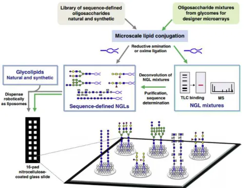
1985). In 2002 studies Wang and colleagues (Wang et al. 2002) showed that polysaccharides and glycoproteins can be immobilized on nitrocellulose by noncovalent interaction. After that, Feizi and colleagues adapted their NGL technology to generate the first microarray system for complex oligosaccharides (Fukui et al. 2002). The technology involves conjugating oligosaccharides by microscale lipid conjugation via reductive amination to an aminolipid, 1,2-dihexadecyl-sn-glycero- 3-phosphoethanolamine (DHPE). The oxime ligation can also be used to conjugate the lipid with the oligosaccharide (Liu et al. 2007). The NGLs can be immobilized on solid matrices, such as nitrocellulose membranes and silica plates, and are also suitable for thin layer chromatography (TLC)-binding experiments in conjunction with mass spectrometry for deconvolution and structural characterization of the probes that are giving binding signals (Chai et al. 2003).
The NGL-based microarrays comprise both NGL (prepared from natural or chemically synthetized oligosaccharides) and glycolipids (natural or synthetic) (Figure 10). These probes are robotically printed onto nitrocellulose-coated glass slides at low femtomol (fmol equivalent to 10-15 mol) levels in a
liposome formulation in the presence of carrier lipids (Liu et al. 2012). This approach promotes a certain level of flexibility and movement of the oligosaccharide being presented to the protein, which could be essential for particular recognition systems. The NGL-technology allows the expansion of the library of
probes by the “designer” microarray approach (Figure 1.10). The term “designer” microarray, refers to
18 1.4.1.3 Biological Applications
Carbohydrate microarrays have become a powerful tool to map out interactions involving carbohydrates in a high-throughput manner and gave a massive contribution to significant advances in glycomics. Nowadays, there are several applications for the carbohydrate microarray technology in health and disease (Palma et al. 2014) but this section is not an exhaustive description of all the applications, instead, will highlight 2 important examples in the context of this Thesis.
The first one was the development of carbohydrate microarrays from plant polysaccharides and derived oligossacharides as a sensitive method for probing microbial CBMs and anti-plant antibodies important for plant biology (Pedersen et al. 2012). The second example was the implementation of the NGL-based microarrays, coupled with mass spectrometry for construction of “designer” microarrays
from plant, fungal and bacterial glycomes (Palma et al. 2015). This microarray has been applied to identify and assign novel ligands for diverse carbohydrate recognition systems, including lectins on the immune system, therapeutic antibodies and CBMs with biological relevance.
The study of CBM-carbohydrate interactions allows the understanding of the divergent evolution of different microorganisms, as exemplified in this Thesis. It is also a means to create new strategies to improve the energy obtained by the microbial degradation of the plant cell wall carbohydrates, as well as understanding the symbiosis among these microorganisms.
19 1.4.2 Other biophysical methods
1.4.2.1 Microscale Thermophoresis
Microscale Thermophoresis (MST) is a recently developed technique by NanoTemper (Germany) that is based on the directed movement of molecules in a temperature gradient (Jerabek-Willemsen et al. 2014). Thermophoresis strongly depends on a variety of molecular properties such as size, charge, hydration shell or conformation. This technique allows a precise quantification of molecular events, independent of the size or nature of the investigated sample, due to high sensitivity (measures dissociation constants down to 1 picomolar). In a MST experiment, a microscopic temperature gradient is induced by an infrared laser, and the directed movement of molecules is detected and quantified using either covalently attached dyes, fluorescent fusion proteins or intrinsic tryptophan fluorescence (Jerabek-Willemsen et al. 2014).
Besides providing a precise determination of binding constants, it can also be used to derive additional information about the molecular mechanism of the investigated interaction. For instance, MST can be used to discriminate between different binding modes, and can also be used to determine interaction stoichiometries. A study from Wong and colleagues showed interactions between a protein called LysM domain with bacterial cell wall fragments and chitin oligomers using MST (Wong et al.
2014).
A typical MST experiment requires: one fluorescent binding partner and one nonfluorescent binding partner (the ligand). The fluorescence can be provided by an extrinsic label (e.g. NT-647 dye) covalently attached to the molecule of interest. The concentration of the fluorescent molecule is kept constant. The non-fluorescence molecule is then varied and 16 samples prepared at different concentrations. This dilution can be done in microtiter plates and a multichannel pipette to prepare multiple dilutions at once. After mixing the interaction partners, the samples are left to reach the equilibrium. The capillaries load themselves through capillary action once placed in the sample and are then loaded into the capillary tray.
20 1.4.2.2 Surface plasmon resonance
Surface plasmon resonance (SPR) was first introduced in early 1990s and is a powerful technique to determine specificity, affinity and kinetic parameters during the binding event of macromolecules. SPR is an optical technique that measures the refractive index changes in the vicinity of thin metal layers, such as gold, in response to biomolecular interactions. For this approach it is necessary to first immobilize the probe on the surface of the SPR chip and then a solution with the testing samples flows across this surface. During binding, changes in the SPR angle can be determined by varying the incidence angle and recording the reflected light intensity (Nguyen et al. 2015).
SPR technology has an important role in biomedical applications such as interaction analyses, conformational change studies, and mutation detection. SPR is widely used to study protein-carbohydrate interactions, including CBM-protein-carbohydrate interactions measuring affinity binding constants between the two molecules (Linman et al. 2009).
1.4.2.3 Isothermal Titration Calorimetry
Isothermal Titration Calorimetry (ITC) is a technique used in quantitative studies of a wide variety of biomolecular interactions.
This technique works by directly measuring the heat that is either released or absorbed during a biomolecular binding event (Freire et al. 1990). The ITC instrument operates on the heat compensation principle where the instrumental response (measured signal) is the amount of power (micro calories per second) necessary to maintain the temperature difference between the reaction and reference cells. ITC is the only technique that can determine all binding parameters in a single experiment (Leavitt & Freire 2001). Requiring no modification of binding partners, either with fluorescent tags or through immobilization, ITC measures the affinity of binding partners in their native states.
A B