Universidade de Lisboa
Faculdade de Farmácia
Targeting Necroptosis in Human Disease:
Potential Novel Modulators
Ana Rita Teixeira Poim
Dissertação orientada pelo Doutor Rui E. Castro e coorientada pela
Professora Doutora Cecília M. P. Rodrigues
Mestrado em Ciências Biofarmacêuticas
Universidade de Lisboa
Faculdade de Farmácia
Targeting Necroptosis in Human Disease:
Potential Novel Modulators
Ana Rita Teixeira Poim
Dissertação orientada pelo Doutor Rui E. Castro e coorientada pela
Professora Doutora Cecília M. P. Rodrigues
Mestrado em Ciências Biofarmacêuticas
The studies presented in this thesis were performed within the Cellular Function and Therapeutic Targeting research group, at the Research Institute for Medicines (iMed.ULisboa), Faculty of Pharmacy, Universidade de Lisboa, under the supervision of Rui E. Castro, PhD. and Cecília M. P. Rodrigues, PhD.
This project has received funding from European Structural and Investment Funds through the COMPETE Programme and from National Funds through FCT – Fundação para a Ciência e a Tecnologia, under the Programme grant SAICTPAC/0019/2015 and by LisbonPH fellowship.
Publications
The studies included in this thesis were presented as a poster communication:
Poim A, Santos SA, Brito H, Paulo A, Castro RE, Moreira R, Rodrigues CMP. Screening and identification of a selective inducer of necroptosis. 9th iMed.ULisboa Postgraduate Students Meeting, Lisbon, Portugal, 2017
Contents
Abstract ... vii
Resumo ... viii
Agradecimentos ... xi
Abbreviations... xiii
1 Introduction and Objectives ... 1
1.1 Necroptosis ... 3
1.1.1 Molecular mechanisms of necroptosis... 3
1.1.2 Necroptosis in inflammation and disease ... 5
1.1.3 Inhibitors of necroptosis ... 8
1.2 Colon Cancer ... 11
1.2.1 Treatment ... 11
1.2.2 Therapeutic resistance ... 13
1.2.3 Necroptosis in colon cancer ... 13
1.3 Objectives ... 15
2 Materials and Methods ... 3
2.1 Reagents ... 19
2.2 Cell Culture ... 19
2.3 Screening for Necroptosis Modulators ... 19
2.4 Viability Assays and Caspase-3/7 Activity ... 19
2.5 Statistical Analysis ... 21
3 Results ... 19
Screening for potential modulators of necroptosis ... 25
Hit validation ... 26
Modulation of TNF-α-induced MLKL phosphorylation by MS-PSR90 ... 27
Determination of SAS9 IC50 in different cell lines ... 28
Modulation of RIP1 expression and MLKL phosphorylation by SAS9 in L929 cells ... 32
4 Discussion ... 25
5 Conclusion and Future Perspectives ... 35
References ... 43
Annex information ... 1
Table of Figures
Figure 1 - TNF-induced apoptotic and necroptotic signaling complexes: ... 5Figure 2 – The pro-necroptotic cancer therapy ... 7
Figure 3 – Stages of colorectal cancer. ... 11
Figure 4 – SAS1, SAS5, SAS28, MS-PSR82, MS-PSR55, MS-PSR90 and CGAboc inhibits necroptosis in L929 cell line and SAS9 increases cell death. ... 25
Figure 5 - MS-PSR90 has the lowest EC50... 27
Figure 6 – MS-PSR90 inhibits TNF-α-induced MLKL phosphorylation levels. ... 28
Figure 7 – SAS9 exhibits higher cytotoxicity towards L929 cells in comparison with primary mouse hepatocytes. ... 29
Figure 8 – SAS9 is cytotoxic in HT29 cells. ... 30
Figure 9 - SAS9 induces both necroptosis and apoptosis. ... 31
Abstract
Necroptosis is an alternative pathway of regulated cell death, occurring independently of caspases, and mediated by receptor interacting serine/threonine-protein kinase-1 (RIP1), RIP3, and mixed lineage kinase domain-like (MLKL) protein. Necroptosis participates in the pathogenesis of several human conditions and activation may be beneficial for treating cancer. Despite this information, there are currently no pharmacological inhibitors or activators of necroptosis available with therapeutic properties. Colon cancer is the second most prevalent cancer worldwide and the third with the highest mortality rate. Cancer therapy has improved in recent years, but pro-apoptotic therapies still face cancer resistance.
In this thesis, we started by screening three libraries of newly synthesized compounds for its ability to modulate necroptosis, using the murine fibrosarcoma L929 cell line as an
in vitro model. We identified 7 compounds with the ability to inhibit TNF-α-induced
necroptosis L929 cells by more than 70%. To further characterize compound activity, the half maximal effective concentration (EC50) for inhibiting necroptosis was determined. The compound with the lowest EC50 - MS-PSR90 - was further shown to reduce TNF-α-induced MLKL phosphorylation. Interestingly, one of the compounds - SAS9 - strongly induced necroptosis in both L929 cells and in human colorectal adenocarcinoma cells HT29; IC50 values were 16.96 and 49.73 µM, respectively. Interestingly, SAS9 concomitantly induced apoptosis in both cell lines, with increased caspase-3/7-like activity. In L929 cells, SAS9-induced necroptosis encompassed MLKL phosphorylation but not increased RIP1 expression.
In conclusion, several novel modulators of necroptosis were identified in our screening, including a potential inducer of necroptosis. A better understanding of its mechanisms of action, as well as the investigation of pharmacokinetic properties, should help to determine their potential utility in cancer treatment.
Resumo
A necroptose é uma via de morte celular regulada alternativa e independente de caspases, mediada pelas proteínas cinase receptor interacting serine/threonine-protein 1 (RIP1), RIP3 e mixed lineage kinase domain-like (MLKL). A necroptose faz parte da patogénese de várias doenças e recentemente foi descoberto que a sua ativação pode ser benéfica no cancro. Apesar da necroptose ser responsável por várias patologias, não existe nenhum inibidor ou indutor farmacológico de necroptose com propriedades terapêuticas. O cancro do colon é o segundo cancro mais prevalente a nível mundial, sendo o terceiro mais mortífero. A terapia contra o cancro é cada vez mais eficaz, mas são vários os casos de resistência à terapia indutora de morte celular por parte das células cancerígenas.
Neste trabalho começámos por estudar três bibliotecas de compostos sintetizados de
novo, usando a linha celular murina de fibrosarcoma L929 como modelo in vitro, para
procurar potenciais moduladores de necroptose. Identificámos 7 compostos com a capacidade de inibir a necroptose pelo TNF-α em células L929 em mais de 70%. Para melhor estudar estes 7 compostos e a sua atividade, determinámos a concentração que induz metade do efeito máximo (EC50) de inibição na necroptose. Desta análise selecionámos o composto com menor valor de EC50 – MS-PSR90 – para averiguar a sua capacidade de reduzir a fosforilação da proteína MLKL, um dos passos fundamentais da necroptose. MS-PSR90 foi capaz de reduzir a fosforilação da proteína MLKL induzida pela necroptose causada pelo TNF-α.
De interesse surgiu outro composto, SAS9, com a capacidade de aumentar a morte celular causada pelo TNF-α em células L929. O SAS9 parece ser capaz de induzir necroptose na linhas celular murina L929 e na linha celular humana HT29 de adenocarcinoma coloretal, apenas quando privilegiámos essa via, com valores de IC50 de 16,96 e 49,73 µM, respetivamente. Verificámos, ainda, que o SAS9 também induz apoptose em ambas as linhas celulares, medida pelo aumento da atividade da caspase-3/7. Estes resultados são particularmente importantes em células resistentes a agentes pro-apoptóticos, onde o SAS9 poderá induzir necroptose. Por outro, em células não possuindo a maquinaria de necroptose, o SAS9 é indutor de apoptose. No caso da linha celular HT29, quando privilegiámos a morte por necroptose, esta parece ser independente da RIP1, visto que a Nec-1, inibidor da RIP1, não consegue reverter a necroptose. Nas células L929, a indução de necroptose pelo SAS9 aumenta a fosforilação de MLKL, comprovando a existência de necroptose, mas sem aumentar a expressão de RIP1.
Em conclusão, vários novos moduladores de necroptose foram identificados no nosso estudo, incluindo um potencial indutor de necroptose. No entanto, é necessário perceber melhor os seus mecanismos de ação, como também as propriedades farmacocinéticas, para determinar o seu potencial e utilidade no tratamento do cancro.
Palavras-chave: Alvos moleculares, Necroptose; Screening de fármacos; Terapêutica do cancro
Agradecimentos
Em primeiro lugar gostaria de agradecer à Professora Cecília por me ter sugerido este grupo de investigação e de me ter integrado nele. Os seus conselhos fizeram-me crescer tanto pessoalmente como profissionalmente. Agradeço também o seu tempo para me ajudar a decicir os próximos passos e auxiliar na escrita e correção da tese. Ao Professor Rui, agradeço pela orientação, críticas e conselhos e especialmente pela paciência que é ouvir-me quando nada parece correr bem. Nesta fase final obrigada pelo tempo todo disponibilizado, apesar de tanto trabalho que tem pela frente. Por fim agradeço a amizade, o apoio e momentos de boa disposição e humor que acabam por tornar aqueles dias de trabalho melhores.
Aos Professores Alexandra Paulo, Ana Ressureição, do grupo de investigação de Química Medicinal e Professor Carlos Afonso, do grupo de investigação Química Bioorgânica, da Faculdade de Farmácia, pela cedência dos compostos testados neste trabalho.
Marta, agora Doutora Marta, obrigada por tudo o que ensinaste e pelas discussões sobre os meus resultados porque sem isto tudo teria sido tão mais difícil compreender e escrever a tese. Vou tentar aproveitar ao máximo todos os teus ensinamentos de “Necroqueen” e também seguir o teu exemplo de pessoa e investigadora espetacular que és.
Obrigada Diane pelo teu apoio e disponibilidade, por estares sempre lá para mim, seja para me ajudares seja para aturares o meu mau feitio de Murta Queixosa.
Pedro Rodrigues, Hugo, André Simão, André Santos, Pedro Dionísio, Alexandra, Tânia, Maria, Vanda, Professora Joana e Professora Susana sem vocês tudo isto não era a mesma coisa. Tantos momentos de piadas e conversas que muitas vezes só alegram e ajudam aqueles dias mais difíceis.
Sara obrigada pela paciência para me ouvires e pelas tantas ajudas que me deste, especialmente nesta reta final.
Às minhas companheiras desta batalha, Mariana, Carolina e Marta, foi dificil mas penso que todas fizemos um bom trabalho e nos tentámos ajudar umas às outras. Houve momentos difíceis mas está a chegar ao fim!
Pedro, Catarina, Rute, Carolina, Zhang e Miguel foram dois anos convosco que estão a terminar e agradeço pela amizade especialmente. Espero que continuem as pessoas e amigos que são.
Sara e Adriana obrigada pela paciência que é por terem de me aturar todos os dias, especialmente naqueles emq ue só quero explodir.
Ao meu grupo de amigos, Cris, Luis, Alex, Miguel, João, Rui, Hugo, Ruben, Zé, Stive e Bruno, obrigada pelo apoio e por me ouvirem, apesar de muitas vezes não perceberem nada do que estava para ali a falar. Chegar ao final do dia e poder falar convosco e aliviar tanto stresse foi essencial para ultrapassar esta fase.
Aos meus músicos e manos preferidos, Carolina e André, o apoio e as piadas tal como aqueles ensaio espetaculares só ajudaram a ultrapassar esta fase complicada.
Ao meu pai e ao meu tio que apesar de muita coisa sei que posso contar com eles. Aos meus avós que são tudo para mim. Isto tudo é por vocês e por estarem comigo ao longo da minha vida toda. Sou o que sou devido a vocês.
Mokas, a minha maninha, sei que às vezes não é fácil lidar comigo diariamente mas obrigada por tudo, por me ajudares quando isto parecia que já não dava mais. Gosto muito de ti e és um orgulho!
Mãe, não foi fácil chegar aqui. Sei que às vezes não pareço justa mas também sei o que fazes por mim. Não foram anos facéis mas estiveste sempre lá para ouvires as minhas teorias e coisas estranhas que eu falo. Obrigada por tudo.
David, estiveste comigo estes 5 anos, sempre ao meu lado, sempre a apoiar-me e a ajudar-me a ultrapassar tantos desafios que apareceram. Sem a tua força não tinha conseguido chegar até aqui. És tu que me fazes avançar e querer lutar. Amo-te muito.
Abbreviations
5DFUR - 5’deoxy-5-fluoroudine 5-FU - 5-fluorouracil
BMDC - Bone-marrow derived cell BSA - Bovine serum albumin
BV6 - Selective inhibitor of IAP proteins
c-FLIP - Cellular FADD-like IL-1β-converting enzyme-inhibitory protein cIAP - Cellular inhibitor of apoptosis protein
CYLD - Cylindromatosis
DAMPs - Damage-associated molecular patterns DC - Dendritic cell
DMSO - Dimethyl sulfoxide Drp1 - Dynamin-related protein 1
dTMP - Deoxythymidine monophosphate DTT - Dithiothreitol
dTTP - Deoxythymidine triphosphate dUMP - Deoxyuridine monophosphate dUTP - Deoxyrubine triphosphate
EC50 - Half maximum effect concentration
EGFR - Epidermal growth factor receptor
FADD - Fas-associated protein with a death domain FdUMP - Fluorodeoxyuridine monophosphate FdUTP - Fluorodeoxyrubine triphosphate FTMP - Fluororidine triphosphate
HCD - High cholesterol diet HDAC - Histone deacetylase
HMGB1 - High mobility group box I protein IC50 - Half maximum inhibitory concentration
IFN - Interferon
IKK - Inhibitor of κB kinase JNK - C-Jun N-terminal kinase
LUBAC - Linear ubiquitin chain assembly complex MAPK - Mitogen-activated protein kinase
MCMV - Murine cytomegalovirus MDR - Multidrug resistance
MTX - Methrotrexate Nec - Necrostatin
NEMO - Nuclear factor-κB essential modulator NF-kB - Nuclear factor-κB
NGEN - Naringenin
NSA - Necrosulphonamide
OCT - Organic cation transporters PGAM-5 - Phosphoglycerate mutase 5
PIGF - Phosphatidylinositol-glycan biosynthesis class F PRPP - Phosphoribosyl pyrophosphate
PS - Phosphatidylserine
RHIM - RIP homotypic interaction motif RIP - Receptor interacting proteins ROS - Reactive oxygen species
RT-PCR - Real time polychain reaction SEM - Standard error of the mean siRNAs - Small interfering RNA
SMAC - Second mitochondria-derived activator of caspase TAK1 - TAB-transforming growth factor-activated kinase 1 TLR - Toll-like receptor
TNFR - TNF-α receptor
TNF-α - Tumor necrosis factor alpha TP - Thymidine phosphorylase
TRADD - TNFR-associated death domain TRAF2 - TNFR-associated factor 2
TRAILR - NF-related apoptosis-inducing ligand receptor TS - Thymidylate synthase
UP - Uridine phosphorylase
VEGF-A - Vascular endothelial growth factor-A VEGFR - VEGF receptor
1.1 Necroptosis
For a long time, apoptosis was regarded as the only form of regulated cell death; cells would either undergo programmed cell death, apoptosis, or die due to chemical or physical insults by necrosis (Weinlich et al., 2016). In 1998, however, two studies demonstrated that mouse L929 fibroblasts could be sensitized to tumor necrosis
factor-α(TNF-α) or Fas ligand- programmed necrosis in the presence of a caspase inhibitor (Vercammen et al., 1998a, 1998b). This form of programmed necrosis was later named necroptosis (Degterev et al., 2005).
Necroptosis can be described as a necrosis-like cell death mechanism which is usually triggered when apoptosis is blocked (Degterev et al., 2005). Apoptosis is the caspase-dependent dominant cell death pathway while necroptotic cell death is largely caspase-dependent on kinase cascades. In that sense, a family of kinase activity proteins known as receptor interacting proteins (RIP) are needed as cell stress sensors, particularly RIP1 and RIP3 (Galluzzi et al., 2009; Holler et al., 2000; Meylan and Tschoopp, 2005; Saeed and Jun, 2014). Still, several studies have shown that necroptosis can occur independently of RIP1 (Upton et al., 2010; Vanlangenakker et al., 2011; Zhang et al., 2009), with RIP1 activation also resulting in nuclear factor-κB (NF-κB) or mitogen-activated protein kinase (MAPK) activation, or even apoptosis (Festjens et al., 2007). The RIP1/RIP3 kinase cascade also regulates mitochondrial oxidative stress through C-Jun N-terminal kinase (JNK) activation (An et al., 2013; Ramachandran et al., 2013; Roychowdhury et al., 2013). In turn, RIP3 is thought to be activated upon increased pro-inflammatory cytokines (Moriwaki and Chan, 2013; Roychowdhury et al., 2013) and inflammasome activation (Moriwaki and Chan, 2013) and is thought to act as a switch between apoptosis and necrosis (Zhang et al., 2009).
1.1.1 Molecular mechanisms of necroptosis
Necroptosis in engaged in response to a wide range of stimuli, like cytokines, viral infection, chemicals, damage-associated molecular patterns (DAMPs) or several forms of physicochemical cellular stress (Vanlangenakker et al., 2012). These necroptotic stimuli can be recognized or sensed by specific sensors or receptors exposed on the cell surface or present intracellularly (Meng et al., 2015). In particular, necroptosis can be triggered by Fas or TNF related apoptosis inducing ligand receptor (TRAILR) (Feoktistova et al., 2011; Geserick et al., 2009); Toll-like receptor-4 (TLR4) and TLR3; double-stranded DNA viruses, such as murine cytomegalovirus (MCMV); interferon-α (IFN-α) and IFN-β; and polynosine-polycytidylic acid (Meng et al., 2015). Nonetheless,
TNF-α-induced necroptosis is the most well characterized molecular pathway of necroptosis (Wu et al., 2012; Zhao et al., 2015a).
Upon TNF-α and TNF-α receptor 1 (TNFR1) ligation, TNFR1 recruits proteins such as TNFR-associated death domain (TRADD), RIP1, and TNFR-associated factor 2 and 5 (TRAF2/5) (Harper et al., 2003; Micheau and Tschoopp, 2003). TRAF2 then binds to cellular inhibitor of apoptosis proteins 1 and 2 (cIAP1/2), forming complex I (Moriwaki and Chan, 2013). RIP1 is then Lys63-linked polyubiquitylated by cIAP ligases (Mahoney et al., 2008; Varfolomeev et al., 2008), allowing the recruitment of the linear ubiquitin chain assembly complex (LUBAC), which supports the recruitment of the TAB-transforming growth factor-activated kinase 1 (TAK1) complex and inhibitor of κB kinase (IKK) subunit NF-κB essential modulator (NEMO), activating the NF-κB pathway (Bertrand et al., 2008; Haas et al., 2009; Wu et al., 2006). After deubiquitination by cylindromatosis (CYLD) (Welz et al., 2011; Wright et al., 2007), RIP1 recruits TRADD, the Fas-associated protein with a death domain (FADD), cellular FADD-like
IL-1β-converting enzyme-inhibitory protein (c-FLIP) and caspase-8, thus forming the TNFR1 complex II (Cho et al., 2009; Dillon et al., 2012; Micheau and Tschoopp, 2003; Wang et al., 2008), which mediates apoptosis. Within TNFR1 complex II, apoptotic proteins FADD, c-FLIP and caspase-8 suppress the induction of necroptosis. Caspase-8 inactivates RIP1 and RIP3 by proteolytic cleavage, after which the proapoptotic activation of caspases takes place (Vanlangenakker et al., 2011). When caspase-8 is inactivated or absent, RIP1 and RIP3 are not cleaved and become phosphorylated (Vandenabeele et al., 2010). In this scenario, the necrosome is formed (Li et al., 2012). RIP1 recruits RIP3 through RIP homotypic interaction motif (RHIM) domain mediated-interactions. This RIP1-RIP3 interaction promotes the recruitment and phosphorylation of mixed-lineage kinase domain-like (MLKL) protein (Z Cai et al., 2014; Li et al., 2012). Phosphorylated MLKL forms tetramers and translocates onto the plasma membrane (Z Cai et al., 2014; Sun et al., 2012), leading to Ca2+ influx (Z Cai et al., 2014). In addition,
RIP3 is reported to activate a number of different downstream signals such as phosphoglycerate mutase 5 (PGAM-5) and dynamin-related protein (Drp1) that induce reactive oxygen species (ROS) production in the mitochondria (Hitomi et al., 2008a). Finally, RIP3-multimers are stabilized by chaperones that are essential for necroptosis (Linkermann et al., 2013).
Figure 1 - TNF-induced apoptotic and necroptotic signaling complexes: Upon TNF ligand
binding, the intracellular tails of TNFR1 recruit multiple proteins to form complex I. Deubiquitination of RIP1 by CYLD or inhibition of cIAP proteins promote the conversion of complex I to complex II and inhibits NF-κB activation. Caspase-8 becomes activated in complex II and initiates apoptosis. When levels of RIP3 are high, RIP3 enters complex II via interaction with RIP1, after stimulation. The RIP3-containning complex is called complex IIb (necrosome). In the presence of cFLIP, caspase-8 is unable to initiate apoptosis but can cleave RIP1 and RIP3 and, thus, inhibit necroptosis. Depletion of FADD or caspase-8, inhibition of caspase-8 or induction of RIP3 can free RIP1-RIP3 from inhibition and initiate necroptosis by mixed-lineage kinase domain-like protein (MLKL) in TNF-treated cells.
1.1.2 Necroptosis in inflammation and disease
Necroptosis exerts proinflammatory effects, being able to initiate a cycle of necroptosis-driven inflammation and inflammation-necroptosis-driven necroptosis (Linkermann et al., 2014). In fact, the necroptotic machinery was shown to contribute to many inflammatory disorders, including Crohn’s disease (Gunther et al., 2011), rheumatoid arthritis (Lawlor et al.,
2015), multiple sclerosis (Ofengeim et al., 2015), TNF-driven autoinflammatory disorders (Berger et al., 2014; Duprez et al., 2011; Huang et al., 2016), severe cutaneous reactions to drugs (Saito et al., 2014), allograft rejection (Lau et al., 2013; Pavlosky et al., 2014), remote injury postemgraftment (Zhao et al., 2015b), cigarette smoke-driven chronic obstructive pulmonary disease (COPD) (Mizumura et al., 2014; Pouwels et al., 2016), acute pancreatitis (He et al., 2009; Karch et al., 2015; Wu et al., 2013) and some manifestations of systemic lupus erythematosus (Fan et al., 2014). Nonetheless, the role of necroptosis-driven inflammation as disease-cause or -consequence is difficult to assess due to the lack of a definitive molecular marker (Kopalli et al., 2016). Studies using the RIP1 inhibitor necrostatin-1 (Nec-1) (Degterev et al., 2005) or RIP3-deficient mice have established a role for necroptosis in the pathophysiology of pancreatitis (He et al., 2009), atherosclerosis (Lin et al., 2013), retinal cell death (Murakami et al., 2013), ischemic organ damage and ischemia-reperfusion injury in both the kidney (Linkermann et al., 2013), the heart (Oerlemans et al., 2012) and the liver (Afonso et al., 2015).In addition, MLKL specific antibodies, that can detect necrotic hepatocytes in drug-induced liver damage, may be able to provide potential molecular markers. Still, more research is needed to underscore its role for detecting necroptosis in inflammatory diseases (Wang et al., 2014).
1.1.2.1 Necroptosis in cancer
Several evidences have shown that necroptosis is involved in the regulation of cancer (Cho and Park, 2014; Fulda, 2014, 2013; Jain et al., 2013; Su et al., 2016; Yu et al., 2013). Tumours are often able to dodge cell death (Hanahan and Weinberg, 2011, 2000). In fact, the most frequent causes of cancer therapeutic resistance relate with cell death deficits, being important to understand and explore cancer cell death to develop strategies to overcome therapeutic resistance (Meng et al., 2015). Because necroptosis follows different execution pathways when compared to apoptosis, cancer cells that are resistant to apoptosis may be sensitive to necroptosis inducers, which would then constitute novel strategies to overcome therapeutic resistance.
Several cancer cell lines undergo necroptosis when exposed to classic necroptosis inducers or chemotherapeutic agents. These cancer cell lines cover most of the common cancer cell types: colorectal cancer, leukemia, multiple myeloma, lung cancer, ovarian cancer, breast cancer, hepatocarcinoma, bladder carcinoma, head and neck carcinoma, glioblastoma, cervical cancer and neuroblastoma (Su et al., 2016). From these, colorectal cancer cells and hematopoietic neoplasm cells are the most sensitive to induction of necroptosis. HT29 colorectal cancer cells, for example, undergo necroptosis
upon stimulation with TNF-α + SMAC mimetics + zVAD-fmk (Zhenyu Cai et al., 2014; Moriwaki et al., 2015).
However, cancer cell lines with intact necroptotic machinery represent only a small part of cancer cells. Many others, like colorectal cancer cells HCT-116, do not have an effective necroptotic machinery, thus not being suitable for necroptotic-based cancer therapy studies (Moriwaki et al., 2015). In addition, there is still limited histological or animal data showing that necroptotic inducers can trigger tumour necroptosis in vivo (Su et al., 2016).
Figure 2 – The pro-necroptotic cancer therapy.
Necroptosis plays an important role in tumorigenesis, backing up cell death mechanism in cancer cells. To identify cellular signalling networks targeted by necroptosis, a genome-wide screen using small interfering RNAs (siRNAs) was performed, from which two suppressor genes were identified, namely CYLD and EDD1, suggesting a possible role of necroptosis in tumorigenesis (Hitomi et al., 2008b). In addition, in colon and lung cancer, the ratio of RIP3-r, a truncated splice variant of RIP3, to RIP3 has been found significantly increased, when compared to normal tissues, showing that RIP3-r may be a major splice form associated with tumorigenesis (Yang et al., 2005). Other evidences further suggest that necroptosis may play a role in tumorigenesis, including mutations in CYLD, a positive regulator of necroptosis that may protect tumour cells from cell death induction (Reed, 2006).
Necroptotic cancer cells can trigger adaptive immune responses. Phagocyte recruitment is accomplished by the release of “find-me” signals from dying cells (Munoz et al., 2010; Peter et al., 2010). In necroptotic cells, the “find-me” signals may be monosodium urate
(MSU) crystals (Shi et al., 2010), high mobility group box I protein (HMGB1) (Urbonaviciute et al., 2008) and annexin A1 (Blume et al., 2012, 2009). When phagocytes find the dying cells, they then recognize “eat-me” signals, exposed on the surface of dying cells. Necroptotic cells expose annexin A1 (Blume et al., 2009; Caipl et al., 2001), phosphatidylserine (PS) (Bottcher et al., 2006; Brouckaert et al., 2004) and the C-type lectin Clec9A (Ahrens et al., 2012; Sancho et al., 2009; Zhang et al., 2012). After dying cells are “eaten”, phagocytic cells (DCs, macrophages, monocytes and neutrophils) produce pro-inflammatory cytokines and chemokines, up-regulate stimulatory molecules and enhance cross-presentation, triggering adaptive immune responses (Biswas and Mantovani, 2010; Steinman, 2007) with anti-tumor activity.
1.1.3 Inhibitors of necroptosis
Few attempts have been made to discover potential therapeutic agents capable of inhibiting necroptosis, likely due to the conservative notion that necroptosis, in opposition to apoptosis, is unregulated (Kopalli et al., 2016). Still, a number of inhibitors have been developed, each displaying major drawbacks and/or side effects.
Necrostatins (Necs) are tryptophan-based small molecules originally found to halt necroptosis by directly inhibiting RIP1 (Degterev et al., 2008, 2005). Several Necs and its analogues have been synthetized and its use as potential necroptosis inhibitors tested in experimental animal models (Degterev et al., 2008; Jagtap et al., 2007; Rosenbaum et al., 2010; Teng et al., 2007, 2005; Wang et al., 2007; Xie et al., 2013). The most used and most well-known Nec is Nec-1 (5-(1H-Indol-3-ylmethyl) - (2-thio-3-methyl) hydantoin). Nec-1 inhibits RIP1 in a T-loop-dependent manner, binding to and stabilizing the inactive conformation of RIP1, thereby shifting the equilibrium towards the inactive state (Degterev et al., 2008). Inhibition of necroptosis by Nec-1 has been shown to reduce infarct volume and ameliorate neurological deficits in a middle cerebral artery occlusion mouse model (Degterev et al., 2005), retina ischemiareperfusion (Rosenbaum et al., 2010), myocardial infarction (Lim et al., 2007), cardiac ischemia-reperfusion injury (Zhang et al., 2014), and traumatic brain injury (You et al., 2008). Some studies, namely in mouse models, have illustrated some of the downsides of Nec-1; its specificity, effective concentration, inadequate pharmacokinetics properties. Further, because necroptosis can occur in a RIP1-independent manner, some concerns were rendered on the likelihood of being used in the clinics (Liao et al., 2014; Tamura et al., 2011; Zhang et al., 2016).
Histone deacetylase (HDAC) inhibitors are used as anticancer agents with anti-inflammatory and neuroprotective effects. Vorinostat (suberoylanilide hydroxamic acid;
SAHA) was approved by the FDA for the treatment of relapsed/refractory cutaneous T Cell Lymphoma. More recently, its anti-necroptotic effects were reported; specifically, murine fibrosarcoma L929 cells pretreated with vorinostat (1 µM) were protected against TNF-α induced necroptosis through mechanisms involving RIPK1-dependent NF-κB and p38 MAPK activation, JNK and Akt kinase inactivation, autophagy initiation, and enhanced cFLIPL expression (Wang et al., 2013).
Ponatinib is an oral multi-targeted tyrosine kinase inhibitor developed for the treatment of chronic myeloid leukemia and Philadelphia chromosome-positive acute lymphoblastic leukemia (Cortes et al., 2012; O’Hare et al., 2009). Pazopanib is also an oral receptor tyrosine inhibitor and is used for the treatment of advanced renal cell carcinoma and soft tissue sarcoma (van der Graaf and Gelderblom, 2012; Ward and Stadler, 2010). Both drugs were highlighted in a study where a group of 268 FDA-approved drugs were screened for their ability to inhibit TNF-α-induced necroptosis (Fauster et al., 2015). Ponatinib and Pazopanib abrogated TNF-α-induced MLKL phosphorylation; still, their mechanisms of action remain unclear and additional studies in animal models covering a variety of necroptosis-associated pathologies is needed.
Necrosulphonamide (NSA) is a direct inhibitor of human MLKL. NSA is considered safe and with low cellular toxicity (Sun et al., 2012). NSA interferes with MLKL oligomerization by targeting Cys86, thus blocking necroptosis. Of note, mouse models lack an orthologous Cys, rendering them inappropriate for pharmacological, pharmacokinetics and toxicity pre-clinical testing (Liao et al., 2014; Sun et al., 2012).
Necroptosis inhibitors from natural products such as G. lucidium are very popular medicinal fungus in traditional Chinese medicine. It is consumed in the form of MAK, a functional food derived from this fungus. MAK chronic consumption has been shown to prevent H/I-induced RIP3 expression and decrease necroptotic cell death (Xuan et al., 2015); in parallel, T. chebula water extract (WETC) antagonizes mitochondrial-derived ROS production, thus inhibiting TNF-induced necroptosis (Lee et al., 2016); Kongensin A, a natural product isolated from Croton kongensis, blocks cell death induced via treatment with a combination of TNF-α, second mitochondria-derived activator of caspase (SMAC) mimetic and zVAD-fmk before or at the RIP3 activation step (Li et al., 2016); Celastrol is a triterpene from the root bark of the Chinese medicinal plant
Tripterygium wilfordii; celastrol was shown to inhibit RIP3 and MLKL and increase
caspase-8, thus preventing necroptosis (Jia et al., 2015); Naringenin (NGEN) is a natural compound belonging to the chemical class of flavones (Ahmed et al., 2014). When co-administered to rats fed a high cholesterol diet, hepatic tissue fibrosis was improved,
providing insights into possible molecular mechanisms underlying NGEN suppression of necroptosis (Chtourou et al., 2015); Finally, curcumin, a bright yellow chemical, is the principal curcuminoid of Curcuma longa (turmeric), a member of the ginger family (Zingiberaceae). Curcumin was shown to decrease the expression of RIP1 in a dose and time-dependent manner, suggesting that its role in protecting primary cortical neurons against iron-induced neurotoxicity is dependent, at least in part, on inhibition of necroptosis (Dai et al., 2013).
1.2 Colon Cancer
Colorectal cancer is the second most prevalent cancer worldwide and the third cancer with the highest mortality rate (Stewart and Kleihues, 2014). Colon cancer usually starts with a growth called polyp on the inner lining of the colon. Nonetheless, not all polyps will develop cancer; adenomatous polyps may change into cancer while hyperplastic polyps and inflammatory polyps usually do not. Tumors usually develop in the innermost layer of the colon - the mucosa. From here, cancer cells can enter blood or lymphatic vessels, and metastasize (Stewart and Kleihues, 2014).
Figure 3 – Stages of colorectal cancer. Stage 0: The cancer is found only in the mucosa of the
colon or rectum. Stage I: The tumor has growth into the inner wall of the colon or rectum. Stage II: The tumor extends more deeply through the wall of the colon or rectum. It can start to invade nearby tissues. Stage III: The cancer has spread to nearby lymph nodes, but not to other parts of the body. Stage IV: The cancer has spread to other parts of the body. From National Institute of Health, 2015
1.2.1 Treatment
Available therapies for colon cancer are surgery and the use of cytotoxic and targeted agents for systemic therapy. In surgery, total resection of the tumor is done, removing lymph nodes and margins of the tumor (Cunningham et al., 2010). Treatment with adjuvant chemotherapeutic agents typically follows aiming to destroy any remaining tumor cells and to reduce the risk of recurrence (Stein et al., 2011). The type and length
of chemotherapy depends on disease stage (Stein et al., 2011; Wilkes and Hartshorn, 2009).
5-fluorouracil (5-FU), an analogue of uracil with a fluorine atom at the C-5 position instead of a hydrogen, is the main choice for adjuvant therapy in colon cancer (Wolpin and Mayer, 2009). Within the cell, 5-FU is converted to three active metabolites: fluorodeoxyuridine monophosphate (FdUMP), fluorodeoxyrubine triphosphate (FdUTP) and fluororidine triphosphate (FTMP). The active metabolites disrupt RNA synthesis and the action of the thymidylate synthase (TS) enzyme, causing DNA damage (Longley et al., 2003) and, ultimately, cell death.
After decades of sole 5-FU-based treatment, both improved 5-FU-based therapies and new and more effective agents are changing the way colon cancer is treated. Strategies to improve the anticancer activity of 5-FU include decreasing 5-FU degradation, increasing 5-FU activation and increasing the TS binding activity of FdUMP (Longley et al., 2003). More recently, monoclonal antibodies have been used as targeted therapy in colon cancer treatment (Kozovska et al., 2014). Bevacizumab is a humanized monoclonal antibody that inhibits blood-vessel formation; it binds to vascular endothelial growth factor-A (VEGF-A), preventing its binding to VEGFR and inhibiting angiogenesis, tumor growth and metastatic development (Recondo et al., 2014) (Segal and Saltz, 2009). It was the first anti-angiogenic drug approved for metastatic colon cancer. Ziv-aflibercept is a recombinant fusion protein that acts as a soluble receptor binding to VEGF-A, VEGF-B and phosphatidylinositol-glycan biosynthesis class F (PIGF) and inhibits them, resulting in decreased neovascularization and decreased vascular permeability (Recondo et al., 2014). Cetuximab is a chimeric monoclonal antibody that binds to the extracellular domain of the epidermal growth factor receptor (EGFR) and blocks ligand-induced receptor signaling (Segal and Saltz, 2009). These anti-EGFR therapies are used in the treatment of stage IV colon cancer (Recondo et al., 2014). Panitumumab is a fully human monoclonal antibody specific to EGFR, similar to Cetuximab (Segal and Saltz, 2009). Regorafenib is an oral tumor deactivation agent that blocks multiple protein kinases, including kinases involved in tumor angiogenesis, oncogenesis and the tumor microenvironment (Recondo et al., 2014).
Altogether, and despite the existence of different therapeutic approaches, colon cancer treatment remains suboptimal. Novel anti-cancer molecules and anti-resistance approaches are needed to improve clinical outcomes in patients.
1.2.2 Therapeutic resistance
Despite advances in cancer therapy, resistance still occurs in about 90% of patients with metastatic cancer (Longley and Johnston, 2005). Tumors may present intrinsic resistance and/or acquired resistance to therapy, each determining specific initial and subsequent lines of treatment (Hammond et al., 2016). Innate resistance is usually noticed during early stages of drug development or early phase clinical trials, but some are only understood during retrospective analysis studies (Bardelli and Siena, 2010; Cunningham et al., 2004). Acquired resistance may develop for each cytotoxic therapy and each targeted pathway and, often, acquired resistance to one drug leads to resistance to other drugs that work by different mechanisms of action. This is called multidrug resistance (MDR). Resistance to traditional cytotoxic therapy may result from decreased drug delivery and/or uptake by the cancer cell, increased efflux out of the cell, or by changes in the enzymes involved in drug metabolism (Hammond et al., 2016). In parallel, resistance can also result from genetic or epigenetic modifications (Gottesman et al., 2002). Resistance to targeted therapies may occur through upregulation, mutation or activation of downstream signaling molecules within specific pathways, pathway bypass mechanisms or increased cross-talk between analogous pathways (Longley and Johnston, 2005; Tejpar et al., 2012). Understanding the mechanisms of drug resistance is essential to develop novel, rational and more effective treatment combinations, helping to guide future therapies.
1.2.3 Necroptosis in colon cancer
As mentioned above, 5-FU remains the main cell death-inducing therapy in colon cancer. However, because cancer cells develop resistance to 5-FU-induced apoptosis, new alternatives are being studied. For instance, HT29 colon cancer cells were sensitized to 5-FU induced cell death in the presence of pan-caspase inhibitor zVAD-fmk, leading to autocrine NF-κB-dependent TNF-α production (Metzig et al., 2016). Similar responses were observed using tissue biopsies from colon cancer patients but not in two additional colon cancer cell lines, HTC116 and SW480, probably due to their lack of RIP3 expression.
In addition, incubation of HT29 cells with cobalt chloride, an hypoxia-inducing agent that mimics features of the tumor microenvironment, also consolidated the idea that necroptosis occurs in tumorigenesis, as cell death was increased in the presence of zVAD-fmk (Cells et al., 2015). Further, necroptosis appeared to occur independently of ROS, which is a key finding because ROS generation in cancer cells may lead to cell proliferation and survival, as well as drug resistance (Saeidnia and Abdollahi, 2013). As
such, the prospect of using pro-necroptotic drugs for treating colon cancer is appealing, although it would be key to avoid unwanted side-effects due to caspase inhibition. Caspase-8 deficiency is frequently a feature of human colon cancers and accumulation of RIP3 has been found in intestinal epithelial caspase-8 deficient cells. In addition, caspase-8 mutations in human colon cancer cells lead to decreased apoptosis (Kim et al., 2003). In line with this results, a SMAC mimetic is able to induce necroptosis in vivo and cause loss of stemness in intestinal and colon cells (He et al., 2017). Also, SMAC mimetic local injection led to a significant tumor reduction in caspase-8 deficient mice. It seems that deficient caspase-8 expression sensitizes mouse colon tumors to SMAC mimetic treatment. The level of RIP3 expression in different types of colon cancer cells appears to relate with susceptibility to SMAC mimetic-induced necroptosis in the presence of zVAD-fmk.
1.3 Objectives
Necroptosis is involved in several physiological processes and is found deregulated in several human diseases, including colon cancer. The overall goal of this study was to identify novel modulators of necroptosis and to elucidate their mechanism(s) of action. The main objectives were:
1. Screen three libraries of compounds for their potential to modulate necroptosis; 2. Validate hits, with assessment of EC50 values, using different cell lines, including human colon adenocarcinoma cells;
3. Explore some of the signaling pathways involved in modulation of cell death by selected hits.
2.1 Reagents
Reagents used were as follows: TNF-α (Prepotech, London, UK); BV6 (CliniSciences, Nanterre, France); Nec-1 and dimethyl sulfoxide (DMSO; Sigma-Aldrich, St. Louis, MO, USA); z-Val-Ala-Asp-fluoromethylketone (zVAD-fmk; Enzo Life Sciences, Farmingdale, NY, USA). A library of 67 small molecules, synthetized and gently provided by Professors Alexandra Paulo, Ana Ressureição (Medicinal Chemistry, iMed.ULisboa) and Carlos Afonso (Bioorganic Chemistry, iMed.ULisboa), were screened for their potential to modulate necroptosis. Compounds were diluted in DMSO at the concentration of 10 mM.
2.2 Cell Culture
The murine fibrosarcoma L929 cell line was cultured in Dulbecco's Modified Eagle Medium (DMEM; GIBCO® Life Technologies, Inc.; Grand Island, USA) supplemented with 10% fetal bovine serum (FBS) and 1% penicillin/streptomycin (GIBCO®). The human colon cancer HT29 cell line was cultured in Roswell Park Memorial Institute 1640 medium (RPMI; GIBCO®), supplemented with 10% FBS and 1% penicillin/streptomycin (GIBCO®). Primary hepatocytes were isolated from male mice by in-situ collagenase liver perfusion, as previously described (Castro et al., 2010) using liver perfusion and liver digest medium (GIBCO®) according to the manufacturer’s protocols with some modifications. After 4 hours of culture, the medium was changed. Cells were maintained at 37°C in a humidified atmosphere of 5% CO2.
2.3 Screening for Necroptosis Modulators
L929 cells were plated in 384-well plates at 1.000 cells/well (15l) for 16 h before incubation with TNF-α and each of the compounds at 30 μM each, in duplicates. Appropriate controls were used, namely DMSO control wells, as well as Nec-1 (30μM) and TNF-α (30μM) isolated wells.
2.4 Viability Assays and Caspase-3/7 Activity
After 6h incubation, general cell death was determined using 17 μL of culture media from each well mixed with 17 μL of bioluminescent cytolysis assay, designed to measure the release of adenylate kinase enzyme from damaged cells (ToxiLight™ BioAssay Kit, Lonza; Basel, Switzerland). The mixture was incubated at room temperature for 5 min. The resulting luminescence was measured using the GloMax-Multi+ Detection System (Promega Corp; Wisconsin, USA).
Half maximum effect concentration (EC50), in inhibiting necroptosis, were determined for select hits, based on dose-response curves in L929 cells. Briefly, L929 cells were plated in 96-well plates (5.000 cells/well) for 16 h and then incubated for 6 h with TNF-α plus selected compounds at 10 different concentration: 0.004 μM; 0.01 μM; 0.04 μM; 0.1 μM; 0.4 μM; 1.0 μM; 4.0 μM; 10 μM; 32 μM and 100 μM. Simultaneous control experiments were run in parallel – cells incubated with TNF-α, Nec-1 (at the same concentrations), and TNF-α and DMSO. Release of adenylate kinase enzyme from damaged cells was measured as previous described. Compound toxicity was also assessed by determining the half maximal inhibitory concentration (IC50) from the dose-response curves using 7 different concentrations: 2.0 μM; 3.0 μM; 5.0 μM; 8.0 μM; 15 μM; 20 μM and 50 μM in L929 cells and primary mouse hepatocytes and 9.0 μM; 15 μM; 25 μM; 40 μM; 70 μM; 120 μM and 200 μM in HT29 cells.
Parallel assays were run incubating cells with selected compounds plus/minus apoptosis inhibitor zVAD-fmk (L929 cells) plus/minus BV6 (HT29 cells). Cells were also incubated with compounds alone and with/without Nec-1 for 6 hours. Release of adenylate kinase enzyme from damaged cells was measured as previous described.
For determining caspase-3-like activity, 75 µL of Caspase-Glo 3/7 reagent was added to each well, and plates were mixed by orbital shaking for 30 s. Subsequently, the mixture was incubated at room temperature for 30 min, leading to complete cell lysis, stabilization of substrate cleavage by caspases, and accumulation of luminescent signal. The resulting luminescence was measured using the GloMax-Multi+ Detection System (Promega).
2.5 Total Protein Extraction
For total protein isolation, L929 cells were plated in 6-well plates at 2.5×10⁵ cells/plate. 16 hours after cell plating, cells were treated with the compounds at their respective EC50 or IC50 concentration. Alternatively, cells were incubated with compounds in the presence or absence of TNF-α. Cells incubated with DMSO, TNF-α or Nec-1 alone were used as controls. After 8h, adherent cells were collected and resuspended in nonyl phenoxypolyethoxylethanol (NP-40) lysis buffer (1% NP-40, 20 mM Tris-HCl pH 7.4, 150 mM NaCl, 5 mM EDTA, 10% Glycerol, 1 mM dithiothreitol (DTT), and 1X proteases and phosphatases inhibitors), followed by sonication and centrifugation at 3200 g during 10 min at 4°C. Total protein extracts were recovered and stored at -80ºC.
2.6 Western Blot Analysis and Densiometric Analysis
Protein concentrations were determined using the Bio-Rad Protein Assay reagent (Bio-Rad), according to the manufacturer’s instructions. BSA (Sigma-Aldrich) was used as standard, and absorbance measurements were performed at 595 nm using microplate reader Model 680 (Bio-Rad). Protein concentrations were interpolated from the BSA standard curve.
For western blot analysis, 20-35 µg of total protein extracts were denatured, electrophoretically resolved on 8% SDS-polyacrylamide gels, and transferred onto nitrocellulose membranes. Uniform protein loading and transfer was confirmed by transient staining with 0.2% Ponceau S (Merck, Darmstadt, Germany). Next, nonspecific binding sites were blocked with a 5% milk solution in Tris-buffered saline (TBS) for 1 h. Membranes were then incubated overnight at 4°C with primary rabbit antibodies reactive to MLKL (#M6697; Sigma Aldrich), p-MLKL (Ser358) (#196436 from Abcam; Cambridge, UK), and RIP1 (#7881 from Santa Cruz Biotechnology; CA, USA). Membranes were then washed three times with TBS containing 0.2% Tween 20 (TBS-T), and incubated with anti-rabbit or anti-mouse secondary antibodies conjugated with horseradish peroxidase (Bio-Rad) for 2h at room temperature. After rinsing three times with TBS-T, the immunoreactive complexes were visualized by chemiluminescence using ImmobilonTM Western (Milipore) or SuperSignal West Femto substrate (Thermo Fisher Scientific, Inc.). β-actin (AC-15; Sigma-Aldrich) was used as loading control. Densitometric analysis was performed with the Image Lab Software Version 5.1 Beta (Bio-Rad).
2.5 Statistical Analysis
All data were expressed as mean ± standard error of mean (SEM) from at least three independent experiments. Analyses and graphical presentation were performed with the GraphPad Prism software version 5 (GraphPad Software, Inc., San Diego, CA, USA) Statistical significance was evaluated using the Student’s t-test. Values of p < 0.05 were considered significant.
Screening for potential modulators of necroptosis
Screenings to discover novel modulators of necroptosis have been published in the literature before (Fauster et al., 2015; Harris et al., 2013; Liao et al., 2014). Still, although some molecules have been found to inhibit or potentiate necroptosis, none has yet transitioned to advanced clinical trials. As such, the discovery of new molecules with the potential to modulate necroptosis is still needed.
Here we used the already established L929 in vitro model of necroptosis to screen for the ability of three libraries of compounds in inhibiting TNF-α-induced necroptosis, as described in Materials and Methods. The list of 67 compounds is described in Table I. Compounds SAS81, SAS103 and L-145 were excluded from the screening due to precipitation issues.
Results showed that TNF-α-induced AK release, an indirect measure of TNF-α-induced necroptosis, was significantly prevented upon co-incubation with Nec-1 (p < 0.01; data not shown). Among the 67 compounds tested, 7 were found to inhibit necroptosis by >70%, compared with Nec-1 at 100% (Figure 4). Interestingly, 4 compounds further increased TNF-α-induced necroptosis, including SAS9, which induced 60% more AK release compared with TNF-α alone.
Figure 4 – SAS1, SAS5, SAS28, MS-PSR82, MS-PSR55, MS-PSR90 and CGAboc inhibits necroptosis in L929 cell line and SAS9 increases cell death. L929 cells were incubated with TNF-α (30 µM), TNF-α together with Nec-1 (30 µM) and TNF-α together with the compounds (30
µM). Cell death was determined by AK release and necroptosis inhibition calculated. Data are presented as the mean value ± SEM of three independent experiments.
Hit validation
To further characterize the activities of SAS1, SAS5, SAS28, MS-PSR55, MS-PSR82, MS-PSR90 and CGAboc, the half maximal effective concentration (EC50) for inhibiting necroptosis was next determined using concentrations ranging from 0.004 µM to 100 µM. Results showed that MS-PSR90 had the lowest EC50 value (9.43 µM), displaying a similar dose-response profile when compared to Nec-1 (Figure 5). Still, Nec-1 displayed a much lower EC50 value (0.22 µM).
Figure 5 - MS-PSR90 has the lowest EC50.L929 cells were incubated with α (30 µM),
TNF-α together with Nec-1 at 0.004 μM; 0.01 μM; 0.04 μM; 0.1 μM; 0.4 μM; 1.0 μM; 4.0 μM; 10 μM; 32 μM and 100 μM and TNF-α together with the chosen compounds at the same concentrations. Cell death was determined by AK release and necroptosis inhibition calculated. Data are presented as the mean value ± SEM of three independent experiments normalized to TNF-α.
Modulation of TNF-α-induced MLKL phosphorylation by MS-PSR90
The compound with the lowest EC50 - MS-PSR90 – was then selected to better characterize its anti-necroptotic activity. To do so, MLKL phosphorylation, a key event during TNF-α-induced was analyzed by western blot analysis. In fact, within the necrossome, RIPK3 phosphorylates the activation loop of MLKL, promoting conformational changes and allowing the formation of MLKL oligomers, which migrate to the plasma membrane and induce its rupture (Rodriguez et al., 2016). L929 cells were incubated with 30 µM TNF-α (or no addition) in the presence or absence of MS-PSR90 for 8 h. Thirty μM Nec-1 was used as a positive control. Results showed that TNF-α-induced MLKL phosphorylation by almost 2.5-fold (Figure 6). Interestingly, TNF-α-induced MLKL phosphorylation was reduced by approximately 60% upon co-incubation of either Nec-1 or MS-PSR90. Still, it should be noted that incubation of L929 cells with
MS-PSR90 alone was slightly toxic (p < 0.05 vs. control) and that its EC50 value is still 9-times superior to that of Nec-1 (Figure 5).
Figure 6 – MS-PSR90 inhibits TNF-α-induced MLKL phosphorylation levels.
L929 cells were incubated with 30 µM TNF-α (or DMSO) in the presence or absence of 30 μM MS-PSR90 for 8 h. Thirty μM Nec-1 was used as a positive control. Total protein fractions were prepared for Western blot analysis of p-MLKL and MLKL. Representative immunoblots are shown. Blots were normalized to Ponceau S staining. Results are presented as the mean value ± SEM for at least three independent experiments. §p < 0.05 vs. DMSO.
Determination of SAS9 IC
50in different cell lines
It has been shown that induction of necroptosis can eliminate tumor cells directly, while also inducing innate immune responses to help in the clearance of tumor cells (Meng et al., 2015). To further characterize the cytotoxic effects of SAS9, its IC50 in inducing necroptosis was next determined using concentrations ranging from 2 to 50 µM, in both L929 cells and primary mouse hepatocytes. The later were used to compare SAS9 cytotoxicity in cancer cells versus non-cancer cells.
Figure 7 – SAS9 exhibits higher cytotoxicity towards L929 cells in comparison with primary mouse hepatocytes.. L929 cells and primary mouse hepatocytes were incubated with SAS9 in
concentrations ranging from 2 to 50 µM. Cell death was determined using the ToxiLight™ BioAssay Kit. Results are presented as the mean value ± SEM for at least three independent experiments performed in duplicate
SAS9-induced AK release in L929 cells was found to exhibit an IC50 of 16.96 (Figure 7). Surprisingly, we were unable to calculate SAS9 IC50 in primary mouse hepatocytes (IC50>50). As such, a larger range of concentrations should be used in the future. While the fact that SAS9 was apparently much more cytotoxic in L929 cells when comparing with primary mouse hepatocytes might point for some selectivity against cancer cells, larger range of concentrations should be used in the future to really ascertain the cytotoxic potential of this compound.
Next, we decided to calculate SAS9 IC50 in HT29 cell line, a human colon cancer cell line that has all the necroptosis machinery, using concentrations ranging from 9 to 200 µM. SAS9-induced AK release in HT29 cells was found to exhibit an IC50 value of 49.73 µM (Figure 8).
Figure 8 – SAS9 is cytotoxic in HT29 cells. HT29 cells were incubated with SAS9 in concentrations ranging from 2 to 50 µM. Cell death was determined using the ToxiLight™ BioAssay Kit. Results are presented as the mean value ± SEM for at least three independent experiments performed in duplicate.
Modulation of necroptosis and apoptosis by SAS9
To better understand the cell death mechanisms induced by SAS9, both L929 and HT29 cell lines were incubated with necroptosis inhibitor Nec-1 and apoptosis inhibitor z-VAD-fmk, in the presence or absence of SAS9. Because zVAD-fmk alone is not sufficient to inhibit apoptosis in HT29 cells, (Wilson and Browning, 2002) SMAC mimetic BV6 was co-incubated with z-VAD-fmk in this cell line. TNF-α was used as a control. In L929 cells, SAS9-induced AK release was comparable to that of TNF-α, with both being almost completely inhibited upon co-incubation with Nec-1 (Figure 9A). Of note, SAS9 was also shown to significantly induce caspase-3/7-like activity (p < 0.01; Figure 9B), suggesting that it may also induce apoptosis. In fact, SAS9-induced AK release was slightly reduced in the presence of zVAD-fmk. Surprisingly, SAS9-induced caspase-3/7-like activity was also slightly reduced in the presence of Nec-1. This could be in line with Nec-1 also being able to partially inhibit apoptotic mechanisms (Wang et al., 2012). In HT29 cells, SAS9-induced AK release was augmented in the presence of zVAD-fmk+BV6, while Nec-1 failed to suppress both SAS-9 induced cell death (Figure 9C) and, unlike in L929 cells, caspase-3/7-like activity (Figure 9D). These results could suggest that SAS9-induced cell death mechanisms in L929 and HT29 cells occur through different pathways; in HT29 cells in particular, it may be that SAS9-induced apoptosis surpasses its necroptotic actions and/or that SAS9-induced necroptosis in HT29 cells is independent of RIP1, as Nec-1 was not able to reverse cell death. In turn, upon inhibition of caspases using caspase inhibitor zVAD-fmk and SMAC mimetic BV6, SAS9-induced cell death appears
to shift to necroptosis. Because the concentration of SAS9 used on each cell line was different, the fact that these results may be concentration-related cannot be ruled out. As such, additional concentrations of SAS9 should be tested in each cell line.
Figure 9 - SAS9 induces both necroptosis and apoptosis. L929 cells were incubated with 17
µM SAS9 in the presence or absence of 30 µM Nec-1 or 20 µM z-VAD-fmk. HT29 cells were incubated with 50 µM SAS9 in the presence or absence of 30 µM Nec-1 or 20 µM z-VAD-fmk plus 1µM BV6. As a control, cells were also incubated with 30 µM TNF-α in the presence or absence of 30 µM Nec-1 or 20 µM z-VAD-fmk plus 1µM BV6. Cell death was determined using the ToxiLight™ BioAssay Kit (A, C) and caspase 3/7 activity assessed using a commercial kit, as described in Materials and Methods (B, D). Results are presented as the mean value ± SEM for at least three independent experiments. (A) and (B) *p< 0.01 and §p< 0.05 vs. control; ‡p<0.01 and †p< 0.05 vs.TNF-α. (C) and (D) *p< 0.01 and §p< 0.05 vs. control; ‡p< 0.01 and †p< 0.05 vs. zVad-fmk+BV6+TNF-α.
C D
Modulation of RIP1 expression and MLKL phosphorylation by SAS9 in
L929 cells
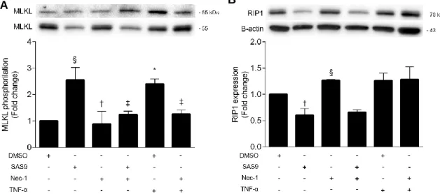
To further dissect the mechanisms responsible for SAS9-induced necroptosis, L929 cells were incubated with 17 µM SAS9 (or no addition) in the presence or absence of 30 μM Nec-1 for 8 h. 30 μM TNF-α was used as a positive control. Results showed that, similarly to TNF-α, SAS9 significantly induced MLKL phosphorylation, compared with control (p < 0.05 for SAS9 and p <0.01 for TNF-α; Figure 10A). In addition, co-incubation with Nec-1 almost completely abrogated this effect (p<0.01), suggesting that SAS9 strongly induces necroptosis in L929 cells. Intriguingly, RIP1 levels were decreased upon incubation of L929 cells with SAS9 alone or in combination with Nec-1. While these results could strengthen the notion that SAS9-induced necroptosis might be RIP1-independent, it is worth to mention that the structure of SAS9 is based on the indole group of Nec-1 and, as such, it could be that SAS9 is interacting with RIP1 and activating its kinase activity. However, the fact that incubation of cells with TNF-α also failed to significantly increase RIP1 protein expression levels (Figure 10B) may also indicate that RIP1 phosphorylation and/or sequestration in the insoluble protein fractions is determining execution of necroptosis. Last but not least, it might also be that SAS9 is inducing such high levels of cell death that protein quantitation through Western blot is underestimated. Additional experiments should be performed to fully assess all of these hypotheses.
Figure 10 - SAS9 increases MLKL phosphorylation in L929 cells but not RIP1 expression.
L929 cells were incubated with 17 µM SAS9 in the presence or absence of 30 μM Nec-1. TNF-α was used as a positive control. Total protein fractions were prepared for Western blot analysis of p-MLKL, MLKL, Rip1 and β-actin. Representative immunoblots are shown. Results are presented as the mean value ± SEM for at least three independent experiments. *p < 0.01 and §p < 0.05 vs. control; ‡p < 001 and †p < 0.05 vs. TNF-α.
The role of necroptosis in different pathologies is becoming increasingly evident. Cell death by necroptosis can contribute to disease, for which pharmacological inhibitors of necroptosis could be developed. In turn, many cancer cell types are resistant to pro-apoptotic chemotherapy and, as such, inducing those cells to undergo necroptosis would be advantageous. This is true for colon cancer, for which current treatments have a low response rate, with patients quickly developing resistance to therapy. Further, colon cancer prevalence is increasing and it currently stands as one of the major causes of cancer mortality around the world.
Despite the current absence of pro-necroptotic therapies in cancer, it is well established that cancer cell lines can undergo necroptosis (Su et al., 2016). Necroptosis is a form of regulated cell death that differs from apoptosis by being independent from caspases (Vercammen et al., 1998a). Most of the investigation surrounding necroptosis has focused on its pathological role (Weinlich et al., 2016), highlighting the potential benefits of its inhibition (Tamura et al., 2011; Zhao et al., 2015a; Zhu et al., 2012). However, several studies show that induction of necroptosis can be used to kill cancer cells or to stimulate an immune response against the tumour (Aaes et al., 2016; Cells et al., 2015; Fulda, 2013). Induction of necroptosis stands as a potential strategy to overcome apoptosis resistance, as the necroptosis machinery largely differs from the apoptotic signalling cascade. Still, several concerns are raised. One of the main concerns relates with the selectivity of necroptosis inducers towards cancer cells and not normal cells. Combining the inducers with tumour-guided drugs or tumour-targeting antibodies might overcome this limitation (Su et al., 2016). Another limitation may be the release of DAMPs by necroptotic cancer cells, which can act as a trigger of inflammation (Pasparakis and Vandenabeele, 2015). In turn, an inflammatory microenvironment may promote tumour development (Mantovani et al., 2008). Nonetheless, several studies have been published describing novel modulators of necroptosis with potential clinical use. Nec-1 was the first identified necroptosis inhibitor, directed towards RIP1 activity (Degterev et al., 2005). Several additional inhibitors of necroptosis were reported afterwards, including RIP3 and MLKL inhibitors (Kopalli et al., 2016). Still, they all possess some limitations. Nec-1 for instance, one of the most effective inhibitors of necroptosis displays inadequate pharmacokinetic properties and appears not to be RIP1-specific (Liao et al., 2014; Tamura et al., 2011; Zhang et al., 2016). NSA would likely constitute a good inhibitor of necroptosis because it specifically inhibits MLKL, one of the most downstream elements of the necroptosis pathway. However, it only acts in
human MLKL, thus precluding preclinical testing (Liao et al., 2014). Altogether, the discovery of novel, specific pharmacological inhibitors of necroptosis is still a priority. In this thesis, we screened a library of 67 compounds for their ability to inhibit TNF-α-induced cell death in L929 cells. L929 cells have been previously demonstrated to undergo necroptosis in the presence of TNF-α alone, thus constituting a simple but reliable model to test necroptosis (Vanlangenakker et al., 2011).
From this screening we undercovered 7 potential necroptosis inhibitors, which inhibited TNF-α induced cell death by more than 70%, when compared with Nec-1 at 100%. We next calculated the EC50 values for these compounds, and obtained numbers ranging from 9.43µM (MS-PSR90) up to >100µM (MS-PSR82 and CGABoc). MS-PSR90 was then confirmed to inhibit necroptosis, as seen by its inhibition of TNF-α-induced MLKL S358 phosphorylation. Despite the fact that none of the compounds was as effective as Nec-1 in inhibiting necroptosis, it might still be worthwhile to design other molecules based on the MS-PSR90 scaffold to investigate whether its anti-necroptotic properties or EC50 improve.
Interestingly, one of the compounds screened - SAS9 - was found to further potentiate TNF-α-induced necroptosis. To further explore the mechanisms behind its cytotoxicity, SAS9 IC50 was calculated in both L929 cells and primary mouse hepatocytes. Because L929 are cancer cells, it was thought that these would be more resistant to SAS9-induced cell death. However, the IC50 for SAS9 in L929 and primary mouse hepatocyte cells was 16.96 µM and >50 µM, respectively, suggesting that mouse primary hepatocytes are somehow more resistant to SAS9-induced cell death. This may relate with the fact that primary mouse hepatocytes do not express all the components of the necroptotic machinery under normal conditions (Afonso et al., 2015). HT29, a human colon cancer cell line, expresses all the necroptosis machinery and has distinct apoptosis and necroptosis cell death processes, although both types of cell death may occur at the same time (Wilson and Browning, 2002). Also due to our interest in testing novel potential modulators of necroptosis for future use in colon cancer, we next calculated SAS9 IC50 in HT29 cells, known to be resistant to many of the current cancer therapies (Hu et al., 2016). Results showed that SAS9 is able to kill HT29 cells with an IC50 value of 49.73 µM. To confirm specificity of SAS9 for necroptosis and its machinery, and/or its targeted cytotoxicity in cancer cells, additional experiments need to be performed using multiple non-cancer cells and cancer cells. Still, we next tried to evaluate whether SAS9-induced cell death also encompassed apoptosis, by co-incubating L929 and HT29 cells with SAS9 plus apoptosis inhibitor z-VAD-fmk.
Indeed, SAS9 appears to induce both apoptosis and necroptosis in L929 cells; Nec-1 almost completely reversed SAS9-induced cell death but not z-VAD.fmk, with SAS9 simultaneously inducing significant caspase-3/7 activity. Intriguingly, SAS9-induced caspase activation was reduced in the presence of Nec-1; in that regard, at least one study has shown that Nec-1 may also reduce apoptotic cell death (Wang et al., 2012), justifying these results. These results are important because in many cases, cancer cells develop resistance to pro-apoptotic agents, in which case SAS9 would still be able to kill cells by necroptosis. In cases where cancer cells do not have necroptotic machinery, SAS9 would be able to kill by apoptosis (Su et al., 2016). Of note, cell death mechanisms induced by SAS9 appear to be distinct in HT29 cells. First off, TNF-α alone is not sufficient to induce necroptosis in HT29 cells (Wilson and Browning, 2002). Only when in the presence of z-VAD-fmk and SMAC mimetic BV6 is cell death induced. Our results suggest that, similarly, SAS9 alone induces apoptosis-like cell death while, in the presence of z-VAD-fmk and BV6, SAS9-induced cell death switch to necroptosis. Of note, Nec-1 fails to completely inhibit SAS9-induced cell death; because Nec-1 is a RIP1 inhibitor, one possible explanation would be that SAS9 activates necroptosis independently of RIP1, as previously shown in different cellular contexts (Moujalled et al., 2013).
To further elucidate this and go deeper on the mechanisms of necroptosis induced by SAS9, immunoblotting of total RIP1 expression levels and MLKL S358 phosphorylation in L929 cells was performed. Although SAS9 was confirmed to induce phosphorylation of MLKL, RIP1 levels were decreased by SAS9 alone or in the presence of Nec-1 when compared with control or TNF-α induced necroptosis. One justification for the fact that RIP1 is downregulated with SAS9 but there is still necroptosis is that necroptosis can happen in a RIP1-independent way, whereas under certain conditions RIP1 can act as an inhibitor of necroptosis (Kearney et al., 2014). Still, further studies are needed to confirm the hypothesis that SAS9 is interacting with RIP1. Knockout of RIP1 can show if SAS9-induced necroptosis is possible in the absence of RIP1.