Avaliação de Tecnologias em
Saúde e o Impacto na
Imagem Médica
Luís Lança, Ph.D.
ESTeSL
Lisboa/Portugal
Sumário
1. Avaliação de Tecnologias em Saúde (ATS)
2. Métodos para a ATS na imagem médica
3. Influência da ATS na imagem médica
Objetivos
– Definir o conceito de Avaliação de Tecnologias em
Saúde (ATS)
– Identificar métodos de pesquisa para ATS no âmbito
da imagem médica
– Entender o valor de revisões sistemáticas e
meta-análise em ATS
– Compreender a influência da ATS na imagem
médica
Introdução
– Os sistemas de saúde em todo o mundo, estão
confrontados com o desafio de como gerir a
prestação de cuidados de saúde, em contexto de
restrição de recursos
– Há uma necessidade de maximizar o impacto
positivo das intervenções de cuidados de saúde na
saúde da população
Prestação de
cuidados de
saúde em
contexto de
restrição de
recursos
Aumento do
número de
exames
radiológicos
Introdução
– O paradigma da ATS surgiu como uma resposta às
perguntas dos decisores sobre a difusão
incontrolada de equipamento médico dispendioso
– A ATS começou no início dos anos 1970, quando a
rápida demanda por tomografia computadorizada
se tornou uma questão de política pública devido
ao seu elevado custo
Jonsson E, Banta D. Management of health technologies: an international view. British Medical Journal, 1999, 319(7220):1293.
Questões
– O que é a Avaliação de Tecnologias em Saúde
(ATS)?
– Que métodos de pesquisa estão disponíveis para a
ATS na imagem médica?
– Como é que a ATS pode influenciar a imagem
médica?
O que é a ATS?
– Qualquer intervenção que pode ser utilizada para
promover a saúde, prevenir, diagnosticar ou tratar a
doença, ou para a reabilitação, ou cuidados de longa
duração
– Engloba os dispositivos médicos, desde os mais simples
aos mais sofisticados sistemas de imagem médica;
medicamentos; procedimentos médicos e cirúrgicos e os
sistemas organizacionais e de suporte, em que esses
cuidados são prestados
O que é a ATS?
– A avaliação sistemática das propriedades, efeitos e/ou impactos
da tecnologia nos cuidados de saúde
– Pode tratar das consequências directas das tecnologias, bem
como as suas consequências não intencionais, indiretas
– O seu principal objetivo é informar acerca da formulação de
políticas relacionadas com a tecnologia na área da saúde
– É conduzida por grupos interdisciplinares utilizando quadros
analíticos explícitos a partir de uma variedade de métodos
Porquê a ATS?
– Pense numa tecnologia de imagem que
teve uma evolução tecnológica
assinalável nos últimos anos:
• Conseguimos entender o que eram a
2014 – TC-CE de perfusão
com mapa de côr
1971: primeira TC diagnóstica:
Atkinson Morley's Hospital
A avaliação de tecnologias em saúde e a
difusão de tecnologias em saúde
19 WHO Medical device technical series
system, and slowing the uptake of
technologies that seem promising but
have persistent uncertainties.
For some, especially industry, HTA is
perceived as a hurdle to the introduction
of innovative technologies into the
health system. In this perspective, HTA
is sometimes classifi ed as the “fourth
hurdle”, after the assessment of safety,
efficacy and quality that are part of
the regulatory requirements in many
countries
(17). This view of HTA as an
additional hurdle is sometimes used as
a synonym for an additional requirement
of demonstrating cost-effectiveness
for coverage decisions
(18). The term
“fourth hurdle” is most commonly used
in relation to the coverage of new drugs,
where it becomes increasingly evident
that not only cost-effectiveness, but also
budget impact is an important dimension
in decision-making
(19). However, it is
becoming increasingly clear that HTA can
be a highly benefi cial additional step for
moving technologies from the laboratory
to the bedside (20).
There is general concern among medical
device manufacturers about the timing
of HTAs in relation to the innovation
process. The 2008 Eucomed position
paper on HTA states that:
Policy makers should consider
the implications of HTA on the
environment needed to foster
innovation of medical devices. If
HTA introduces significant new
challenges to market entry then there
is a potential that this may impact on
the rate of innovation in the device
sector which already faces a number
of challenges. Intellectual property
associated with medical devices
is less well protected than patents
on new medical compounds. In
addition to this, medical device
development is characterized by
iterative improvement of technologies
resulting in a more rapid life-cycle
and increased competition
(21).
Indeed, industry is one of the main
contributors to innovation in health
technology. However, the innovation
process goes beyond the development
of a new device for regulatory approval.
Innovation comprises both invention and
exploitation, meaning an invention only
Figure 6. Health technology assessment and diffusion of health technologies
A figura mostra o ciclo de vida natural das tecnologias em saúde
Qual é a função da ATS?
– Tecnologia em Saúde é tudo aquilo que pode ser utilizado em
procedimentos e processos médicos
• i.e. medicamentos, dispositivos, equipamentos e acessórios,
procedimentos médicos e cirúrgicos, sistemas de apoio e sistemas
organizacionais e de gestão
Qual é a função da ATS?
– Inclui a avaliação da eficácia e eficiência dos equipamentos e
das técnicas
• Eficácia - obtenção da melhoria da saúde pela aplicação da ciência e da
tecnologia nas condições mais favoráveis (controladas)
• Eficiência - Capacidade em reduzir os custos dos cuidados, sem diminuir
a efectividade destes
– Compreende vários métodos para avaliar tecnologias em
saúde
Probst, H., & Brealey, S. (2010). Health technology assessment. In A. Ramlaul (Ed.), Medical Imaging and Radiotherapy Research - Skills and Strategies. London: Churchill Livingstone - Elsevier.
Technical performance
• Does MRI reliably result in good quality images which
are anatomically representative?
Diagnostic performance
• Do the images produced allow accurate diagnoses to
be made?
Diagnostic impact
• Does MRI change diagnostic confidence and displace
other investigations?
Therapeutic impact
• Do the results of MRI contribute to planning and
delivery of therapy?
Patient outcome
• Does the use of MRI contribute to the improved health
of a patient?
Societal
• Is the cost (borne by the society as a whole) of MRI
acceptable?
Hierarquia para
avaliação da eficácia
das tecnologias de
diagnóstico:
exemplo da RM
Adapted from:Probst, H., & Brealey, S. (2010). Health technology assessment. In A. Ramlaul (Ed.), Medical Imaging and Radiotherapy Research - Skills and Strategies. London: Churchill Livingstone - Elsevier.
Métodos para ATS em imagem
médica
– Tipologias
• Testes de diagnóstico
• Ensaios clínicos aleatórios
• Avaliação económica
• Revisões sistemáticas e meta-análise
Adapted from:Probst, H., & Brealey, S. (2010). Health technology assessment. In A. Ramlaul (Ed.), Medical Imaging and Radiotherapy Research - Skills and Strategies. London: Churchill Livingstone - Elsevier.
Testes de
diagnóstico
COMPUTED TOMOGRAPHY
Comparing five different iterative reconstruction algorithms
for computed tomography in an ROC study
Kristin Jensen&Anne Catrine T. Martinsen&
Anders Tingberg&Trond Mogens Aaløkken&Erik Fosse
Received: 15 January 2014 /Revised: 1 July 2014 / Accepted: 8 July 2014 # European Society of Radiology 2014
Abstract
Objectives The purpose of this study was to evaluate lesion conspicuity achieved with five different iterative reconstruc-tion techniques from four CT vendors at three different dose levels. Comparisons were made of iterative algorithm and filtered back projection (FBP) among and within systems. Methods An anthropomorphic liver phantom was examined with four CT systems, each from a different vendor. CTDIvol
levels of 5 mGy, 10 mGy and 15 mGy were chosen. Images were reconstructed with FBP and the iterative algorithm on the system. Images were interpreted independently by four observers, and the areas under the ROC curve (AUCs) were calculated. Noise and contrast-to-noise ratios (CNR) were measured.
Results One iterative algorithm increased AUC (0.79, 0.95, and 0.97) compared to FBP (0.70, 0.86, and 0.93) at all dose levels (p<0.001 and p=0.047). Another algorithm increased AUC from 0.78 with FBP to 0.84 (p=0.007) at 5 mGy. Differences at 10 and 15 mGy were not significant (p-values:
0.084–0.883). Three algorithms showed no difference in AUC compared to FBP (p-values: 0.008–1.000). All of the algo-rithms decreased noise (10–71 %) and improved CNR. Conclusions Only two algorithms improved lesion detection, even though noise reduction was shown with all algorithms. Key Points
• Iterative reconstruction algorithms affected lesion detection differently at different dose levels.
• One iterative algorithm improved lesion detectability com-pared to filtered back projection.
• Three algorithms did not significantly improve lesion detectability.
• One algorithm improved lesion detectability at the lowest dose level.
Keywords Computed tomography . Image reconstruction . Radiological phantom . Liver
Introduction
In order to improve visualization of pathology without in-creasing radiation exposure to the patient, CT vendors have developed new reconstruction techniques such as iterative reconstruction algorithms. According to the vendors, iterative reconstruction improves image quality, and thereby radiation dose can be reduced compared to the standard reconstruction technique, filtered back projection (FBP), [1–4]. The new reconstruction techniques may have an effect on image texture and diagnostic image quality [5–8], however, and there may be inter-vendor differences. Therefore, it is important to test new techniques before implementing them in clinical routine. FBP has been the primary image reconstruction technique in CT [9,10]. Simplifications in technique have improved speed and reduced power consumption, but artefacts and
K. Jensen (*)
:
A. C. T. Martinsen:
E. FosseThe Intervention Centre, Rikshospitalet, Postboks 4950, Nydalen, 0424 Oslo, Norway
e-mail: uxjekr@ous-hf.no K. Jensen
:
A. C. T. Martinsenlnstitute of Physics, University of Oslo, 0027 Oslo, Norway A. Tingberg
Department of Medical Radiation Physics, Lund University, Skåne University Hospital, 205 02 Malmö, Sweden
T. M. Aaløkken
Department of Radiology and Nuclear Medicine, Rikshospitalet, Postboks 4950, Nydalen, 0424 Oslo, Norway
E. Fosse
lnstitute of Clinical Medicine, University of Oslo, 0027 Oslo, Norway
Eur Radiol
DOI 10.1007/s00330-014-3333-4
COMPUTED TOMOGRAPHY
Comparing five different iterative reconstruction algorithms
for computed tomography in an ROC study
Kristin Jensen
&
Anne Catrine T. Martinsen
&
Anders Tingberg
&
Trond Mogens Aaløkken
&
Erik Fosse
Received: 15 January 2014 /Revised: 1 July 2014 / Accepted: 8 July 2014
# European Society of Radiology 2014
Abstract
Objectives The purpose of this study was to evaluate lesion
conspicuity achieved with five different iterative
reconstruc-tion techniques from four CT vendors at three different dose
levels. Comparisons were made of iterative algorithm and
filtered back projection (FBP) among and within systems.
Methods An anthropomorphic liver phantom was examined
with four CT systems, each from a different vendor. CTDI
vollevels of 5 mGy, 10 mGy and 15 mGy were chosen. Images
were reconstructed with FBP and the iterative algorithm on
the system. Images were interpreted independently by four
observers, and the areas under the ROC curve (AUCs) were
calculated. Noise and contrast-to-noise ratios (CNR) were
measured.
Results One iterative algorithm increased AUC (0.79, 0.95,
and 0.97) compared to FBP (0.70, 0.86, and 0.93) at all dose
levels (p<0.001 and p=0.047). Another algorithm increased
AUC from 0.78 with FBP to 0.84 (p=0.007) at 5 mGy.
Differences at 10 and 15 mGy were not significant (p-values:
0.084–0.883). Three algorithms showed no difference in AUC
compared to FBP (p-values: 0.008–1.000). All of the
algo-rithms decreased noise (10–71 %) and improved CNR.
Conclusions Only two algorithms improved lesion detection,
even though noise reduction was shown with all algorithms.
Key Points
• Iterative reconstruction algorithms affected lesion detection
differently at different dose levels.
• One iterative algorithm improved lesion detectability
com-pared to filtered back projection.
• Three algorithms did not significantly improve lesion
detectability.
• One algorithm improved lesion detectability at the lowest
dose level.
Keywords Computed tomography . Image reconstruction .
Radiological phantom . Liver
Introduction
In order to improve visualization of pathology without
in-creasing radiation exposure to the patient, CT vendors have
developed new reconstruction techniques such as iterative
reconstruction algorithms. According to the vendors, iterative
reconstruction improves image quality, and thereby radiation
dose can be reduced compared to the standard reconstruction
technique, filtered back projection (FBP), [
1
–
4
]. The new
reconstruction techniques may have an effect on image texture
and diagnostic image quality [
5
–
8
], however, and there may
be inter-vendor differences. Therefore, it is important to test
new techniques before implementing them in clinical routine.
FBP has been the primary image reconstruction technique
in CT [
9
,
10
]. Simplifications in technique have improved
speed and reduced power consumption, but artefacts and
K. Jensen (*)
:
A. C. T. Martinsen
:
E. Fosse
The Intervention Centre, Rikshospitalet, Postboks 4950, Nydalen,
0424 Oslo, Norway
e-mail: uxjekr@ous-hf.no
K. Jensen
:
A. C. T. Martinsen
lnstitute of Physics, University of Oslo, 0027 Oslo, Norway
A. Tingberg
Department of Medical Radiation Physics, Lund University, Skåne
University Hospital, 205 02 Malmö, Sweden
T. M. Aaløkken
Department of Radiology and Nuclear Medicine, Rikshospitalet,
Postboks 4950, Nydalen, 0424 Oslo, Norway
E. Fosse
lnstitute of Clinical Medicine, University of Oslo, 0027 Oslo,
Norway
Eur Radiol
DOI 10.1007/s00330-014-3333-4
Jensen, K., Martinsen, A. C. T., Tingberg, A., Aaløkken, T. M., & Fosse, E. (2014). Comparing five different iterative reconstruction algorithms for computed tomography in an ROC study. European Radiology. doi:10.1007/s00330-014-3333-4
COMPUTED TOMOGRAPHY
Comparing five different iterative reconstruction algorithms
for computed tomography in an ROC study
Kristin Jensen&Anne Catrine T. Martinsen&
Anders Tingberg&Trond Mogens Aaløkken&Erik Fosse
Received: 15 January 2014 /Revised: 1 July 2014 / Accepted: 8 July 2014 # European Society of Radiology 2014
Abstract
Objectives The purpose of this study was to evaluate lesion conspicuity achieved with five different iterative reconstruc-tion techniques from four CT vendors at three different dose levels. Comparisons were made of iterative algorithm and filtered back projection (FBP) among and within systems. Methods An anthropomorphic liver phantom was examined with four CT systems, each from a different vendor. CTDIvol
levels of 5 mGy, 10 mGy and 15 mGy were chosen. Images were reconstructed with FBP and the iterative algorithm on the system. Images were interpreted independently by four observers, and the areas under the ROC curve (AUCs) were calculated. Noise and contrast-to-noise ratios (CNR) were measured.
Results One iterative algorithm increased AUC (0.79, 0.95, and 0.97) compared to FBP (0.70, 0.86, and 0.93) at all dose levels (p<0.001 and p=0.047). Another algorithm increased AUC from 0.78 with FBP to 0.84 (p=0.007) at 5 mGy. Differences at 10 and 15 mGy were not significant (p-values:
0.084–0.883). Three algorithms showed no difference in AUC compared to FBP (p-values: 0.008–1.000). All of the algo-rithms decreased noise (10–71 %) and improved CNR. Conclusions Only two algorithms improved lesion detection, even though noise reduction was shown with all algorithms. Key Points
• Iterative reconstruction algorithms affected lesion detection differently at different dose levels.
• One iterative algorithm improved lesion detectability com-pared to filtered back projection.
• Three algorithms did not significantly improve lesion detectability.
• One algorithm improved lesion detectability at the lowest dose level.
Keywords Computed tomography . Image reconstruction . Radiological phantom . Liver
Introduction
In order to improve visualization of pathology without in-creasing radiation exposure to the patient, CT vendors have developed new reconstruction techniques such as iterative reconstruction algorithms. According to the vendors, iterative reconstruction improves image quality, and thereby radiation dose can be reduced compared to the standard reconstruction technique, filtered back projection (FBP), [1–4]. The new reconstruction techniques may have an effect on image texture and diagnostic image quality [5–8], however, and there may be inter-vendor differences. Therefore, it is important to test new techniques before implementing them in clinical routine. FBP has been the primary image reconstruction technique in CT [9,10]. Simplifications in technique have improved speed and reduced power consumption, but artefacts and
K. Jensen (*)
:
A. C. T. Martinsen:
E. FosseThe Intervention Centre, Rikshospitalet, Postboks 4950, Nydalen, 0424 Oslo, Norway
e-mail: uxjekr@ous-hf.no K. Jensen
:
A. C. T. Martinsenlnstitute of Physics, University of Oslo, 0027 Oslo, Norway A. Tingberg
Department of Medical Radiation Physics, Lund University, Skåne University Hospital, 205 02 Malmö, Sweden
T. M. Aaløkken
Department of Radiology and Nuclear Medicine, Rikshospitalet, Postboks 4950, Nydalen, 0424 Oslo, Norway
E. Fosse
lnstitute of Clinical Medicine, University of Oslo, 0027 Oslo, Norway
Eur Radiol
ATS com ensaios clínicos aleatórios
– Definem geralmente:
• O objetivo do estudo
• Como os participantes são expostos à intervenção
• O número de participantes
• Como a intervenção é avaliada
Probst, H., & Brealey, S. (2010). Health technology assessment. In A. Ramlaul (Ed.), Medical Imaging and Radiotherapy Research - Skills and Strategies. London: Churchill Livingstone - Elsevier.
Sierink J, Saltzherr TP, et al, (2012) A multicenter, randomized controlled trial of iimmediate total-body CT scanning in trauma patients (REACT-2). Emergency Medicine, 12:4,
ATS com
ensaios
clínicos
aleatórios
Avaliação económica
– Lidar com os custos e resultados das
atividades
– A finalidade básica de uma avaliação
económica é identificar, medir, avaliar e
comparar os custos e as alternativas
Probst, H., & Brealey, S. (2010). Health technology assessment. In A. Ramlaul (Ed.), Medical Imaging and Radiotherapy Research - Skills and Strategies. London: Churchill Livingstone - Elsevier.
Avaliação
económica
(exemplo)
Westwood, M., Al, M., Burgers, L., Redekop, K., Lhachimi, S., Armstrong, N., … Kleijnen, J. (2013). A systematic review and economic evaluation
of new-generation computed tomography scanners for imaging in coronary artery disease and congenital heart disease: Somatom Definition Flash, Aquilion ONE, Brilliance iCT and Discovery CT750 HD. Health Technology Assessment, 17(9), 1–243.
NIHR Journals Library
vi Abstract
97.7% [95% confidence interval (CI) 88.0% to 99.9%], 97.7% (95% CI 93.2% to 99.3%) and 96.0% (95% CI 88.8% to 99.2%) for patients with arrhythmias, high heart rates and previous stent, respectively. The corresponding estimates of specificity were 81.7% (95% CI 71.6% to 89.4%), 86.3% (95% CI 80.2% to 90.7%) and 81.6% (95% CI 74.7% to 87.3%), respectively. In patients with high coronary calcium scores, previous bypass grafts or obesity, only per-segment or per-artery data were available. Sensitivity estimates remained high (> 90% in all but one study). In patients with suspected CAD, the NGCCT-only strategy appeared most cost-effective; the incremental cost-effectiveness ratio (ICER) of NGCCT–ICA compared with NGCCT only was £71,000. In patients with known CAD, the most cost-effective strategy was NGCCT–ICA (highest cost saving, dominates ICA only). The ICER of NGCCT only compared with NGCCT–ICA was £726,230. For radiation
exposure only, the ICER for NGCCT compared with 64-slice CT in congenital heart disease ranged from £521,000 for the youngest patients to £90,000 for adults.
Limitations: Available data were limited, particularly for obese patients and patients with previous bypass grafts. All studies of the accuracy of NGCCT assume that the reference standard (ICA) is 100% sensitive and specific; however, there is some evidence that ICA may sometimes underestimate the extent and severity of stenosis. Patients with more than one criterion that could contribute to difficulty in imaging were often excluded from studies; the effect on test accuracy of multiple difficult to image criteria remains uncertain.
Conclusions: NGCCT may be sufficiently accurate to diagnose clinically significant CAD in some or all difficult-to-image patient groups. Economic analyses suggest that NGCCT is likely to be considered cost-effective for difficult-to-image patients with CAD, at current levels of willingness to pay in the NHS. For patients with suspected CAD, NGCCT only would be most favourable; for patients with known CAD, NGCCT–ICA would be most favourable. No studies assessing the effects of NGCCT on therapeutic decision making, or subsequent patient outcomes, were identified. The ideal study to address these questions would be a large multi-centre RCT. However, one possible alternative might be to establish a multicentre tracker study. High-quality test accuracy studies, particularly in obese patients, patients with high coronary calcium, and those with previous bypass grafts are needed to confirm the findings of our systematic review. These studies should include patients with multiple difficult to image criteria.
Funding: The National Institute for Health Research Health Technology Assessment programme. This project was funded by the HTA programme, on behalf of NICE, as project number 10/107/01.
DOI 10.3310/HTA17090
HEALTH TECHNOLOGY ASSESSMENT
VOLUME 17 ISSUE 9 MARCH 2013 ISSN 1366-5278
A systematic review and economic evaluation of
new-generation computed tomography scanners for
imaging in coronary artery disease and congenital
heart disease: Somatom Definition Flash,
Aquilion ONE, Brilliance iCT and Discovery CT750 HD
M Westwood, M Al, L Burgers, K Redekop, S Lhachimi, N Armstrong,
H Raatz, K Misso, J Severens and J Kleijnen
Chapter 6 Conclusions
Implications for service provision Suggested research priorities
Acknowledgements Contributions of authors
References
Appendix 1
Literature search strategies
Clinical effectiveness search strategies Electronic searching of conference abstracts Cost-effectiveness search
Guidelines search
Appendix 2
Study-specific guide to completion of QUADAS-2 Domain 1: patient selection
Domain 2: index test
Domain 3: reference standard Domain 4: flow and timing
Appendix 3
Quality assessment: QUADAS-2 results Study ID: Alkadhi 200841
Study ID: Brodoefel 200846
Study ID: Brodoefel 200842
Study ID: de Graaf 201040
Study ID: LaBounty 201038
Study ID: Leber 200743
Health Technology Assessment 2013; Vol. 17: No. 9
DOI 10.3310/HTA17090
HEALTH TECHNOLOGY ASSESSMENT
VOLUME 17 ISSUE 9 MARCH 2013 ISSN 1366-5278
A systematic review and economic evaluation of
new-generation computed tomography scanners for
imaging in coronary artery disease and congenital
heart disease: Somatom Definition Flash,
Aquilion ONE, Brilliance iCT and Discovery CT750 HD
M Westwood, M Al, L Burgers, K Redekop, S Lhachimi, N Armstrong,
H Raatz, K Misso, J Severens and J Kleijnen
Chapter 6 Conclusions
Implications for service provision Suggested research priorities
Acknowledgements Contributions of authors
References
Appendix 1
Literature search strategies
Clinical effectiveness search strategies Electronic searching of conference abstracts Cost-effectiveness search
Guidelines search
Appendix 2
Study-specific guide to completion of QUADAS-2 Domain 1: patient selection
Domain 2: index test
Domain 3: reference standard Domain 4: flow and timing
Appendix 3
Quality assessment: QUADAS-2 results Study ID: Alkadhi 200841
Study ID: Brodoefel 200846
Study ID: Brodoefel 200842
Study ID: de Graaf 201040
Study ID: LaBounty 201038
Study ID: Leber 200743
Revisões sistemáticas e
meta-análise
– O que é uma revisão sistemática?
– O que é uma meta-análise?
Hierarquia de tipos de estudo
Meta-analysis
Systematic review
Randomized controlled trial
Cohort study
Case control study
Case series/case reports/expert opinion
Animal research
In vitro research
Hierarquia da evidência
Adapted from:
Marshall, G., & Sykes, A. E. (2011). Systematic reviews: A guide for radiographers and other health care professionals. Radiography, 17(2), 158–164. Haidich, A.-B. (2010). Meta-analysis in medical research. Hippokratia, 14(Suppl 1), 29–37.
Revisões sistemáticas e
meta-análise – princípios básicos comuns
- Identificar literatura relevante - selecione artigos
relevantes
- Avaliar criticamente os artigos
- Identificar padrões gerais de resultados
- Identificar discordâncias cruciais e controvérsias
- Propor explicações válidas para as discordâncias
- Fornecer um resumo claro sobre o estado-da-arte
Revisões sistemáticas e
meta-análise – princípios
básicos comuns
• Identificar literatura
relevante -
selecione artigos
relevantes
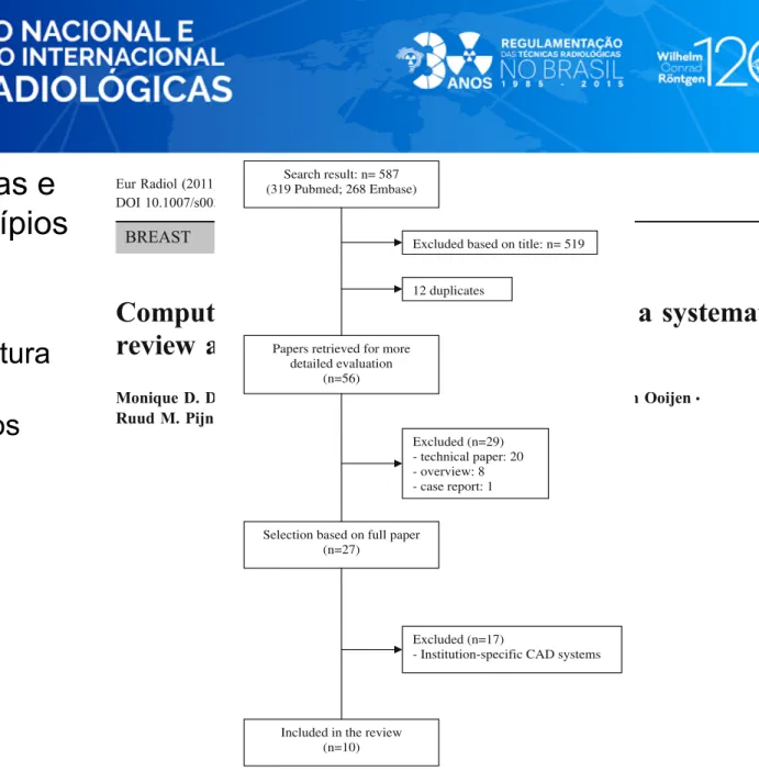
Dorrius, M. D., Jansen-van der Weide, M. C., van Ooijen, P. M. a, Pijnappel, R. M., & Oudkerk, M. (2011). Computer-aided detection in breast MRI: a systematic review and meta-analysis. European Radiology, 21(8), 1600–8.
BREAST
Computer-aided detection in breast MRI: a systematic
review and meta-analysis
Monique D. Dorrius&Marijke C. Jansen-van der Weide&Peter M. A. van Ooijen&
Ruud M. Pijnappel&Matthijs Oudkerk
Received: 10 September 2010 / Revised: 1 December 2010 / Accepted: 12 January 2011 / Published online: 15 March 2011 # The Author(s) 2011. This article is published with open access at Springerlink.com
Abstract
Objectives To evaluate the additional value of computer-aided detection (CAD) in breast MRI by assessing radiol-ogists’ accuracy in discriminating benign from malignant breast lesions.
Methods A literature search was performed with inclusion of relevant studies using a commercially available CAD system with automatic colour mapping. Two independent researchers assessed the quality of the studies. The accuracy of the radiologists’ performance with and without CAD was presented as pooled sensitivity and specificity.
Results Of 587 articles, 10 met the inclusion criteria, all of good methodological quality. Experienced radiologists reached comparable pooled sensitivity and specificity before and after using CAD (sensitivity: without CAD: 89%; 95% CI: 78–94%, with CAD: 89%; 95%CI: 81–94%) (specificity: without CAD: 86%; 95% CI: 79–91%, with CAD: 82%; 95% CI: 76–87%). For residents the pooled sensitivity increased from 72% (95% CI: 62–81%) without CAD to 89% (95% CI: 80–94%) with CAD, however, not significantly. Concerning specificity, the results were similar (without CAD: 79%; 95% CI: 69–86%, with CAD: 78%; 95% CI: 69–84%).
Conclusions CAD in breast MRI has little influence on the sensitivity and specificity of experienced radiologists and therefore their interpretation remains essential. However, residents or inexperienced radiologists seem to benefit from CAD concerning breast MRI evaluation.
Keywords Magnetic resonance imaging . Breast . Computer aided detection . CAD . Meta-analysis Introduction
Dynamic contrast-enhanced Magnetic Resonance Imaging (MRI) is increasingly used to evaluate pathological features of the breast. Applications for MRI of the breast include diagnostic and screening indications [1–6]. Image analysis is based on the enhancement pattern of lesions in dynamic breast MRI and on morphological characteristics [7–9]. Using those two criteria for the interpretation of the images, breast MRI has a very high sensitivity, which usually exceeds 90% [10–12] and a negative breast MRI shows a sufficient high negative predictive value (NPV) (97%) to safely rule out malignancy [13–15]. However, breast MRI has several limitations, the overall reported specificity varies between 67% and 72%, which therefore results in a high number of false-positive results [10,12,16]. Further-more, MRI requires significant time for image acquisition, processing and interpretation [17, 18]. In order to try to overcome those limitations, Computer Aided Detection (CAD) programs for MR imaging of the breast have been developed [18]. In general, CAD software was developed to identify suspect features on the image and bring them to the attention of the radiologist, in order to decrease false-negative readings [19]. However, in breast MRI, most lesions were regarded as having already been detected by the radiologist. Therefore, the primary aim to develop CAD for breast MRI was not to identify lesions, but to assist the radiologist in determining which lesions are benign and which are malignant.
Computer-aided detection systems automate many pro-cessing and analysis functions, which would normally have
M. D. Dorrius (*)
:
M. C. J.-v. der Weide:
P. M. A. van Ooijen:
R. M. Pijnappel:
M. OudkerkDepartment of Radiology, Center for Medical Imaging, University Medical Center Groningen,
Hanzeplein 1, PO box 30.001, 9700 RB Groningen, the Netherlands
e-mail: m.d.dorrius@rad.umcg.nl Eur Radiol (2011) 21:1600–1608 DOI 10.1007/s00330-011-2091-9
summary sensitivity and specificity were calculated, and a summary ROC curve was drawn (with AUC and confi-dence intervals). A forest plot was generated containing the individual study sensitivities and specificities with 95% confidence intervals (CI) and the pooled sensitivity and specificity estimates.
A test for heterogeneity was applied, using the I2statistic
[32]. This statistic calculates the percentage of total variation across studies that can be attributed to inter-study heterogeneity, ranging from 0 (no heterogeneity) to 100% (all variance due to heterogeneity). The presence of publication bias was visually assessed by producing a funnel plot. In STATA linear regression was performed of log odds ratios on the inverse root of effective sample sizes as a test for funnel plot asymmetry. The log odds ratios are defined as the log transformed diagnostic odds ratios, which are needed for the performance of linear regression.
Publication bias was considered present if there was a significant non-zero slope coefficient, (p<0.10), suggesting that only the small studies reporting a high sensitivity with CAD had been published, whereas the small studies reporting a lower sensitivity had not been published. Data were analysed in SPSS 16.0 (SPSS, Chicago, IL, USA), Meta Disc [33] and STATA SE version 11.0 (STATA, College Station, TX, USA).
Results
Study descriptives
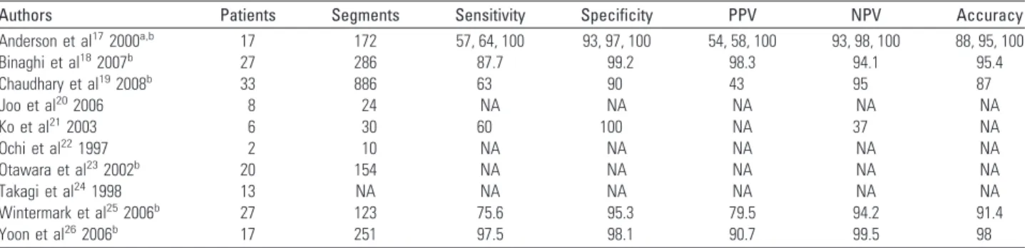
The 10 studies included a total of 895 patients (range 29– 329) with a total of 1264 breast lesions (range 33–469) of which 606 were classified as malignant (range 9–279) and 658 as benign (range 22–190) [20–29].
In 5 [23, 24, 26–28] studies a selection was made of patients with suspect findings based on mammography and ultrasound examinations. In the other 5 studies [20–22,25, 29] patients with a suspect lesion on MRI were included. One of these 5 studies retrospectively searched the database of an ongoing MRI screening study of patients at high risk of breast cancer for BIRADS 3–5 lesions that were detected with MRI [22], and 2 studies included lesions that were not palpable and were not visible on mammography or ultrasound [20, 21]. In all 10 studies histology was used as the gold standard. In 4 studies a follow-up MRI after 6 or 24 months was performed [23,25,28,29]; in the case of positive findings biopsy provided further histological assessment.
Mean study quality was 12.6, ranging from 10 to 14. Four studies were of maximum quality (Table1) [20,21,26,27]. CAD systems
In all 10 studies the CAD systems (CADstream, DynaCAD, Fulltime point, 3-Time-Point Method and CAD-Gaea) incor-porated precontrast medium (unenhanced) images and 2 (immediate and delayed) or all postcontrast medium (en-hanced) images. The CAD systems compared pixel intensity values on the precontrast medium and immediated postcontrast medium series. If a pixel value increased above a user-specified minimum enhancement threshold, such as a 50 or 100% increase in enhancement, the pixel was regarded as meeting threshold enhancement. Once a pixel was identified as enhancing above the established threshold, the CAD systems compared pixel signal intensity values on the immediate and delayed postcontrast medium series to indicate washout enhancement, plateau enhancement or persistent enhancement. A specific colour or colour intensity was assigned to each pixel for different types of tissue enhancement. The end result of all CAD systems was a colour overlay on each MRI slice
Search result: n= 587 (319 Pubmed; 268 Embase)
Excluded based on title: n= 519
Papers retrieved for more detailed evaluation
(n=56)
Selection based on full paper (n=27) Excluded (n=29) - technical paper: 20 - overview: 8 - case report: 1 Excluded (n=17)
- Institution-specific CAD systems
Included in the review (n=10)
12 duplicates
Fig. 1 Flow chart of search results, with reasons for exclusion and the total number of studies included
Revisão sistemática
– Revisão formal da evidência sobre um determinado tópico
com uma
pergunta de partida específica
, com uma
estratégia de pesquisa detalhada que permita a replicação
– Envolve a
seleção de aspetos-chave
das publicações,
tais como metodologia, características dos sujeitos,
medidas utilizadas em outros estudos (avaliação
qualitativa)
Probst, H., & Brealey, S. (2010). Health technology assessment. In A. Ramlaul (Ed.), Medical Imaging and Radiotherapy Research - Skills and Strategies. London: Churchill Livingstone - Elsevier.
Revisão sistemática
Exemplo
The patient experience of high technology medical imaging: A systematic review
of the qualitative evidence
qZachary Munn*, Zoe Jordana
The Joanna Briggs Institute, Faculty of Health Sciences, The University of Adelaide, Adelaide, South Australia 5005, Australia
a r t i c l e i n f o
Article history: Received 1 May 2011 Received in revised form 22 June 2011 Accepted 26 June 2011 Available online 18 July 2011 Keywords: Qualitative Systematic review Patient experience MRI CT Meta-synthesis a b s t r a c t
Background:When presenting to an imaging department, the person who is to be imaged is often in a vulnerable state, and can experience the scan in a number of ways. It is the role of the radiographer to produce a high quality image and facilitate patient care throughout the imaging process. A qualitative systematic review was performed to synthesise the existent evidence on the patient experience of high technology medical imaging. Only papers relating to Magnetic Resonance Imaging (MRI) and Computed Tomography (CT) were identified.
Inclusion criteria:Studies that were of a qualitative design that explored the phenomenon of interest, the patient experience of high technology medical imaging. Participants included anyone who had under-gone one of these procedures.
Methods:A systematic search of medical and allied health databases was conducted. Articles identified during the search process that met the inclusion criteria were then critically appraised for methodo-logical quality independently by two reviewers.
Results:During the search and inclusion process, 15 studies were found that were deemed of suitable quality to be included in the review. From the 15 studies, 127 findings were extracted from the included studies. These were analysed in more detail to observe common themes, and then grouped into 33 categories. From these 33 categories, 11 synthesised findings were produced. The 11 synthesised findings highlight the diverse, unique and challenging ways in which people experience imaging with MRI and CT scanners.
Conclusion:The results of the review demonstrate the diverse ways in which people experience medical imaging. All health professionals involved in imaging need to be aware of the different ways each patient may experience imaging.
! 2011 The College of Radiographers. Published by Elsevier Ltd. All rights reserved.
Introduction
Medical imaging is an ever changing and advancing field, with significant advancements in imaging techniques and technologies over the years. The amount of imaging and the subsequent costs associated with it have been rising rapidly in many parts of the world for the last 30 years,1,2leading to a larger percentage of people being exposed to these different imaging modalities.3 However, these advancements in imaging technology do not necessarily guarantee a similar advance in patient care.4 High
technology imaging in particular, such as Computed Tomography (CT), Magnetic Resonance Imaging (MRI), Positron Emission Tomography (PET) and Single Photon Emission Computed Tomog-raphy (SPECT), have seen significant increases in their use.1These scans are more complex than some other medical imaging proce-dures, and can be more difficult to conduct, which may have an effect on the holistic care of the patient, as it distances the radi-ographer from the patient.5Consequently, the patient and patient care can often be ignored or overlooked, as the focus of the radi-ographer can be directed largely towards the technology and not the patient.6Healthcare professionals involved in patient care may unwittingly objectify patients, and not necessarily see them as people in pain or distress, but as problems needing solving.7As imaging is essentially a ‘hit and run’ process,8the patient can sometimes be seen as just a translucent screen upon which the health professional peers to find a diagnostic entity within,9and the body is perceived differently by the patient and the practi-tioner.7It is however imperative that imaging staff remember that q This is an abridged report of a full systematic review located in the Joanna
Briggs Library of Systematic Reviews, accessible online at: http://connect.
jbiconnectplus.org/JBIReviewsLibrary.aspx.
*Corresponding author. Tel.: þ61 8 8303 4770; fax: þ61 8 8303 4881.
E-mail addresses:Zachary.munn@adelaide.edu.au(Z. Munn),zoe.jordan@adelaide.
edu.au(Z. Jordan).
aTel.: þ61 8 8303 3893, þ61 0408 825 516 (mobile); fax: þ61 8 8303 4881.
Contents lists available atScienceDirect
Radiography
j o u r n a l h o m e p a g e : w w w . e l s e v i e r . c o m / l o c a t e / r a d i
1078-8174/$ e see front matter! 2011 The College of Radiographers. Published by Elsevier Ltd. All rights reserved.
doi:10.1016/j.radi.2011.06.004
Radiography 17 (2011) 323e331
they meet patients at a critical time in their life,10and through studies such as those included in this review, gain a better under-standing of the experience of their patients, with the hope of improving practice.
Research in imaging
In the past, there has been an emphasis on quantitative research designs in medical imaging, resulting in a significant lack of liter-ature on the experience of the individual that is undergoing examination.11Research in medical imaging largely stems from the positivist paradigm, where hypotheses are tested through quanti-tative research designs.12This may stem from the historical domi-nance of the medical profession in medical imaging.13,14However, quantitative research designs are not always suitable to answer all questions generated from the medical imaging process, as they are limited to observations and data that can be measured and ana-lysed mathematically using statistical methods.15For questions looking at experience, perception, meanings, understanding and acceptance of imaging, qualitative methodologies are the most appropriate approach for enquiry.6,15It is thought that the infor-mation generated from qualitative studies will support health professionals when dealing with patients, and assist in improving communication with the patient and in understanding and addressing their concerns, making the procedure more acceptable to the patient.15In recent times it has been identified that there is a role for qualitative research in medical imaging, as the paradigm shifts from technology-focussed to patient-focussed research.16,17 The aim of this research is to ‘more clearly define what radiogra-phers do and how they do it’6(p. 194). Equally important, and also included in the aims of this project, is the need to identify issues relating to the patient in medical imaging, and the need to highlight the patient’s experience and perspective of health care.6 The qualitative systematic review
Interest in practising evidence-based medicine (a term first coined in 1992),18and evidence-based healthcare, has increased exponentially since the 1990s. Evidence-based medicine has been defined as ‘the conscientious, explicit, and judicious use of current best evidence in making decisions about the care of individual patients.19’Systematic reviews can be seen as the pillar on which evidence-based healthcare rests, as they provide health profes-sionals with a comprehensive synthesis of the existent literature on a certain healthcare topic.19e21Evidence-based organisations such as the Cochrane Collaboration and the Joanna Briggs Institute, both established in the 1990s, have been set up to develop methodolo-gies and guidance on the process of systematic reviews. The applicability, importance and need for systematic reviews for medical imaging professionals has been stressed in previous arti-cles,20,22,23and there does exist published examples in radiog-raphy24and radiotherapy.25However; the focus of these reviews are all quantitative in nature. Qualitative systematic reviews also play an important role in evidence-based healthcare, to inform healthcare professionals regarding issues that are not conducive to empirical research methods.21
The patient experience
When presenting to an imaging department, the person who is to be imaged is often in a vulnerable state, and out of their comfort zone. It is the role of the radiographer to produce a high quality image and facilitate patient care throughout the imaging process. Qualitative research is necessary to better inform the radiographer and to help them to understand the experience of the person being
imaged.11Some issues that have been identified in the literature include fear,26 claustrophobia,27 dehumanisation,28 and an uncomfortable or unusual experience.29There is now a small but worthwhile qualitative literature base focussing on the patient experience in medical imaging. As far as we are aware, there is no other qualitative synthesis of the literature on the patient experi-ence in medical imaging. It is therefore timely and worthwhile to produce a systematic review to identify and summarise the existent literature exploring the patient experience of diagnostic imaging. Methods
A qualitative systematic review was conducted according to the methodology of the Joanna Briggs Institute. The Joanna Briggs Institute is a not-for-profit research institute committed to the translation of evidence into practice.30The Joanna Briggs Institute’s approach was chosen as it is a leader in conducting systematic reviews, particularly those of qualitative evidence.30Prior to con-ducting the review, a protocol was developed outlining the scope and methods of the review which was submitted to the Joanna Briggs Institute; the protocol was then approved and the review conducted. Review objectives
The objective of this systematic review was to identify and describe from the available literature the experience and percep-tions of people undergoing diagnostic imaging with high technol-ogies, such as Magnetic Resonance Imaging, Computed Tomography, and Nuclear Medicine procedures. The question that the researchers were seeking to answer was: ‘How do patients/ clients being scanned with high technology imaging experience the imaging procedure?’
Criteria for considering studies for this review Types of studies
This review considered studies that focused on qualitative data or included a qualitative aspect, including, but not limited to, designs such as phenomenology, ground theory, ethnography, action research, qualitative descriptive studies, and feminist research. These were limited to English language studies, with no time limit.
Types of participants
This review included publications that included persons of any age who had undergone high technology medical imaging. These participants may have received medical imaging for a wide range of indications, and may have any pre-existing condition or disability. Phenomena of interest
This review considered studies that investigated the patient experience of diagnostic imaging using high technology imaging, and the meaningfulness of that experience. Advanced, high tech-nology imaging procedures are increasingly prevalent scans and there is rapid growth in these imaging modalities.3MRI, CT, PET and SPECT are the procedures considered high technology or advanced medical imaging,1,3,26and were searched for particularly. Standard digital radiography was not included. All diagnostic imaging proce-dures included in this review were non-invasive or minimally inva-sive. Interventional diagnostic procedures were not included, as their experience may be considerably different due to the invasive nature. Search strategy
The search strategy aimed to find both published studies and grey literature, as advised by the Joanna Briggs Institute,30and was
Z. Munn, Z. Jordan / Radiography 17 (2011) 323e331 324
they meet patients at a critical time in their life,
10and through
studies such as those included in this review, gain a better
under-standing of the experience of their patients, with the hope of
improving practice.
Research in imaging
In the past, there has been an emphasis on quantitative research
designs in medical imaging, resulting in a significant lack of
liter-ature on the experience of the individual that is undergoing
examination.
11Research in medical imaging largely stems from the
positivist paradigm, where hypotheses are tested through
quanti-tative research designs.
12This may stem from the historical
domi-nance of the medical profession in medical imaging.
13,14However,
quantitative research designs are not always suitable to answer all
questions generated from the medical imaging process, as they are
limited to observations and data that can be measured and
ana-lysed mathematically using statistical methods.
15For questions
looking at experience, perception, meanings, understanding and
acceptance of imaging, qualitative methodologies are the most
appropriate approach for enquiry.
6,15It is thought that the
infor-mation generated from qualitative studies will support health
professionals when dealing with patients, and assist in improving
communication with the patient and in understanding and
addressing their concerns, making the procedure more acceptable
to the patient.
15In recent times it has been identified that there is
a role for qualitative research in medical imaging, as the paradigm
shifts from technology-focussed to patient-focussed research.
16,17The aim of this research is to ‘more clearly define what
radiogra-phers do and how they do it’
6(p. 194). Equally important, and also
included in the aims of this project, is the need to identify issues
relating to the patient in medical imaging, and the need to highlight
the patient’s experience and perspective of health care.
6The qualitative systematic review
Interest in practising evidence-based medicine (a term first
coined in 1992),
18and evidence-based healthcare, has increased
exponentially since the 1990s. Evidence-based medicine has been
defined as ‘the conscientious, explicit, and judicious use of current
best evidence in making decisions about the care of individual
patients.
19’
Systematic reviews can be seen as the pillar on which
evidence-based healthcare rests, as they provide health
profes-sionals with a comprehensive synthesis of the existent literature on
a certain healthcare topic.
19e21Evidence-based organisations such
as the Cochrane Collaboration and the Joanna Briggs Institute, both
established in the 1990s, have been set up to develop
methodolo-gies and guidance on the process of systematic reviews. The
applicability, importance and need for systematic reviews for
medical imaging professionals has been stressed in previous
arti-cles,
20,22,23and there does exist published examples in
radiog-raphy
24and radiotherapy.
25However; the focus of these reviews
are all quantitative in nature. Qualitative systematic reviews also
play an important role in evidence-based healthcare, to inform
healthcare professionals regarding issues that are not conducive to
empirical research methods.
21The patient experience
When presenting to an imaging department, the person who is
to be imaged is often in a vulnerable state, and out of their comfort
zone. It is the role of the radiographer to produce a high quality
image and facilitate patient care throughout the imaging process.
Qualitative research is necessary to better inform the radiographer
and to help them to understand the experience of the person being
imaged.
11Some issues that have been identified in the literature
include fear,
26claustrophobia,
27dehumanisation,
28and an
uncomfortable or unusual experience.
29There is now a small but
worthwhile qualitative literature base focussing on the patient
experience in medical imaging. As far as we are aware, there is no
other qualitative synthesis of the literature on the patient
experi-ence in medical imaging. It is therefore timely and worthwhile to
produce a systematic review to identify and summarise the existent
literature exploring the patient experience of diagnostic imaging.
Methods
A qualitative systematic review was conducted according to the
methodology of the Joanna Briggs Institute. The Joanna Briggs
Institute is a not-for-profit research institute committed to the
translation of evidence into practice.
30The Joanna Briggs Institute’s
approach was chosen as it is a leader in conducting systematic
reviews, particularly those of qualitative evidence.
30Prior to
con-ducting the review, a protocol was developed outlining the scope and
methods of the review which was submitted to the Joanna Briggs
Institute; the protocol was then approved and the review conducted.
Review objectives
The objective of this systematic review was to identify and
describe from the available literature the experience and
percep-tions of people undergoing diagnostic imaging with high
technol-ogies, such as Magnetic Resonance Imaging, Computed
Tomography, and Nuclear Medicine procedures. The question that
the researchers were seeking to answer was: ‘How do patients/
clients being scanned with high technology imaging experience the
imaging procedure?’
Criteria for considering studies for this review
Types of studies
This review considered studies that focused on qualitative data
or included a qualitative aspect, including, but not limited to,
designs such as phenomenology, ground theory, ethnography,
action research, qualitative descriptive studies, and feminist
research. These were limited to English language studies, with no
time limit.
Types of participants
This review included publications that included persons of any
age who had undergone high technology medical imaging. These
participants may have received medical imaging for a wide range of
indications, and may have any pre-existing condition or disability.
Phenomena of interest
This review considered studies that investigated the patient
experience of diagnostic imaging using high technology imaging,
and the meaningfulness of that experience. Advanced, high
tech-nology imaging procedures are increasingly prevalent scans and
there is rapid growth in these imaging modalities.
3MRI, CT, PET and
SPECT are the procedures considered high technology or advanced
medical imaging,
1,3,26and were searched for particularly. Standard
digital radiography was not included. All diagnostic imaging
proce-dures included in this review were non-invasive or minimally
inva-sive. Interventional diagnostic procedures were not included, as their
experience may be considerably different due to the invasive nature.
Search strategy
The search strategy aimed to find both published studies and
grey literature, as advised by the Joanna Briggs Institute,
30and was
Z. Munn, Z. Jordan / Radiography 17 (2011) 323e331 324
Munn, Z., & Jordan, Z. (2011). The patient experience of high technology