MOLECULAR TYPING OF PORTUGUESE CLINICAL ISOLATES OF
Salmonella spp., S. Enteritidis AND S. Typhimurium
Thesis presented to Escola Superior de Biotecnologia of the Universidade Católica Portuguesa to fulfill the
requirements of Master of Science degree in Food Inovation
by
Diana Luísa Campos Ferreira Ramos
TIPAGEM MOLECULAR DE ISOLADOS CLÍNICOS PORTUGUESES DE
Salmonella spp., S. Enteritidis E S. Typhimurium
Tese apresentada à Escola Superior de Biotecnologia da Universidade Católica Portuguesa para obtenção do grau de Mestre em Inovação Alimentar
por
Diana Luísa Campos Ferreira Ramos
MOLECULAR TYPING OF PORTUGUESE CLINICAL ISOLATES OF
Salmonella spp., S. Enteritidis AND S. Typhimurium
Thesis presented to Escola Superior de Biotecnologia of the Universidade Católica Portuguesa to fulfill the requirements of Master of Science degree in Food Inovation
by
Diana Luísa Campos Ferreira Ramos
under the supervision of
Prof. Doutora Paula Teixeira / Doutora Joana Silva
TIPAGEM MOLECULAR DE ISOLADOS CLÍNICOS PORTUGUESES DE
Salmonella spp., S. Enteritidis E S. Typhimurium
Tese apresentada à Escola Superior de Biotecnologia da Universidade Católica Portuguesa para obtenção do grau de Mestre em Inovação Alimentar
por
Diana Luísa Campos Ferreira Ramos
sob orientação de
Prof. Doutora Paula Teixeira / Doutora Joana Silva
1
Resumo
A Salmonella spp. é um importante patogénico associado a doença humana. Permanece
uma das maiores causas de surtos de origem alimentar em todo o mundo. Na Europa, dois serotipos permanecem no topo da lista de número de casos por ano, Enteritidis e Typhimurium. A salmonelose é usualmente uma doença manifestada por diarreia e que dispensa tratamento médico, embora a terapia com antibióticos seja necessária em casos de doença invasiva ou infeções com complicações associadas. A resistência a antibióticos tem sido um problema crescente mundialmente. Salmonella enterica proveniente de uma variedade de alimentos e animais, tem sido extensamente estudada e caracterizada quanto a fenótipos de resistência e vários serotipos revelaram resistência a múltiplos antibióticos.
Na Europa, o ECDC (European Center of Disease Control) é o responsável, desde 2007, pela rede de vigilância de infeções gastrointestinais humanas. Em Portugal o INSA (Instituto Nacional de Saúde Dr. Ricardo Jorge) foi designado como laboratório de referência para fornecer dados epidemiológicos ao ECDC.
Neste estudo 216 isolados clínicos de Salmonella, provenientes do Hospital de São João no Porto (HSJ) e Hospital de São Marcos em Braga (HSM) ambos na região Norte de Portugal, foram testados quanto à resistência a antibióticos e analisados por PFGE, como implementado pelo CDC nos Estados Unidos. A amostra é constituída por isolados de S. Enteritidis e S. Typhimurium, assim como isolados identificados como Salmonella spp..
A análise de suscetibilidade a antibióticos confirmou as tendências Europeias, baixa resistência de isolados de S. Enteritidis exceto ao ácido nalidíxico, e elevada resistência em isolados de S. Typhimurium à maioria dos antibióticos testados. Alguns dos isolados de S. Typhimurium testados neste estudo também foram testados em trabalhos prévios no laboratório quanto a resistência a sulfonamidas, a maioria mostrou ter um perfil de resistência típico da Salmonella DT104 (ACSSuTe).
A análise de PFGE dos isolados permitiu a diferenciação de Enteritidis e Typhimurium assim como a identificação do serotipo para a maior parte dos isolados identificados como
Salmonella spp. Um isolado de Salmonella spp. não pertence a nenhum dos outros serotipos. .
As amostras provenientes dos dois hospitais têm uma similaridade elevada entre si. A discriminação dos isolados de Enteritidis provou ser baixa usando o enzima XbaI levando à conclusão de que um painel diferente de enzimas poderia ajudar. Os isolados de Typhimurium foram bem discriminados pelo PFGE porém não foi possível relacionar essa discriminação quer com a resistência a antibióticos quer com a informação disponível dos isolados.
2
Abstract
Salmonella spp. is an important pathogen associated with human disease. It remains one
of the major causes of food-borne outbreaks all over the world. In Europe two serovars are at the top of the list in number of cases per year, Enteritidis and Typhimurium. Salmonellosis is usually a self-limiting diarrheal disease requiring little or no medical intervention. However, in cases of invasive disease or infections with added complications, antimicrobial treatment may be required. Resistance to antimicrobials has been an increasing problem worldwide.
Salmonella enterica from a variety of food and animal sources has been extensively
characterized in terms of resistance phenotypes and numerous serotypes revealed multiple antimicrobial resistance determinants (strains resistant to two or more antimicrobials).
In Europe the ECDC (European Center of Disease Control) has been in charge of the international surveillance network for human gastrointestinal infections since 2007. In Portugal the INSA (Instituto Nacional de Saúde Dr Ricardo Jorge) was the designated reference laboratory to report the epidemiological data, and though an effort was made still the actual data is not available or clearly reported.
In this study 216 clinical Salmonella isolates, from Hospital de São João in Oporto (HSJ) and Hospital de São Marcos in Braga (HSM) both from the Northern region of Portugal, were tested for antimicrobial resistance and by PFGE analysis, as implemented by the CDC in the USA. The sample contained Salmonella Enteritidis, Salmonella Typhimurium and Salmonella spp.
The antimicrobial susceptibility analysis to a panel of antimicrobials has confirmed European tendencies, low resistance in S. Enteritidis isolates except for nalidixic acid, and high resistance in S. Typhimurium isolates to most of the tested antimicrobials. Some of the S. Typhimurium isolates tested in this study were also tested in previous works in the lab for sulphonamides resistance and proved to have the typical DT104 resistance profile (ACSSuT).
The PFGE analysis allowed the differentiation of Enteritidis and Typhimurium isolates as well as the identification of the serotype for most isolated identified has Salmonella spp. One Salmonella spp. Isolate didn’t belong to any of the two serotypes. The samples from the two hospitals showed high similarity between themselves. The discrimination of Enteritidis isolates was insufficient using XbaI enzyme leading to the conclusion that it would benefit with the analysis using a different panel of enzymes. Typhimurium isolates were well discriminated with PFGE though no correlation was possible with the resistance profile or the isolates available information.
3
Acknowledgments
I would like to thank my advisors, Prof. Doctor Paula Teixeira and Doctor Joana Silva for their guidance in the lab and in the development of this study. To Prof. Doctor Paul Gibbs for the review of this thesis and the enjoyable lunch talks.
Thanks to the CINATE lab for the PFGE equipment and Gel Compar software availability.
Thanks to all the colleagues in the department that gave me great support and made the everyday in the lab a fun experience, specially Ricardo Freixo for the support in the analysis of the data.
Very special thanks to Maria Gonzalez my supervisor and friend who made it possible to coordinate my work with my thesis writing. You are the great supporter of my career.
Thanks to my family for their constant support. Thanks dad for teaching me how fun and interesting science can be, and for being my partner in crazy experiments.
4
Index
Resumo ... 1 Abstract ... 2 Acknowledgments ... 3 Index ... 4 Introduction ... 5 Salmonella spp. classification ... 5Salmonella spp. Transmission and Disease ... 8
Salmonella spp. Evolution and Pathogenecity ... 9
Antimicrobial treatment and resistance mechanisms in Salmonella ... 10
Molecular typing – Pulsed Field Gel Electrophoresis ... 15
Objectives ... 19
Materials and Methods ... 20
Bacterial Strains ... 20
Antimicrobial susceptibility testing ... 22
Pulsed-field gel electrophoresis (PFGE) ... 24
Results ... 26
Antimicrobial susceptibility testing ... 26
Discussion ... 30 Conclusion ... 32 Future work ... 33 References ... 34 Annex ... 37
5
Introduction
Salmonella spp. classification
Salmonella spp. are Gram-negative, rod-shaped bacteria belonging to the
Enterobacteriaceae family (Le Minor and Popoff, 1987; Darwin and Miller, 1999).
Salmonella was named after an American bacteriologist, Daniel E. Salmon, who first isolated Salmonella Choleraesuis from porcine intestine in 1884 (Su, 2007). The genus Salmonella comprises a large and closely related population of medically important pathogens and consists of three species, Salmonella bongori, Salmonella enterica (Su et al., 2007) and
Salmonella subterranean, the last recognized in 2005 (Tankouo-Dandjong, 2007). Salmonella enterica is divided into the following six subspecies: S. enterica subsp. enterica (I), S. enterica subsp. salamae (II), S. enterica subsp. arizonae (IIIa), S. enterica subsp. diarizonae
(IIIb), S. enterica subsp. houtenae (IV) and S. enterica subsp. indica (VI) (Grimont and Weill, 2007).
More than 2,500 serovars of zoonotic Salmonella exist and the prevalence of the different serovars changes over time (EFSA, 2009). Salmonella serotypes are closely related evidencing their clonal origin, and based on the degree of sequence divergence, it can be estimated that a common ancestor of the genus existed about 25 to 40 million years ago (Bäumler et al., 1998).
While some serovars of Salmonella such as S. Typhi and S. Pullorum have a restricted host range, most serovars infect a broad range of warm-blooded animals and are capable of causing disease in humans. The majority that causes disease in humans belong to subgroup I (Darwin and Miller 1999).
Salmonella is one of the most extensively studied bacterial genera in terms of its
physiology, genetics, cell structure, and development (Darwin and Miller, 1999). The reasons for this are that they cause significant morbidity and mortality worldwide; they have broad host ranges, infections result in different diseases in different hosts; they are able to establish persistent infections, which serve as reservoirs for transmission/shedding; and they are increasingly resistant to many antibiotics (Boyle et al., 2007).
Even though more than 2,500 serovars of Salmonella enterica have thus far been recognized, few appear to cause most foodborne illnesses (Gebreyes et al., 2006). Food-borne outbreaks are infections or intoxications in humans caused by the consumption of a common contaminated food (EFSA, 2009).
6
In 2007, a total of 5,609 food-borne outbreaks were reported by the European Union Member States (EU MSs) of which 38.7% were verified and where 1,475 people were
affected, hospitalizing 55 and causing 5 deaths. Salmonella was as in previous years, one of the most commonly reported cause of food-borne outbreaks in the EU, second only in numbers caused in 2007 by Campylobacter. A total of 155,540 confirmed cases of human salmonellosis were reported via TESSy (The European Surveillance System) from 30 countries, including the 27 EU MSs and three non-MSs, and directly to the European Food Safety Agency (EFSA) from Switzerland. The number of confirmed cases in the EU has decreased since 2005 (fig 1). Overall, total case counts of salmonellosis have decreased since 2004.
Figure 1 - Notification rates of reported confirmed cases of human salmonellosis in the EU, 2004-2007
(in EFSA. 2009).
Since the EFSA was established in 2002, no data was found on Salmonella for the years 2002 and 2003. General Salmonella Quarterly reports can be found on the ECDC’s Enter-net (Enter- Net 2002-2003 http://ecdc.europa.eu/en/publications/surveillance_reports/fwd)
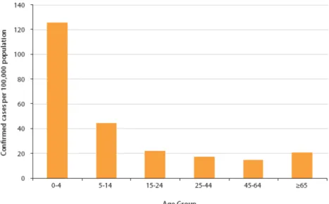
The age distribution of Salmonella cases in 2007 closely parallels those seen in 2006. The distribution by age group is depicted in the following figure (Fig.2). The highest rate was observed for 0 to 4 years old group (EFSA, 2009). Children especially those <1 year of age , and adults over 60, are most susceptible to disease and tend to have more severe infections (Darwin and Miller, 1999).
7
Figure 2 - Age distribution of reported confirmed cases of human salmonellosis in the EU, 2004-2007; data from all Member States, N=131,229. (in EFSA, 2009)
A seasonal effect is notable in the numbers of reported cases of salmonellosis throughout the year, usually higher in the summer and autumn and with a rapid decline in winter months. This pattern supports the influence of temperature and behavior, and has been observed in earlier reports (EFSA, 2002, 2007). When further analyzed by specific serovar counts per month, serovar Enteritidis shows a more prominent summer/autumn peak than other serovars. The two most common Salmonella serovars are Enteritidis and Typhimurium, representing 81%, in 2007, of all identified types in human cases (fig 3).
8
Salmonella spp. Transmission and Disease
Transmission of Salmonella to humans is usually by consumption of contaminated food, but human-to-human transmission and direct animal-to-human transmission can occur (Darwin, 1999). The common reservoir of Salmonella is the intestinal tract of a wide range of domestic and wild animals which results in a variety of foodstuffs of animal and plant origin as sources of infections (EFSA, 2009). The foodstuffs most frequently associated with
Salmonella outbreaks in humans are meat, especially poultry, eggs and egg products.
Human S. Enteritidis cases are most commonly associated with the consumption of contaminated eggs and broiler meat, while S. Typhimurium cases are most often associated with the consumption of contaminated pig, poultry and bovine meat. Transmission often occurs when organisms are introduced into food preparation areas and are allowed to multiply in food, e.g. due to inadequate storage temperatures, inadequate cooking or cross-contamination of ready-to-eat food (EFSA, 2009).
Salmonella enterica serovar Enteritidis is the cause of the foodborne salmonellosis
pandemic in humans, in part because it has the unique ability to contaminate eggs without causing discernible illness in the birds infected (Guard-Petter, 2001). Salmonella serovar Enteritidis is the only human pathogen that contaminates eggs routinely, even though the on-farm environment of the chicken is a rich source of a number of Salmonella serotypes. Human illness caused by infection with Salmonella enterica serovar Enteritidis increased worldwide beginning as early as the mid-1970s and, by 1990, this serovar displaced Salmonella enterica serovar Typhimurium as the primary cause of salmonellosis in the world (Guard-Petter, 2001).
Salmonella is capable of causing a variety of disease syndromes: enteric fever,
bacteremia, enterocolitis, and focal infections. Enterocolitis is by far the most common manifestation of disease caused by Salmonella, but bacteremia and focal infections can accompany or follow enterocolitis (Darwin, 1999). Salmonellosis has also been associated with long-term and sometimes chronic sequelae, e.g. reactive arthritis. The incubation period is from 6 to 72 hours, usually about 12-36 hours (EFSA, 2009). It is estimated from volunteer studies that 105 to 1010 bacteria are required to initiate an infection, but the exact amount needed varies with the strain, what is consumed with the bacteria, and the physiological state of the host (Darwin, 1999). Salmonellosis is usually a self-limiting diarrhoeal disease requiring little or no medical intervention. S. enterica is a facultative intracellular pathogen which preferentially resides inside macrophages, yet requires both antibodies and a cellular
9
immune response for clearance (Kaufmann, 2001). However, in cases of invasive disease or infections with added complications, such as at the extremities of age or in the presence of underlying disease, antimicrobial treatment may be required.
Salmonella spp. Evolution and Pathogenecity
It has been postulated that in the genus Salmonella, virulence evolved in three phases. The first phase involved acquisition of Salmonella pathogenicity island 1 (SPI1) by plasmid or phage mediated horizontal transfer (Marcus et al., 2000). Pathogenicity islands are distinct regions of the chromosome where virulence functions are clustered; they are characteristically unstable and have a high deletion frequency (Maskell, 2006). SPI1 was likely obtained by a lineage ancestral to all Salmonella serotypes, since it is present in all phylogenetic lineages of the genus Salmonella but absent from Escherichia coli and other related organisms. SPI1 encodes virulence factors that mediate mechanisms used by Salmonella serotypes during the intestinal phase of infection, including invasion of intestinal epithelial cells, induction of neutrophil recruitment, and secretions of intestinal fluid (Bäumler, 1998).
The formation of the two species S. enterica and S. bongori could be considered a second phase in the evolution of virulence in the genus Salmonella, since it involved not only divergence of their lineages by point mutation but also acquisition of new virulence determinants by horizontal gene transfer. Serovars belonging to S. enterica possess a second pathogenicity island, designated SPI2, which is not present in S. bongori serotypes (Bäumler, 1998).
The formation of S. enterica subspecies I involved a dramatic expansion in host range: while S. bongori and S. enterica subspecies II, IIIa, IIIb, IV, and VII are mainly associated with cold-blooded vertebrates, members of S. enterica subspecies I are most frequently isolated from avian and mammalian hosts. The host adaptation of S. enterica subspecies I to warm-blooded vertebrates characterized a third phase in the evolution of virulence in the genus Salmonella. In addition to horizontal gene transfer, deletion events and sequence divergence by point mutation likely contributed to changes in the host ranges of S. enterica serovars (Bäumler, 1998).
Salmonella serotypes initiate infection by attaching to the intestinal mucosa of the host.
Upon penetration of the intestinal mucosa of mammals, Salmonella serotypes are confronted by an effective barrier to further spread, namely macrophages that line the lymphatic sinuses of regional lymph nodes. In mammals this host defense mechanism can successfully limit
10
bacterial expansion to the intestine, the gut-associated lymphoid tissue, and the mesenteric lymph node. It has therefore been speculated that Salmonella serotypes evolved in the alimentary tract of reptiles, where they developed from pathogens into commensal organisms. The ability of S. enterica serotypes to cause systemic disease is directly related to the capability to withstand an assault by the macrophages of a given host (Bäumler, 1998). Pathogens that lack host specificity, such as S. enterica serotype Typhimurium and S. enterica serotype Enteritidis, tend to be more frequently associated with disease in young animals than in adults, suggesting that they are not optimally adapted to cope with a fully mature immune system.
Non-typhoidal salmonellosis has been characterized by a pattern of dissemination of clonal Salmonella enterica. Epidemic clones may remain largely restricted to the animal reservoir for years before their incidence rises among human infections (Davis et al., 2007).
Antimicrobial treatment and resistance mechanisms in Salmonella
Food animals have been worldwide exposed to antimicrobials to treat or prevent infectious diseases or to promote growth. Many of these antimicrobials are similar or identical to the ones used to treat infections in humans. As a consequence, antimicrobial resistance has emerged in zoonotic enteropathogens (e.g. Salmonella spp., Campylobacter spp.), commensal bacteria (e.g. E. coli, enterococci), and bacterial pathogens of animals (e.g. Pasteurella,
Actinobacillus spp.), with variable prevalence. Resistance to antimicrobials emerges from the
use in animals and subsequent transfer of resistance genes and bacteria among animals and animal products and the environment (McEwen et al. 2002). Antimicrobial resistance is the best-known example of rapid adaptation of bacteria to a new ecosystem (Carattoli et al., 2003). In 1969, the Swann Committee was asked by the UK Government to report on antimicrobial use in both human and veterinary practice. The Committee concluded that there was a significant problem and made several recommendations. For example, animal feed antibiotics should not be those used in human medicine as this could compromise efficacy in man. Swann also recommended that the UK Government establish a committee that should have overall responsibility for the whole field of antibiotic use (Wise, 2007). In 1986 Sweden was the first country to ban all the food animal growth promoting antibiotics. Other European countries followed this decision and in 1997 and 1999 the European Union banned the remaining four growth promoting antibiotics under the “Precautionary Principle”. This action posed many questions about how to deal with the antimicrobial resistant bacteria that are
11
already in the environment and whether the removal of the antimicrobial agents could have adverse effects (Casewell et al., 2003). To help the European Countries tackle this antimicrobial resistance problem the World Health Organization published a booklet in 2011. This booklet contains instructions and recommendations destined to European Member States to consult in order to establish an “intersectoral and multifaceted approach with effective coordination of action and exchange of information among the agricultural, food, veterinary and health sectors” (WHO, 2011).
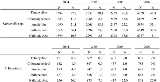
Antimicrobial treatment of salmonellosis is rare and only required in cases of generalized and invasive infection with added complications. Surveillance data of antimicrobial resistance in Salmonella was reported by European Member States for the 2004-2007 periods. In general, in Salmonella susceptibility tested isolates, resistance percentages range from 8 to 42%. Nalidixic acid resistance increased in 2007 as well as ampicillin resistance; on the other hand chloramphenicol resistance has decreased to the level observed in 2005. In S. Enteritidis isolates, low levels of resistance were reported for most antimicrobials tested; however, the percentages of resistant isolates reported for nalidixic acid were at a higher level and varied from 7 to 26%. For S. Typhimurium isolates, medium to high levels of resistance were reported for all antimicrobials tested except for nalidixic acid, during the reporting years (EFSA, 2010).
Salmonella enterica from a variety of food and animal sources has been extensively
characterized in terms of resistance phenotypes and numerous serotypes revealed multiple antimicrobial resistance determinants. Multidrug-resistant (MDR) strains are defined as resistant to two or more antimicrobial agents. MDR S. enterica strains may carry the resistance determinants on chromosomal locations and/or on resistance plasmids.
Resistance to broad-spectrum cephalosporin ceftriaxone, and to the quinolone nalidixic acid, are of particular importance to the medical community since they are the drug of choice for the treatment of pediatric and adult salmonellosis, respectively (Brichta-Harhay, 2011).
12
In this work we determined the resistance to a panel of commonly tested antimicrobials, namely tetracycline, chloramphenicol, ampicillin, streptomycin, nalidixic acid, and trimethoprim-sulphamethoxazole, having different modes of action and therefore subject of different resistance mechanisms. The compiled resistance percentages reported for some antimicrobials in the European Member states in the years 2004 to 2007 are depicted in table 1. No data is shown for the years 2002 and 2003 because they were not available at the EFSA. Quarterly reports from ECDC only contain general numbers for all Salmonella isolates (ECDC 2004).
Table 1 – Resistance (%) to tetracycline, chloramphenicol, ampicillin, sulfonamide and nalidixic acid among
tested Salmonella spp. isolates , 2004 to 2007 (EFSA 2010)
2004 2005 2006 2007 N % N % N % N % Salmonella spp. Tetracycline 1694 37,8 2357 28,4 2463 40,9 6769 42,3 Chloramphenicol 1683 11,6 2285 8,4 2439 15,8 6669 10,9 Ampicillin 1690 21,1 2066 18,4 2137 25,2 5974 33,1 Sulfonamide 1565 36,1 2294 25,0 2330 36,0 6549 38,5 Nalidixic Acid 1599 10,5 2292 8,8 2375 13,6 6787 18,1 2004 2005 2006 2007 N % N % N % N % S. Enteritidis Tetracycline 181 0,0 869 0,0 437 2,0 808 2,0 Chloramphenicol 182 1,0 867 0,0 437 1,0 783 0,0 Ampicillin 183 4,0 842 1,0 432 4,0 685 2,0 Sulfonamide 167 3,0 866 1,0 436 6,0 685 2,0 Nalidixic Acid 156 26,0 875 7,0 417 22,0 808 22,0 2004 2005 2006 2007 N % N % N % N % S. Typhimurium Tetracycline 1342 27,4 1779 25,2 1557 63,1 1941 58,8 Chloramphenicol 1340 24,8 1800 27,5 1557 35,0 1939 28,5 Ampicillin 1326 38,7 1761 46,6 1522 38,2 1920 52,3 Sulfonamide 1260 36,5 1801 54,8 1557 63,4 1923 58,8 Nalidixic Acid 1341 1,3 1800 3,0 1557 2,0 1943 4,3
Ampicillin is a β-lactam antibiotic with action against Gram negative and Gram positive bacteria. It is a peptidoglycan synthesis inhibitor and has a bacteriolytic effect only in
13
growing bacteria or in hypotonic environment. The β-lactam antibiotics stop the parietal synthesis (bacteriostatic effect) and act by activation of the bacterial endogenous autolytic system. These antibiotics bind the cellular membrane through the Penicillin-binding-proteins (PBPs). The bacterial resistance is mediated by the hydrolysis of the lactam ring by the β-lactamase enzyme. These enzymes are of chromosomal or plasmid origin and are excreted to the environment by Gram-positive bacteria and to the periplasm by Gram-negative bacteria.
Chloramphenicol is considered a broad spectrum antibiotic with effective action against Gram-positive and Gram-negative bacteria. It is used in the therapy of meningitis, typhoid fever and eye infections, among others, and has a high penetration rate of the blood-brain barrier. It is a protein synthesis inhibitor that binds to the 50S ribosomal subunit resulting in a bacteriostatic effect. Several Gram-positive and Gram-negative bacterial strains are resistant to chloramphenicol and its derivatives; these strains often show cross resistance to fusidic acid. High-level resistance is conferred by the cat-gene. A mechanism of low-level resistance is the reduction of membrane permeability by alteration of available porin 1α channels. Rare resistance mechanisms to chloramphenicol are the mutation of the ribosomal target, and the activation of efflux-pumps in E.coli strains. The cat-gene, responsible for acetyltransferase production, is located in plasmids or in chromosomal DNA and is expressed constitutively. The resistance plasmids can be co-transferred to other bacterial species by genetic recombination (i.e. chloramphenicol resistance transference from S. typhi to E. coli).
Streptomycin is an aminoglycoside antibiotic and was the first antibiotic remedy for tuberculosis. It is effective against Gram-positive and Gram-negative bacteria and therefore considered a broad spectrum antibiotic. It is a protein synthesis inhibitor that crosses the bacterial envelope altering the permeability of the cell and inhibits the DNA replication. It has a bactericidal and bacteriolytic effect in Gram-positive bacteria. For these antibiotics to be effective they have to accumulate inside the cell in its native form, after crossing the cellular wall and membrane. The selective pressure, especially in hospital environments, has lead to the emergence of resistant bacterial strains. The bacterial resistance is mediated by plasmids, transposons and integrons, which disseminate easily between bacterial strains of the same and different species. The most frequently observed resistance mechanism in clinical isolates is the enzymatic modification of the -NH2 groups, and the –OH groups, among others. Other
described resistance mechanisms are the mutation of ribosomal protein genes and the alteration of cell wall permeability.
14
Tetracycline is a broad spectrum antibiotic with bacteriostatic effect, and bactericidal in high concentrations. It is a protein synthesis inhibitor, acting in the 30S ribosomal subunit. As other protein synthesis inhibitors, tetracycline has to cross the outer membrane through porin channels, in order to reach the periplasm and the cytoplasmic membrane of the bacterial cell. Tetracycline also binds to the DNA, proteins and tRNA. The incidence of tetracycline resistance is very high and broad in bacterial strains. The genetic determinants responsible for the resistance can be of chromosomal origin, the most frequently found is in plasmids or transposons. The resistance mechanisms to tetracycline, in a general way, are due to: outer membrane impermeabilization in Gram-negative bacteria; ribosome mutation; low intracellular concentration due to efflux of the antibiotic; ribosomal protection. The most frequent mechanism is mediated by efflux pumps that keep the tetracycline intracellular concentration low. The strong efflux system is determined by genes tet, which codify the TET proteins that actively transport the tetracycline out of the cell.
Nalidixic acid is a first generation antibiotic of the quinolone group with activity against Gram-negative bacteria, and is usually used in urinary infection treatment. The first generation quinolones are DNA gyrase inhibitors. The inhibition of the DNA gyrase will affect the DNA coiling and consequently the replication and transcription. Therefore quinolones have a bactericidal effect. The resistance to quinolones can be due to the impermeabilization of the outer membrane, antibiotic efflux or mutation of the target enzymes.
Trimethoprim-sulfamethoxazole is a combination of two broad spectrum antibiotics, active against Gram-positive and negative bacteria. When trimethoprim is used in association with sulfonamids they can have a synergistic bactericidal effect, not being linear for all associations. The trimethoprim-sulfamethoxazole association decreases mostly the development of bacterial resistance, compared to monotherapy. Different mechanisms can explain the bacterial resistance to these antibiotics. The permeability barrier against sulfonamides and trimethoprim is generally associated with energy dependent efflux pumps. Chromosomal mutations in the dhps gene also confer resistance to sulfonamides. Point mutations in the dhfr gene can cause trimethoprim resistance. The resistance can also be acquired in plasmids that codify the DHPS and DHPR production, resistance to sulfonamides and trimethoprim, respectively. The sulfonamide resistance in Gram-negative enteric bacteria is generally mediated by horizontal gene transfer of external sul genes.
15
Molecular typing – Pulsed Field Gel Electrophoresis
Typing methods fall into two broad categories; phenotypic methods and genotypic methods.
Phenotypic methods are those that characterize the products of gene expression in order to differentiate strains. Properties such as biochemical profiles, bacteriophage types, antigens present on the cell’s surface, and antimicrobial susceptibility profiles, all are examples of phenotypic properties that can be determined in the laboratory. Because they involve gene expression, these properties all have a tendency to vary, based on changes in growth conditions, growth phase, and spontaneous mutation. Phenotypic methods, such as antimicrobial susceptibility profiles, biochemical profiles, bacteriophage susceptibility patterns, multilocus enzyme electrophoresis (MLEE) profiles, and immunoblot fingerprinting, have allowed investigators to describe the epidemiology of some nosocomial and community acquired infections. However, the phenotypic methods discriminate poorly among strains, they frequently require intensive labour, long elapsed times, and they often produce variable results (Pfaller, 1999).
Genotypic methods are those that are based on the analysis of the genetic structure of an organism and include polymorphisms in DNA restriction patterns based on cleavage of the chromosome by enzymes that cleave the DNA into hundreds of fragments (frequent cutters), or into 10 to 30 fragments (infrequent cutters), and the presence or absence of extra-chromosomal DNA. Genotypic methods are less subject to natural variation, although they can be affected by insertions or deletions of DNA in the chromosome, the gain or loss of extra-chromosomal DNA, or random mutations that may create or eliminate restriction endonuclease sites (Tenover et al., 1997).
In the characterization of methods, four criteria must be considered: rapidity, reproducibility, ease of use, and the ability to differentiate strains with similar phenotypes which are genetically unrelated (Lim et al., 2005).
Salmonella comprises a large genus, and serotyping is widely used to classify isolates
into serogroups according to their surface antigen variability. Serotyping is based on the immunological classification of the lipopolysaccharide (O antigen), the flagellar protein (H antigen), and the capsular polysaccharide moieties (Vi antigen). The Kauffmann-White scheme, generally used for the classification of Salmonella serotypes, recognizes 2523 characterized serotypes (Kaufmann, 1952; Tankouo-Sandjong et al., 2007). However, the
16
complexity of the system and difficulty of laboratory to laboratory comparison of results limit the application of serotyping to reference laboratories (Lim et al., 2005).
Among the most frequently isolated serovars are S. enterica subsp. enterica serovars Typhimurium and Enteritidis. Serotyping is not efficient enough when trying to track the source of common outbreaks of these two serotypes, as well as other frequently seen serovars. To further discriminate within S. Typhimurium and S. Enteritidis, phage typing is the primary sub-typing technique (Torpdahl et al., 2005). Phage typing requires a well-maintained phage-library, precise methodology, and experience in interpretation of results (Ross and Heuzenroeder, 2005). Phage typing of Salmonella can be subjective and fail to provide sufficient discrimination between isolates and it does not necessarily reflect a close genetic relationship between isolates (Ross and Heuzenroeder, 2005).
Several DNA-based typing methods have been developed in an attempt to improve the reproducibility and discriminatory ability in typing of Salmonella (Torpdahl et al., 2005). The most widely used molecular typing methods are the DNA-based methods, such as plasmid profiling, restriction endonuclease analysis of plasmid and genomic DNA, Southern hybridization analysis using specific DNA probes, and chromosomal DNA profiling using either pulsed-field gel electrophoresis or polymerase chain reaction-based methods. The various molecular typing methods may be applied to the investigation of outbreaks of infections or may be used in the context of epidemiological surveillance. For outbreak investigation, typing methods are used to compare isolates from a suspected outbreak to delineate clonally related and unrelated strains with the goal of short-term control of transmission. In the context of epidemiological surveillance, molecular typing methods may be used to monitor geographic spread and prevalence shifts of epidemic and endemic clones with the goal of long-term evaluation of preventive strategies or for the detection and monitoring of emerging and re-emerging infections (Pfaller, 1999).
Investigators have used a variety of DNA-based methods to genotype microbial pathogens. All these methods use electric fields to separate DNA, either restriction endonuclease digestion fragments, amplified DNA fragments, or whole chromosomes or plasmids, into unique patterns or fingerprints that are visualized by staining the DNA with ethidium bromide or by nucleic acid hybridization. Advances in genomic analysis, such as automated nucleic acid sequencing and the use of DNA chip technology, offer the potential
17
for extremely sensitive, high throughput analysis of microorganisms that may be applicable to simultaneous detection and characterization of organisms in clinical material (Pfaller, 1999).
In 1996, the Centers for Disease Control and Prevention established the PulseNet National Database (USA), consisting of pulsed-field gel Electrophoresis (PFGE) patterns obtained from isolates of food-borne pathogens and textual information about the isolates. Though many molecular methods are available, macrorestriction analysis by PFGE has been shown to be particularly useful for the clustering and differentiation of many bacterial pathogens. Although the sensitivity and discriminatory power of PFGE depend on the organism being sub-typed and the restriction enzyme used, its high epidemiological relevance, has made this technique the “gold standard” for sub-typing food-borne bacterial pathogens.
Pulsed-field gel electrophoresis was first described in 1984 as a tool for examining the chromosomal DNA of eukaryotic organisms. Subsequently, PFGE has proven to be a highly effective molecular typing technique for many different bacterial species (Tenover et al., 1995). In this method, the bacterial genome, typically 2000 to 5000 kb pairs in size, is digested with a restriction enzyme that has relatively few recognition sites generating approximately 10 to 30 restriction fragments ranging from 10 to 800 kb. The fragments can be resolved as a pattern of distinct bands using a contour-clamped homogeneous electric field (CHEF). The CHEF apparatus consists of an array of electrodes positioned around the gel (on a contour) and clamped to specific voltages to produce a nearly homogeneous electric field inside the contour. CHEF was developed to bypass two problems of the PFGE apparatus existing then, one being the inhomogeneous electric fields that caused the DNA molecules to migrate with curvilinear, arc-like, or even wavelike trajectories. The second was the non-linear shapes made it difficult to compare samples across the gel (Levene, 1992). The CHEF produces an electric field that causes DNA to wiggle through the gel and the alternation of pulses from the three sets of electrodes produces a back and forward movement that results in a higher level of fragment resolution.
All species are typable by PFGE, although the isolation of intact chromosomal DNA is technically difficult for some species. In general, PFGE is one of the most reproducible and highly discriminatory typing techniques available and is currently the typing method of choice for many species. The major difficulties associated with PFGE relate to the technical demands of the procedure and initial cost of the equipment. However, once the method is operational in
18
a laboratory, it can be applied readily to a wide range of species with only minimal modifications. The interpretation of PFGE is relatively straightforward, and consensus guidelines for correlating variations in restriction profiles with epidemiological relatedness were published (Tenover et al., 1997). Patterns that are distinctly different from the outbreak patterns are considered unrelated types. Patterns that differ from the outbreak pattern by two or three fragment differences are considered to be subtypes of the outbreak pattern (Tenover
19
Objectives
The aim of this work was to characterize Salmonella spp. clinical isolates collected in two Portuguese hospitals from geographically distinct areas. Characterization was based on susceptibility of the isolates to a panel of common antimicrobials, and genetic profiling of a smaller group by PFGE DNA macrorestriction analysis. Correlation of resistance profiles with source of sample, sex and age of patient was assessed and how they relate with trends described by the EFSA for the time period of the sample collection was described. Comparison of PFGE patterns from the two areas was used to assess the clonality of
20
Materials and Methods
Bacterial Strains
This study included 216 Salmonella isolates (35 Salmonella spp., 147 S. Enteritidis and 34 S. Typhimurium) collected and primarily identified in two hospitals, Hospital de São João – Porto (HSJ) and Hospital São Marcos – Braga (HSM). The isolates were collected in the period of 2002 to 2007 from HSM patients, and only in 2007 from HSJ patients; a great percentage of the isolates came from young patients (97 from 0 to 4 years, 95 from > 4 to 15 years). Most of the isolates were collected from faeces samples (197) and some (14) were collected from blood samples. The isolates were transferred from the hospitals and were stored in Tryptone Soy Broth (TSB – LabM, Lancashire, UK) with 30% glycerol at -20 ˚C. Prior to the testing the isolates were always streaked on Tryptone Soy Agar (TSA) and incubated at 37 ˚C for 24 h.
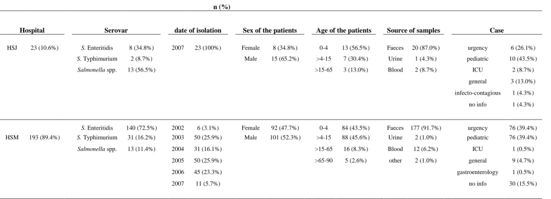
Analyzing the sample per hospital, the isolates retrieved from the HSJ hospital were in the majority identified only as Salmonella spp. whereas the isolates retrieved from HSM the majority was identified as serovar Enteritidis. Though the sample from HSJ is visibly smaller than from HSM the tendencies are a match. The distribution of the sample according to sex shows a higher number in male isolates than female; the majority of isolates are from children age 0-4 or 4-15; around 90% of both samples are isolated from faeces, isolates from blood are present in both samples; most isolates come from urgency and pediatric patients.
The presence of both serovars in blood samples is not unexpected as all Salmonella spp. can cause blood stream infections although the main invasive organisms are S. enterica serovar Typhi, Paratyphi, Choleraesuis and Dublin. The occurrence of blood stream infection caused by Salmonella spp. is more common in the elderly, the very young and in immunocompromised patients (Stephen et al, 2002). In our sample the blood isolates are evenly distributed in all age ranges, but because we don’t have access to the full medical history of each patient we cannot state that it does not follow the expected trend.
21
Table 2 – Distribution of the 216 isolates per hospital, strain, date of isolation, sex and age of the patients, and source of sample, in percentage.
n (%)
Hospital Serovar date of isolation Sex of the patients Age of the patients Source of samples Case
HSJ 23 (10.6%) S. Enteritidis 8 (34.8%) 2007 23 (100%) Female 8 (34.8%) 0-4 13 (56.5%) Faeces 20 (87.0%) urgency 6 (26.1%) S. Typhimurium 2 (8.7%) Male 15 (65.2%) >4-15 7 (30.4%) Urine 1 (4.3%) pediatric 10 (43.5%) Salmonella spp. 13 (56.5%) >15-65 3 (13.0%) Blood 2 (8.7%) ICU 2 (8.7%)
general 3 (13.0%)
infecto-contagious 1 (4.3%)
no info 1 (4.3%)
S. Enteritidis 140 (72.5%) 2002 6 (3.1%) Female 92 (47.7%) 0-4 84 (43.5%) Faeces 177 (91.7%) urgency 76 (39.4%) HSM 193 (89.4%) S. Typhimurium 31 (16.2%) 2003 50 (25.9%) Male 101 (52.3%) >4-15 88 (45.6%) Urine 2 (1.0%) pediatric 76 (39.4%) Salmonella spp. 13 (11.4%) 2004 31 (16.1%) >15-65 16 (8.3%) Blood 12 (6.2%) ICU 1 (0.5%) 2005 50 (25.9%) >65-90 5 (2.6%) other 2 (1.0%) general 9 (4.7%)
2006 45 (23.3%) gastroenterology 1 (0.5%)
2007 11 (5.7%) no info 30 (15.5%)
22
Antimicrobial susceptibility testing
The Minimal Inhibitory Concentration (MIC) of six antimicrobial agents: ampicillin (A), chloramphenicol (C), streptomycin (S), tetracycline (T) and nalidixic acid (Nx) were determined by the agar dilution method as described by the Clinical Laboratory Standards Institute (CLSI, 2007), and of trimethoprim/sulfamethoxazole determined by E-test (ABBiodisk/ BioMérieux, France).
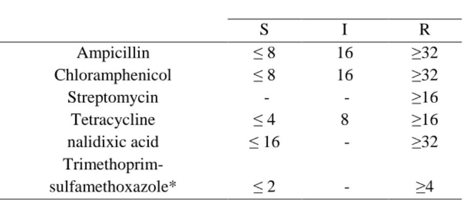
The CLSI manual defines three categories for the results, as quoted:
Susceptible – this implies that isolates are inhibited by the usually achievable concentrations of antimicrobial agent when the recommended dosage is used for the site of infection;
Intermediate – this category includes isolates with antimicrobial agent MICs that approach usually attainable blood and tissue levels and for which response rates may be lower than for susceptible isolates. The intermediate category implies clinical efficacy in body sites where the drugs are physiologically concentrated or when a higher than normal dosage of a drug can be used.
Resistant – this category implies that isolates are not inhibited by the usually achievable concentrations of the agent with normal dosage schedules and/or that demonstrate MICs that fall in the range where specific microbial resistance mechanisms are likely, and clinical efficacy of that agent against the isolate has not been reliably shown in treatment studies.
Table 3- MIC Interpretative standards according to CLSI/NCCLS manual.
MIC (µg/ml) S I R Ampicillin ≤ 8 16 ≥32 Chloramphenicol ≤ 8 16 ≥32 Streptomycin - - ≥16 Tetracycline ≤ 4 8 ≥16 nalidixic acid ≤ 16 - ≥32 Trimethoprim-sulfamethoxazole* ≤ 2 - ≥4 * for E-test was considered resistant if MIC ≥32
The isolates were streaked on TSA plates and incubated for 24 hours at 37 ˚C. Cell suspensions were prepared in sterile Ringer’s solution (LabM) and adjusted to the 0.5 McFarland standard. The suspensions were then spotted, using a calibrated 1µl loop, on
23
Mueller-Hinton agar (MHA; BioMerieux, Marcy l’Etoile, France) plates containing serial dilutions of all antibiotics, except ampicillin that was in Muller-Hinton agar cation-adjusted plates. The plates were incubated for 18 hours at 37 °C and the results were recorded in terms of the highest concentration with bacterial growth (CLSI/NCCLS, 2007).
The E-test was performed according to the manufacturer’s instructions. The E-test strip contained a serial dilution of the antimicrobial; a single organism suspension was used to inoculate a Mueller-Hinton agar plate, on which the e-test strip was placed. The cultures were incubated at 37 °C for 18 hours. For all isolates the breakpoints used were those defined by the CLSI for Enterobacteriaceae (Table 2). Staph. aureus ATCC 29213 was used as the control strain.
24
Pulsed-field gel electrophoresis (PFGE)
Sixty five isolates were subjected to DNA macro-restriction analysis by pulsed-field gel electrophoresis, following XbaI digestion, according to the CDC PulseNet one-day standardized laboratory protocol for molecular subtyping of E.coli O157:H7, non-typhoidal
Salmonella serotypes, and Shigella sonnei (PulseNet, 2004).
The Salmonella isolates were grown on Trypticase Soy Agar with 5% (v/v) defibrinated Sheep blood (Liofilchem, Teramo, Italy) at 37 °C overnight. A bacterial suspension was prepared in 3 ml of Cell Suspension Buffer (100 mM Tris: 100 mM EDTA pH8) using a sterile cotton swab, cell density was adjusted to 1.35 OD610 nm in a heλios spectrophotometer
(Spectronic Unicam, Leeds, UK). 400 µl of adjusted suspension were transferred to a 2 ml microcentrifuge tube and 20 µl of Proteinase K (PK; BIORON GmbH, Ludwigshafen Germany) solution (20 mg/ml) was added, then mixed by pipetting up and down. A volume of 400 µl of melted 1% w/v SeaKem® Gold agarose (Lonza, Rockland, ME USA) 1% w/v Sodium Dodecyl Sulfate (Bio-Rad, CA USA) prepared in sterile TE buffer and maintained at 55 to 60 °C, was added to the cell suspension and mixed gently by pipetting up and down. The mixture was immediately dispensed into plug mold wells (sample reusable plug mold Bio-Rad) and allowed to solidify for 10 to 15 min at room temperature. The agarose plugs were transferred to 15 ml tubes containing 5 ml of Cell Lysis Buffer (50 mM Tris 50 mM EDTA pH8, 1% w/v Sarcosyl, 0.1mg/ml PK) and incubated at 54 °C for 2hours at 170 rpm. After proteolysis the CLB was removed and the plugs were washed twice with 15 ml of sterile ultrapure water at 50 °C (water bath) for 10 min each wash, followed by four washes with 15 ml of TE buffer at the same temperature for 15 min. After the TE wash, the plugs were sliced (2 mm: 6 mm), on a microscope glass slide using a sterile blade, and stored in 1.5 ml TE at 4 °C until restriction. The agarose embedded DNA plug slices were restricted with 50 U of XbaI (Fermentas, Ontario Canada) at 37 °C for 5 hours.
The digested DNA plug slices were separated by PFGE through 1% SeaKemGold agarose in 0.5X TBE buffer (44.5 mM Tris, 44.5 mM boric acid, 1 mM EDTA) at 14 °C using CHEF DR-III system (Bio-Rad Laboratories). The electrophoresis conditions used were as follows: initial switch time 2.2 s; final switch time, 63.8 s; voltage, 6 V; included angle, 120°; run time, 18 to 19 hours. Genomic DNA from Salmonella serotype Braenderup H9812 was also restricted with XbaI and used as a molecular size marker. After electrophoresis the gel was stained with ethidium bromide (40 µl of [1 mg/ml] in 400 ml of deionized water) for 15
25
min and washed twice for 30 min in water (PulseNet, 2004). The gel was then visualized and recorded using Gel-Doc 2000 (Bio-Rad laboratories) under UV transillumination. Band patterns were analyzed with Gelcompar software (Applied Maths, Kortijk, Belgium) using Dice similarity coefficients with 1.5% band position tolerance. Isolates relatedness was determined by the unweighted-pair group method using average linkages (UPGMA).
26
Results
Antimicrobial susceptibility testing
The frequency of resistance and susceptibility of the isolates to individual antimicrobial agents is shown in Table 4. The highest resistance percentage observed was of 42% for nalidixic acid, resistance percentages of 20 to 25 were observed for ampicillin, tetracycline and streptomycin. Most of the isolates were sensitive to trimethoprim-sulfamethoxazole, chloramphenicol, streptomycin, tetracycline and ampicillin.
Table 4- Susceptibility to antibiotics of the Salmonella isolates tested.
Antibiotic %S* %I** %R*** Ampicillin 73 1 25 Chloramphenicol 84 0 15 Streptomycin 79 0 20 Tetracycline 76 0 23 Nalidixic acid 52 0 42 Trimethoprim-sulfamethoxazole 95 0 3
*,Sensible; **, Intermediate; ***,Resistant
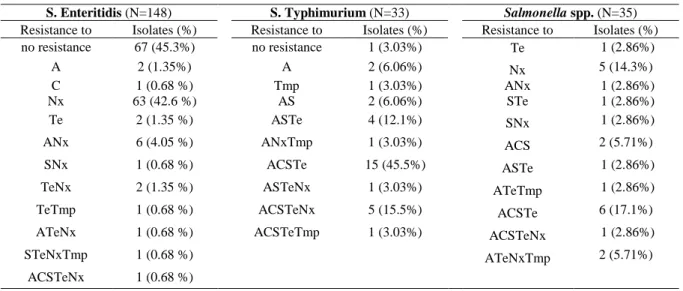
Of the isolates displaying resistance, 44% showed resistance to more than one antibiotic of which 42% were resistant to four and 27% to two. The resistance profiles found in the tested isolates are presented in Table 5. MIC50 and MIC90 values by serovar per year are presented in Table 6 and Figure4, 5, and 6.
Table 5- Frequency of Salmonella isolates resistance patterns by serovar.
S. Enteritidis (N=148) S. Typhimurium (N=33) Salmonella spp. (N=35)
Resistance to Isolates (%) Resistance to Isolates (%) Resistance to Isolates (%)
no resistance 67 (45.3%) no resistance 1 (3.03%) Te 1 (2.86%)
A 2 (1.35%) A 2 (6.06%) Nx 5 (14.3%)
C 1 (0.68 %) Tmp 1 (3.03%) ANx 1 (2.86%)
Nx 63 (42.6 %) AS 2 (6.06%) STe 1 (2.86%)
Te 2 (1.35 %) ASTe 4 (12.1%) SNx 1 (2.86%)
ANx 6 (4.05 %) ANxTmp 1 (3.03%) ACS 2 (5.71%)
SNx 1 (0.68 %) ACSTe 15 (45.5%) ASTe 1 (2.86%)
TeNx 2 (1.35 %) ASTeNx 1 (3.03%) ATeTmp 1 (2.86%)
TeTmp 1 (0.68 %) ACSTeNx 5 (15.5%) ACSTe 6 (17.1%)
ATeNx 1 (0.68 %) ACSTeTmp 1 (3.03%) ACSTeNx 1 (2.86%)
STeNxTmp 1 (0.68 %) ATeNxTmp 2 (5.71%)
ACSTeNx 1 (0.68 %)
27 0,031 0,063 0,125 0,250 0,500 1,000 2,000 4,000 8,000 16,000 32,000 64,000 128,000 256,000 2002 2003 2004 2005 2006 2007 2007 HSJ µ g /m l S. Enteritidis MIC50
ampicillin chloramphenicol streptomycin
tetracyclin nalidixic acid trimethropim-sulfametoxazole
0,031 0,063 0,125 0,250 0,500 1,000 2,000 4,000 8,000 16,000 32,000 64,000 128,000 256,000 2002 2003 2004 2005 2006 2007 2007 HSJ µ g / m l S. Enteritidis MIC90
ampicillin chloramphenicol streptomycin
tetracyclin nalidixic acid trimethropim-sulfametoxazole
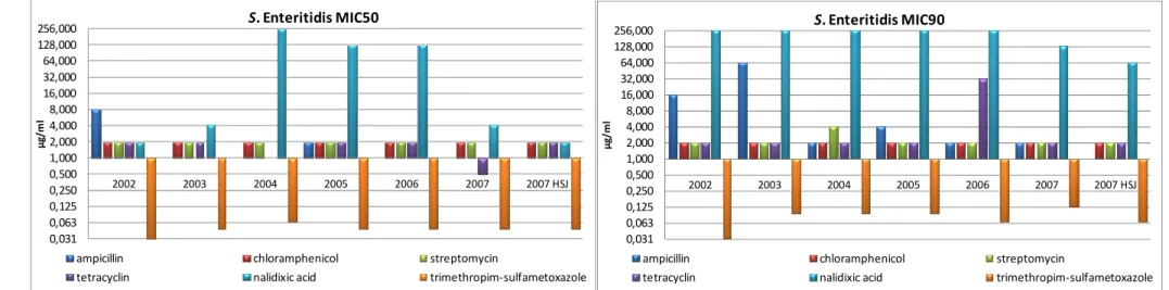
Figure 4 - Graphic representation of the MIC50 and MIC90 for S. Enteritidis per year.
0,063 0,125 0,250 0,500 1,000 2,000 4,000 8,000 16,000 32,000 64,000 128,000 256,000 512,000 2002 2003 2004 2005 2006 2007 2007 HSJ µ g /m l S. Typhimurium MIC90
ampicillin chloramphenicol streptomycin
tetracyclin nalidixic acid trimethropim-sulfametoxazole
0,031 0,063 0,125 0,250 0,500 1,000 2,000 4,000 8,000 16,000 32,000 64,000 128,000 256,000 512,000 2002 2003 2004 2005 2006 2007 2007 HSJ µ g / m l S. Typhimurium MIC50
ampicillin chloramphenicol streptomycin
tetracyclin nalidixic acid trimethropim-sulfametoxazole
28 0,031 0,063 0,125 0,250 0,500 1,000 2,000 4,000 8,000 16,000 32,000 64,000 128,000 256,000 512,000 2003 2004 2005 2006 2007 HSJ µ g /m l Salmonella spp. MIC50
ampicillin chloramphenicol streptomycin
tetracyclin nalidixic acid trimethropim-sulfametoxazole
0,063 0,125 0,250 0,500 1,000 2,000 4,000 8,000 16,000 32,000 64,000 128,000 256,000 512,000 2003 2004 2005 2006 2007 HSJ µ g /m l Salmonella spp. MIC90
ampicillin chloramphenicol streptomycin
tetracyclin nalidixic acid trimethropim-sulfametoxazole
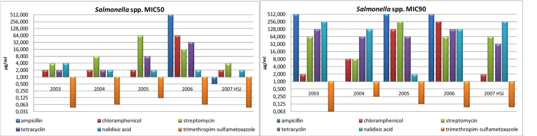
Figure 6 - Graphic representation of the MIC50 and MIC90 for Salmonella spp. per year.
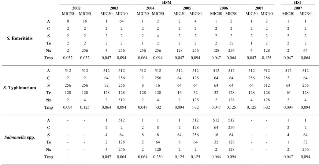
For S. Enteritidis the MIC50 values for all the tested antimicrobials have in the majority maintained. The MIC50 for Nalidixic acid which have increased significantly from 2004 to 2006 has in 2007 decreased to the value observed in 2003. Values from 2007 for the HSJ isolates are similar to the ones observed for the HSM isolates. The MIC90 values for this serovar are mostly stable, ampicillin and tetracycline values oscillate more though the tendency is to decrease. The MIC90 values for Nalidixic acid have been fairly stable until 2006 and show a decrease in 2007.
For S. Typhimurium the MIC50 and MIC90 values for ampicillin are the same for all years. The values for the other antimicrobials show a steady increase. Trimethropim-sulphametoxazole MIC90 values show higher values for 2004, 2005 and 2007. The MIC values for HSJ isolates are mainly lower than for HSM isolates.
29
Pulsed-field gel electrophoresis (PFGE)
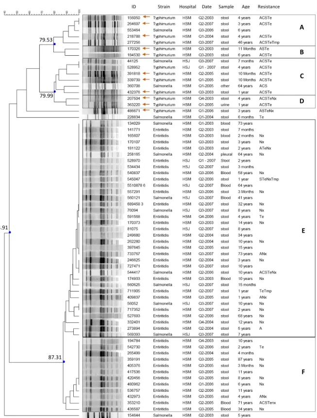
Clusters were defined using an 80% similarity index according to Foley et al. (2006). Using restriction enzyme XbaI, six clusters were found, A to E (fig. 7).
Figure 7 - Dendrogram of similarity illustrating the genetic relationships between the XbaI profiles using the Dice coefficient, and clustering by UPGMA. (←: isolates tested positive for sulphonamides resistance in Freixo et al., 2011)
ID Strain Hospital Date Sample Age Resistance Dice (Opt:1.38%) (Tol 0.9%-0.9%) (H>0.0% S>0.0%) [0.0%-100.0%]
79.53 79.99 43.91 87.31 A B C D E F
30
Discussion
Analyzing the frequency of resistance to 1 up to 5 antimicrobials, there is a noted difference between the tested serovars. S. Enteritidis isolates are either sensitive to all the antimicrobials tested (45.3%) or resistant to just one (45.98%), mainly Nalidixic acid. The frequency of multiresistant Enteritidis isolates is very low, 1.35% are resistant to four or more antimicrobials. On the other hand, S. Typhimurium shows opposite results, 63.4% of the Typhimurium isolates, in this study, are resistant to 4 or more antimicrobials. This is a higher number than the 39.7% reported by the EFSA for the year 2006, but no data from Portugal isolates was available and variability between countries is expected (EFSA,2010). The
Salmonella spp. isolates show mixed results, the majority of these isolates are either sensitive
to all the antimicrobials or secondly, resistant to 4 of the antimicrobials tested. Since these isolates were only identified as Salmonella spp., and no further strain identification was performed, some diversity is expected and therefore the results are diverse.
The analysis of the PFGE profiles of a small panel of the isolates resulted in six clusters when a cut-off of around 80% similarity is applied. Some of the samples only primarily identified as Salmonella spp. are distributed in Enteritidis and Typhimurium clusters as well as unique clones. This validates the assay demonstrating that the discriminatory power of the PFGE tool allows us to differentiate the two serotypes and further identify the samples. Also, isolate 154644 identified as Salmonella spp. was neither similar to Enteritidis or Typhimurium isolates (43.91% similarity), wich can be seen as a control that the assay had no identification bias toward the other two serotypes. The samples from both hospitals are mixed indicating high similarity between isolates from the two areas (Figure 7).
Salmonella serovar Enteritidis shows a general predominant resistance to nalidixic acid
(63 out of 81 resistant isolates). Multidrug-resistant isolates are rare and most are resistant to a second antimicrobial in combination with the resistance to nalidixic acid. The PFGE analysis shows two large groups (E and F) with high similarity (87.31%). Samples from HSJ are all present in cluster E. No differentiation of isolates with different resistance patterns was possible with the PFGE using the XbaI enzyme. Enforcing the evident need to use a different restriction approach. Other works with PFGE profiling of S. Enteritidis show the same difficulty in discrimination power, this can be overcome by the use of a second or third enzyme (Zheng et al., 2007). For some of the isolates extra bands are visible, this can be explained by the acquisition of genetic material containing an extra XbaI restriction site or by point mutation. An explanation to the high similarity can be that the genetic variation may not impact the restriction fragments in a visible way with restriction using XbaI enzyme.
31
Salmonella serovar Typhimurium isolates are mainly resistant to two or more
antimicrobials. The most frequent resistance profile is ACSTe counting for 45% of the isolates. The sample analyzed by PFGE profiling showed high diversity. The isolates are grouped in four clusters, A, B, C and D. Though the discrimination is higher than for the Enteritidis isolates, it is difficult to establish correlation between the isolates of the same group based solely on the isolation information and resistance profile to a small panel of antimicrobials. The different clusters do not present specific characteristics and the available information is fairly distributed. Isolate 486671 presents a very distinctive PFGE profile , with 58% similarity to the rest of the isolates. Also isolate 228834, identified as Salmonella spp. has a very distinct PFGE profile and is resistant only to Te, of the tested panel. The isolates marked with the orange arrow in figure 7 were also tested for sulphadiazine resistance in previous works in the laboratory, and were positive (Freixo et al., 2011) thus displaying the resistance profile of Salmonella Typimurium DT104. Two isolates present the ACSSUTe (ampicillin, chloramphenicol, streptomycin, sulphadiazine and tetracycline) resistance profile in combination to nalidixic acid resistance. The added resistance to a sixth antimicrobial is not uncommon for DT104 isolates and has been observed for gentamicin, trimethoprim and fluoroquinolones (Baggensen et al. 2000). The Typhimurium isolates from the different hospitals are similar; the only two isolates from HSJ are 100% similar in PFGE profile, resistance profile, isolation date and source and present 90% similarity to the rest of the isolates in the same cluster.
32
Conclusion
In this study, two hundred and sixteen isolates identified as Salmonella spp., Salmonella Enteritidis and Typhimurium, were tested for antimicrobial resistance against a panel of five antibiotics and a smaller panel was characterized using PFGE. Results showed that resistance to five antimicrobials is commonly found in Typhimurium isolates while Enteritidis shows a specific emergent resistance to nalidixic acid. High resistance is distributed in the isolates from the younger and older age groups (babies, children and the elderly), as has been described by the EFSA (2009). A panel of sixty-five isolates was subjected to PFGE analysis with XbaI restriction enzyme to assess the clonality. The analysis revealed that the discriminatory power of PFGE is higher for Typhimurium isolates than for Enteritidis. While PFGE is considered the “gold standard” molecular typing method, if genetic variation does not significantly impact the size or mobility of a specific restriction fragment during electrophoresis, then the change may not be identified as a separate pulsotype. This limitation can be overcome by the use of a second enzyme, at least,or even a different panel of restriction enzymes for the analysis. Though the discriminatory power was higher for Typhimurium isolates it is hard to elaborate on the clonallity of the isolates since the available information is not sufficient to establish a correlation between isolates from the same cluster and between different clusters.
The two analyses performed with this set of isolates are only part of a more extensive characterization usually performed on outbreak isolates. To complete the information on this library of Portuguese clinical isolates, we would need access to epidemiological data for the two regions to establish correlations, and the availability of resources to extend the characterization and construct a database.
This work allowed the “Laboratório de Bactérias Lácticas e Pescado” of the Superior School of Biotechnology, of the Portuguese Catholic University, to implement and optimize the PFGE Standard Protocol used by the CDC and to further characterize the existing library of clinical isolates of Salmonella.
33
Future work
The antimicrobial susceptibility testing was limited to the antimicrobials available, more antimicrobials typically tested in Salmonella, like sulphonamides, could be used in further works.
The PFGE analysis of the isolates could be improved with the use of an additional panel of restriction enzymes to further differentiate the S. Enteritidis isolates. Also since only a small sample of the isolates were able to be tested by PFGE, this analysis could be applied to a larger sample in order to verify the tendencies observed.
Salmonella isolates are target of much analysis like phage typing and gene profiling for virulence and resistance, an interesting follow up would be to build a database for these isolates so that all the information collected in the ESB-UCP research projects could be searched and correlated easily. This could be a useful tool for matching new isolates with previously tested ones in order to assess the recurrence of these isolates in food quality control tests and clinical outbreaks.
34
References
Baggesen, D.L., Sandvang D., Aarestrup, F.M. 2000. Characterization of Salmonella
enterica serovar Typhimurium DT104 isolated from Denmark and comparison with
isolates from Europe and the United States. J. Clin. Microbiol. 38:1581–1586.
Bäumler, A.J., Tsolis, R.M., Ficht, T.A., Adams, L.G. 1998. Evolution of host adaptation in Salmonella enterica. American Society for Microbiology, Infection and Immunity vol.66 10:4579-4587.
Boyle, E.C., Bishop, J.L., Grassl, G.A., Finlay, B.B 2007. Salmonella: From pathogenesis to therapeutics. J Bacteriol. 189:1489–1495.
Brichta-Harhay, D.M., Arthur, T.M., Bosilevac, J.M., Kalchayanand, N., Shackelford, S.D., Wheeler, T.L., Koohmaraie, M. 2011. Diversity of Multidrug-Resistant
Salmonella enterica Strains Associated with Cattle at Harvest in the United States. Appl. Environ. Microbiol. 77: 1783-1796.
Carattoli, A. 2003. Plasmid-Mediated Antimicrobial Resistance in Salmonella enterica.
Curr. Issues Mol. Biol. 5:113-122
Casewell, M., Friis, C., Marco, E., McMullin, P. and Phillips, I. 2003. The European ban on growth-promoting antibiotics and emerging consequences for human and animal health. Journal of Antimicrobial Chemotherapy 52:159–161.
McEwen, S.A., Fedorka-Cray, P.J. 2002. Antimicrobial use and resistance in animals. Clin
Infect. Dis. 34:S93-S106.
Clinical and Laboratory Standards Institute/NCCLS. 2007. Performance standards for antimicrobial susceptibility testing: 17th ed. CLSI/NCCLS M100-S17. Clinical and
Laboratory Standards Institute, Wayne, PA
Darwin, K.H. and Miller, V. I. 1999. Molecular basis of the interaction of Salmonella with the intestinal mucosa. Clin. Microbiol. Rev. 12:405-428.
Davis, M.A., Besser, T.E., Eckmann, K., MacDonald, J.K., Green, D., Hancock, D.D. 2007. Multidrug-resistant Salmonella Typhimurium, Pacific Northwest, United States. Emerg. Infect. Dis. vol. 13 10:1583-1586.
EFSA 2009. The Community Summary Report on Food-borne Outbreaks in the European Union in 2007. The EFSA Journal 271.
EFSA 2010. The Community Summary Report on antimicrobial resistance in zoonotic and indicator bacteria from animals and food in the European Union in 2004-2007. EFSA