Desulfovibrio marrakechensis sp. nov., a
1,4-tyrosol-oxidizing, sulfate-reducing bacterium
isolated from olive mill wastewater
Fatima Chamkh,
1Cathrin Spro¨er,
2Paulo Costa Lemos,
3Stephane Besson,
3,4Abdel-Ghani El Asli,
5Rhizlane Bennisse,
1Marc Labat,
6Maria Reis
3and Abdel-Illah Qatibi
1Correspondence Abdel-Illah Qatibi
1Anaerobic Microbiology Team (E02B26), Sciences and Techniques Faculty, Cadi Ayyad University,
PO Box 549, 40 000 Marrakech, Morocco
2DSMZ – Deutsche Sammlung von Mikroorganismen und Zellkulturen GmbH, Inhoffenstraße 7B,
D-38124 Braunschweig, Germany
3REQUIMTE/CQFB, Departamento de Quı´mica, FCT/UNL, 2829-516 Caparica, Portugal 4Unidade de Investigac¸a˜o em Biodiversidade e Desenvolvimento, Universidade Luso´fona de
Humanidades e Tecnologias, Campo Grande 376, 1749-024 Lisboa, Portugal
5School of Science and Engineering, Al Akhawayn University, PO Box 1846, Ifrane, Morocco 6IRD, UMR180 LMBEC, Universite´ de Provence et de La Me´diterrane´e, ESIL, Case 925, 163
Avenue de Luminy, F-13288 Marseille, France
A novel mesophilic sulfate-reducing bacterium, EMSSDQ4T, was isolated from olive mill
wastewater in the semi-arid region of Morocco (Marrakech). Cells were Gram-negative, catalase-positive, straight rods that were non-motile and non-spore-forming and contained cytochrome c3
and desulfoviridin. The DNA G+C content was 65.1 mol%. Phylogenetic analysis based on 16S rRNA gene sequences revealed that the isolate was a member of the genus Desulfovibrio with Desulfovibrio carbinoliphilus D41T, Desulfovibrio alcoholivorans SPSNT, Desulfovibrio
fructosivorans JJTand Desulfovibrio carbinolicus EDK82Tas the most closely related strains with validly published names. In addition to the classical substrates used by Desulfovibrio species, the isolate oxidized 1,4-tyrosol, one of the most abundant phenolic compounds occurring in olive mill wastewater, to 4-hydroxyphenylacetate without ring cleavage. D. alcoholivorans SPSNTwas also found to carry out this reaction. Under air, strain EMSSDQ4Texhibited limited growth on lactate
and yeast extract in the absence of sulfate. On the basis of genotypic and phenotypic characteristics, it is proposed that the isolate represents a novel species, Desulfovibrio marrakechensis sp. nov. The type strain is EMSSDQ4T(5DSM 19337T5ATCC BAA-1562T).
Olive oil manufacturing results annually in the production of more than 36107m3 black olive mill wastewater (OMW), which has a high polluting organic load due to the high content of organic substances, including sugars, tannins, polyphenols, polyalcohols, pectins, lipids and a
wide variety of simple aromatic compounds, resulting from olive cell-wall degradation during the oil extraction process (Labat et al., 2000; Lesage-Meessen et al., 2001). OMW is frequently discharged into evaporation ponds, dumped directly in rivers or spread on soil, resulting in a major environmental problem in the main olive-producing countries of the Mediterranean region, mainly those in the south, such as Morocco. Microbiological studies on OMW and digesters treating these effluents have revealed the presence of metabolically diverse anaerobic bacteria (Mechichi et al., 2000; Chamkha et al., 2001; Thabet et al., 2004), all of which have been identified as anaerobic species belonging to the genus Clostridium.
In the course of a study on microbiological biodiversity in an aeration basin (a very large pond used formerly as an
Abbreviation: OMW, olive mill wastewater.
The authors dedicate this paper to the memory of Professor Norbert Pfennig (1925–2008).
The GenBank/EMBL/DDBJ accession number for the 16S rRNA gene sequence of strain EMSSDQ4Tis AM947130.
Photomicrographs of cells of strain EMSSDQ4Tand growth curves of
strain EMSSDQ4Tand related strains cultivated in air in the absence of
sulfate are available as supplementary material with the online version of this paper.
underground gallery for water for irrigation of the desert) located 10 km north-east of Marrakech city in Morocco (31u389 520 N 7u549 7.30 W) and used to treat OMW by natural evaporation, several aerobic and anaerobic bacteria that are resistant to the simple aromatic compounds present in these effluents have been isolated and char-acterized. One of them has been identified recently as belonging to a new genus (Liebgott et al., 2008) and is a Gram-positive, anaerobic, rod-shaped, spore-forming bac-terium that is unable to carry out dissimilatory sulfate reduction. In the present paper, strain EMSSDQ4T, a novel
Gram-negative, anaerobic, straight-rod-shaped, non-spore-forming, sulfate-reducing bacterium that oxidizes 1,4-tyrosol to 4-hydroxyphenylacetate, which was isolated from the same OMW evaporation pond, is described. To our knowledge, this is the first bacterium to be described that is capable of performing this metabolic process. 1,4-Tyrosol is one of the major simple aromatic compounds present in OMW (Fernandez-Bolanos et al., 1998; Mulinacci et al., 2001) and exhibits toxicity towards several micro-organisms (Capasso et al., 1995; Sayadi et al., 2000). It has been reported that only aerobic bacteria under oxic conditions are able to oxidize 1,4-tyrosol (Allouche et al., 2004; Abdelkafi et al., 2005; Liebgott et al., 2007). The basal medium used consisted of (l21 distilled water): 0.5 g K2HPO4, 0.5 g KH2PO4, 0.5 g NH4Cl, 0.4 g NaCl,
0.05 g CaCl2. 2H2O, 0.3 g MgCl2. 6H2O, 0.1 g KCl, 0.25 g
cysteine hydrochloride, 0.1 g yeast extract (Difco), 1 ml trace element mineral solution (Widdel & Pfennig, 1981) modified by Imhoff-Stuckle & Pfennig (1983) and 1 ml 0.1 % (w/v) resazurin. The pH was adjusted to 7.2 with 10 M KOH. Unless otherwise indicated, aliquots of 9 ml were dispensed into Hungate tubes under a stream of O2
-free N2/CO2 (80 : 20, v/v) gas, closed with butyl rubber
stoppers and subsequently autoclaved. Prior to inoculation, NaHCO3(30 mM), Na2S . 9H2O (1.7 mM) and 1 ml
filter-sterilized vitamin solution l21 (Balch et al., 1979) were added. For solid roll-tube media, 2 % (w/v) agar was added.
Samples of OMW (total content of phenols 2770–2820 mg l21), collected as described by Liebgott et al., (2008) during the 2004–2005 harvest season at a depth of 60 cm, were inoculated (10 %, v/v) into basal medium containing 10 mM lactate and 20 mM sulfate and then incubated for 2–3 weeks at 37 uC. After several transfers under the same conditions and using repeated agar serial dilutions, brown colonies, approximately 1–2 mm in diameter, were observed within 1 week of incubation in agar roll-tubes (Hungate, 1969). Well-separated colonies were picked from the highest dilutions (10210) and transferred to liquid basal medium containing 10 mM lactate and 20 mM sulfate. The purity of the culture was checked routinely by phase-contrast microscopy and by performing growth tests in basal medium containing 1 % (w/v) glucose, 1 % (w/v) yeast extract and 1 % (w/v) biotrypticase. Seven pure strains were obtained and subsequently shown to be similar by amplified rRNA gene restriction analysis profiles (data
not shown). One strain, designated EMSSDQ4 T
, was studied in detail. All physiological and metabolic tests were performed in triplicate at 37uC (except for temper-ature experiments) using cells growing on lactate and sulfate. Unless otherwise indicated, positive growth was determined by monitoring changes in OD580 and by
production of sulfides (Cord-Ruwisch, 1985), compared with negative controls.
Cells of strain EMSSDQ4Twere non-motile, straight rods
with rounded ends, approximately 1.3–1.6 mm in diameter and 3–4 mm in length, occurring either singly or in pairs. Motility was not observed by optical microscopy, even in young cells during their first step of exponential growth. The isolate did not possess a flagellum, as clearly shown by electron microscopy (Supplementary Fig. S1, available in IJSEM Online). Cells were Gram-negative, oxidase-nega-tive and catalase-posioxidase-nega-tive. Spores were not observed either by microscopic examination or in heat-resistance experi-ments (Liebgott et al., 2007).
Optimal growth conditions were determined in basal medium containing 10 mM lactate and 20 mM sulfate as described by Qatibi et al. (1991). Optimum growth temperature was estimated to be 37 uC. No growth was observed at temperatures above 50 uC or below 20uC. Growth was optimal at pH 7, with growth occurring between pH 6.5 and 8.5. When tested at various NaCl concentrations, growth was observed between 0 and 35 g l21, with optimum growth in the absence of NaCl. Reduction of different electron acceptors was performed in basal medium with 10 mM lactate and supplied with distinct electron acceptors: 20 mM sulfate, 10 mM sulfite, 10 mM thiosulfate, 1 % (w/v) elemental sulfur, 5 mM nitrate, 5 mM nitrite and 10 mM fumarate. Positive growth with the latter three electron acceptors was estimated by monitoring changes in optical density. In addition to sulfate, sulfite, thiosulfate, elemental sulfur and fumarate also served as electron acceptors. The isolate did not reduce nitrate or nitrite.
Because several aerobic bacteria were also isolated (see for example Liebgott et al., 2007) from the same site as strain EMSSDQ4T, the capacity of the isolate to tolerate oxygen
(air) was assessed; cysteine hydrochloride and sulfides were omitted from the basal medium for these experiments. Cultivation was carried out in tubes containing 4.5 ml basal medium with 10 mM lactate under an air atmosphere corresponding to two-thirds of the culture volume (the calculated amount of oxygen in the headspace was 80 mmol) and incubated horizontally using 10 % (v/v) inoculum growing in basal medium containing 10 mM sulfate and 20 mM lactate (to ensure that sulfate derived from the inoculum was completely consumed); after inoculation, the sulfide concentration was approximately 0.5 mM. Positive growth was determined by monitoring changes in optical density. In the presence of air, some limited growth (OD580¡0.3) was observed in the absence
rate and the doubling time were approximately 0.008 h21 and 90 h, respectively. Increased growth (OD580¡0.47,
4.65 mM sulfide) of the isolate on 10 mM lactate in the presence of 20 mM sulfate was observed as it shifted back to anaerobic conditions; the maximum growth rate and the doubling time were approximately 0.16 h21 and 5 h, respectively. Under aerobic conditions, cells became elongated (results not shown) and growth was poor or absent after a second transfer. From these preliminary tests, it can be assumed that the isolate was at least able to survive oxygen exposure. It would be interesting to investigate more fully whether oxygen served as a direct or indirect electron acceptor by the isolate.
Aromatic compounds (filter-sterilized) were tested at a final concentration of 5 mM in modified basal medium with increased yeast extract (0.05 %, w/v) and 20 nM sodium selenite; non-inoculated tubes were observed under the same conditions to verify that aromatic compounds were not partially transformed abiotically. In addition to sulfide production and optical density, positive growth was also determined by analysis of degradation products. Aromatic compounds were analysed by HPLC using a Waters apparatus composed of a 1525 binary pump, a 2996 diode array detector, a Rheodyne injector (model 7725i) fitted with a 20 ml loop, a temperature control system and an in-line degasser. Separations were carried out on a Symmetry C18 reversed-phase column (4.66150 mm,
ODS2, 5 mm particle size). Elution was performed at 30uC with a flow rate of 0.8 ml min21 using a linear gradient of acetonitrile (A) in water acidified with 1 % (v/v) acetic acid (B) in two steps: the first step was from 5 to 20 % A for 25 min and the second step was from 20 to 100 % A for 5 min (total 30 min). Aromatic compounds were visualized at 280 nm and total spectra from each peak were analysed from 200 to 400 nm. Peaks were determined by comparing their retention times and respective UV spectra online (diode array detection) with commercial standards, except for o-methylgallate, which is not available commercially and was synthesized according to Scheline (1966).
Utilization of 21 simple aromatic compounds present in OMW in the presence of sulfate as electron acceptor was tested. Strain EMSSDQ4T, Desulfovibrio alcoholivorans
SPSNT, Desulfovibrio fructosivorans JJT and Desulfovibrio carbinolicus EDK82T were negative for utilization of phenol, catechol, resorcinol, 4-hydroxyphenylacetate, benzoate, 4-hydroxybenzoate, 3-hydroxybenzoate, proto-catechuate, syringate, gallate, o-methylgallate, veratrate, p-anisate, cinnamate, p-, o- and m-coumarate, vanillate, ferulate and caffeate; the phenolic compound 1,4-tyrosol was utilized by strain EMSSDQ4T and D. alcoholivorans
SPSNT only (Table 1). 1,4-Tyrosol was oxidized by both these strains to 4-hydroxyphenylacetate at 100 % recovery leaving the aromatic ring intact (5 mmol 1,4-tyrosol was oxidized by strain EMSSDQ4T and D. alcoholivorans
SPSNTresulting in production of approximately 3.1 mmol and 2.47 mmol sulfide, respectively). These observations
strongly suggest the following oxidation equation:
C8O2H10+0.5SO422AC8O3H8+0.5S22+H2O
4-Hydroxyphenylacetate was not metabolized further by either strain even after more than 1 month of incubation. Under fermentation conditions, both strains were unable to utilize 1,4-tyrosol. It is interesting to note that, in the presence of sulfate, both strains were unable to metabolize other phenolic compounds such as, for example, unsub-stituted phenol, 2-hydroxyphenol (catechol), 3-hydroxy-phenol (resorcinol), 4-hydroxycinnamate, 4-hydroxybenzo-ate and 4-hydroxyphenylacet4-hydroxybenzo-ate, indicating that the composition and the position of the side chain were determinants for substrate specificity. A primary alcohol side chain in the para position to the hydroxyl group was required for the transformation of aromatic substrates. When phylogenetic analysis revealed that strain EMSSDQ4
T
was related to some Desulfovibrio species (see below), other simple substrates commonly used by these bacteria were tested. Substrate utilization was tested in basal medium in the presence of 20 mM sulfate (except for fermentation tests) containing different electron donors at a final concentration of 10 mM. Formate, methanol and hydrogen (H2/CO2 at 80 : 20 %, v/v) were tested in the absence or
presence of 5 mM acetate as carbon source. Yeast extract, peptone, Casamino acids and biotrypticase were tested at a final concentration of 0.5 g l21. Lactate and acetate were analysed by HPLC using a Bio-Rad Aminex HPX-87H column, with 0.005 M sulfuric acid as eluent (elution rate of 0.6 ml min21) and an operating temperature of 50uC. A UV detector (Merck) set at 210 nm was used. Growth of the isolate occurred in the absence of vitamins or yeast extract after three transfers. Several non-aromatic substrates were used in the presence of sulfate including ethanol, 2-methoxyethanol, 1-propanol, 2-propanol, 1-butanol, 1,2-propanediol, 1,3-1,2-propanediol, 1,4-butanediol, hydrogen, formate, lactate, pyruvate, fumarate and malate. With the latter three substrates, small amounts of sulfide were produced (¡1.5 mM). When sulfate was replaced by sulfite (10 mM), which is the energetically more favourable electron acceptor, two to three times more sulfide was produced. Lactate was oxidized to acetate and presumably carbon dioxide. Growth with molecular hydrogen and formate required acetate as carbon source. Growth with formate, 1,4-butanediol and 2-propanol was weak. In the absence of sulfate, fumarate, malate and pyruvate could be fermented. No growth was observed on acetate, propionate, butyrate, methanol, 2-butanol, glycerol, ethylene glycol, 2,3-butanediol, crotonate, alanine, glycine, arginine, lysine, choline, lactose, sucrose, glucose, fructose, maltose, man-nose, ribose, xylose, yeast extract, biotrypticase, Casamino acids or peptone.
Pigments were assayed as described previously (Qatibi et al., 1991), except that cells were disrupted by ultrasound cycles and the resulting cell-free extract was concentrated in an Amicon ultrafiltration cell (10 kDa pore size) before
examination for cytochrome c3 and desulfoviridin. The
spectrum of the oxidized extract showed a characteristic absorption band at 629 nm, indicating the presence of desulfoviridin. When reduced by sodium dithionite, the spectrum of cell-free extracts exhibited the characteristic absorption bands of cytochrome c3, with maxima at 415.0,
522.5 and 553.0 nm.
The DNA G+C content of strain EMSSDQ4T, determined
by HPLC (Mesbah et al., 1989) at the Identification Service of the DSMZ (Deutsche Sammlung von Mikroorganismen und Zellkulturen, Braunschweig, Germany), was 65.1 mol%, which is within the range reported for Desulfovibrio species (48–65 mol%).
Phylogenetic analysis was carried out as follows. DNA from strain EMSSDQ4T was extracted using the DNeasy tissue
kit according to the manufacturer’s protocol (Qiagen). Purified PCR product was sequenced using the CEQTMDTCS-Quick Start kit (Beckmann Coulter) as directed in the manufacturer’s protocol. Sequence reac-tions were electrophoresed using the CEQTM 8000 genetic analysis system. Primers F (59-GAGTTTGATCCTGG-CTCA-39) and R (59-AGAAAGGAGGTGATCCAGCC-39) were used to amplify the 16S rRNA gene. The nucleotide sequence was aligned manually using the sequence alignment editor ae2 (Maidak et al., 2001). Sequences used in the phylogenetic analysis were obtained from the RDP (Maidak et al., 2001) and GenBank (Benson et al., 1999).
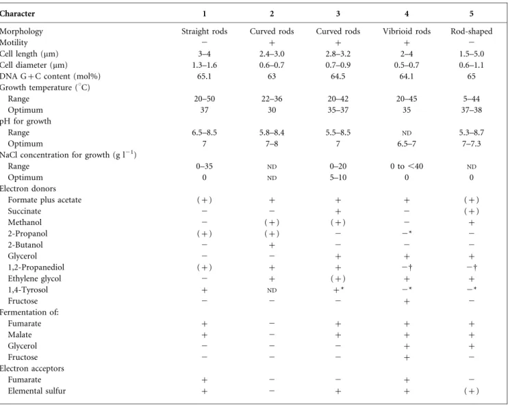
Table 1. Differential characteristics of strain EMSSDQ4 T
and type strains of related Desulfovibrio species
Strains: 1, D. marrakechensis sp. nov. EMSSDQ4T(data from this study); 2, D. carbinoliphilus D41T(Allen et al., 2008) 3, D. alcoholivorans SPSNT
(Qatibi et al., 1991); 4, D. fructosivorans JJT(Ollivier et al., 1988; Qatibi et al., 1991); 5, D. carbinolicus EDK82T(Nanninga & Gottschal, 1987; Qatibi et al., 1991).+, Positive; 2, negative; (+), weakly positive;ND, no data available.
Character 1 2 3 4 5
Morphology Straight rods Curved rods Curved rods Vibrioid rods Rod-shaped
Motility 2 + + + 2
Cell length (mm) 3–4 2.4–3.0 2.8–3.2 2–4 1.5–5.0
Cell diameter (mm) 1.3–1.6 0.6–0.7 0.7–0.9 0.5–0.7 0.6–1.1
DNA G+C content (mol%) 65.1 63 64.5 64.1 65
Growth temperature (uC)
Range 20–50 22–36 20–42 20–45 5–44
Optimum 37 30 35–37 35 37–38
pH for growth
Range 6.5–8.5 5.8–8.4 5.5–8.5 ND 5.3–8.7
Optimum 7 7–8 7 6.5–7 7–7.3
NaCl concentration for growth (g l21)
Range 0–35 ND 0–20 0 to ,40 ND
Optimum 0 ND 5–10 0 0
Electron donors
Formate plus acetate (+) + + + (+)
Succinate 2 2 + 2 (+) Methanol 2 (+) (+) 2 + 2-Propanol (+) (+) 2 2* 2 2-Butanol 2 + 2 2 2 Glycerol 2 2 + + + 1,2-Propanediol (+) + + 2D 2D Ethylene glycol 2 + (+) + + 1,4-Tyrosol + ND +* 2* 2* Fructose 2 2 2 + 2 Fermentation of: Fumarate + 2 + + + Malate + 2 + + + Glycerol 2 2 2 + + Fructose 2 2 2 + 2 Electron acceptors Fumarate + 2 2 + 2 Elemental sulfur + 2 + + (+)
*Tested in the present study.
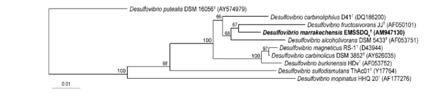
Pairwise evolutionary distances were calculated using the method of Jukes & Cantor (1969). A dendrogram was constructed using the neighbour-joining method (Saitou & Nei, 1987). Confidence in the tree topology was deter-mined using 1000 bootstrapped trees (Felsenstein, 1985). Analysis of the complete sequence (1539 bp) of the 16S rRNA gene of strain EMSSDQ4T revealed that the isolate
was related to species of the genus Desulfovibrio in the family Desulfovibrionaceae in the Deltaproteobacteria. It represents a new sublineage (supported by a bootstrap value of 100 %) within the genus Desulfovibrio (Fig. 1) and, in particular, forms a subline with a subcluster of species consisting of Desulfovibrio carbinoliphilus D41T (97.5 % sequence similarity with the type strain; Allen et al., 2008), D. alcoholivorans SPSNT (97.4 %; Qatibi et al., 1991), D. fructosivorans JJT (97.2 %; Ollivier et al., 1988), D. carbinolicus EDK82T (96.7 %; Nanninga & Gottschal, 1987), Desulfovibrio burkinensis HDvT (96.0 %; Ouattara et al., 1999), Desulfovibrio magneticus RS-1T (95.6 %; Sakaguchi et al., 2002) and Desulfovibrio sulfodismutans ThAc01T(94.2 %; Bak & Pfennig., 1987) Strain EMSSDQ4T
shared a branching node with D. fructosivorans JJT, although this relationship was not supported by a statistically significant bootstrap value (67 %), demonstrat-ing that this is not a particularly specific relationship. A 16S rRNA gene sequence similarity of 97 % is commonly considered as the upper limit for the definition of separate species (Stackebrandt & Goebel, 1994). Although more than 97 % similarity indicates that strains may belong to the same species, it is now generally acknowledged that this rule does not always apply for several Desulfovibrio species. Furthermore, Table 1 shows that strain EMSSDQ4T
exhibited several characteristics that clearly distinguish it from closely related species with validly published names. Strain EMSSDQ4T differed from D. carbinoliphilus D41T
(isolated from a gas condensate-contaminated aquifer), D. alcoholivorans SPSNT(isolated from a bioethanol produc-tion plant) and D. fructosivorans JJT (isolated from estuarine sediment) in its cell shape and motility; unlike these three motile Desulfovibrio species, cells of strain EMSSDQ4
T
were non-motile, straight rods. Two species of non-motile, straight rods, D. carbinolicus and Desulfovibrio
piger (Loubinoux et al., 2002), isolated from an anaerobic purification plant and human faeces, respectively, have already been included in the genus Desulfovibrio. Moreover, in contrast to the three motile Desulfovibrio species, the isolate did not possess a flagellum. It is improbable that the isolate was motile with a corkscrew-like motion as has been observed in some spirilloid Desulfovibrio species. Strain EMSSDQ4Talso differed from
D. carbinoliphilus D41T in its DNA G+C content, its temperature range and optimum temperature for growth, its inability to grow with methanol and 2-butanol and its ability to use fumarate and elemental sulfur as electron acceptors. Additionally, in contrast to D. carbinoliphilus D41T, the isolate fermented fumarate and malate. Strain EMSSDQ4T differed from D. alcoholivorans SPSNT by its
temperature range and optimum salinity for growth, its inability to grow with succinate, methanol and glycerol and its ability to grow on 2-propanol and to use fumarate as electron acceptor. The isolate also differed from D. fructosivorans JJT by its inability to use fructose and glycerol (in the presence or absence of sulfate) and its ability to use 1, 2-propanediol, 1,4-tyrosol and 2-propanol. Unlike the non-motile D. carbinolicus EDK82T, strain EMSSDQ4T was unable to use methanol, glycerol (in the
presence or absence of sulfate) and succinate, was able to use 1,2-propanediol, 1,4-tyrosol and 2-propanol as sub-strates and was able to use fumarate as electron acceptor. The temperature growth range also differed for the two strains. On the other hand, it is important to note that, even though D. alcoholivorans SPSNT, D. carbinolicus EDK82T, D. fructosivorans JJT (present study) and Desulfovibrio magneticus RS-1T (Sakaguchi et al., 2002) were also catalase-positive, they could not be cultivated in the presence of oxygen (air). This enzyme was supposed to be one of the two enzymes responsible for hydrogen peroxide elimination in Desulfovibrio species (for reviews see Dolla et al., 2006 and references therein).
Our data strongly indicate that strain EMSSDQ4T
repre-sents a novel species belonging to the genus Desulfovibrio and classification of this isolate as a representative of a novel species, Desulfovibrio marrakechensis sp. nov., is therefore proposed.
Fig. 1. Unrooted phylogenetic dendrogram, based on 1445 unambiguous base pairs of 16S rRNA gene sequence data, indicating the position of Desulfovibrio marrakechensis strain EMSSDQ4Tamongst the most closely related strains of the genus
Desulfovibrio. GenBank accession numbers are given in parentheses. Bootstrap values, expressed as percentages of 1000 replications, are shown at branching points. Bar, 1 difference per 100 nucleotide positions.
Description of Desulfovibrio marrakechensis sp. nov.
Desulfovibrio marrakechensis[mar.ra.kech.en9sis. N.L. masc. adj. marrakechensis pertaining to Marrakech, in south-west Morocco, the source of isolation of the type strain; also referring to the first sulfate-reducing bacterium isolated from Morocco (Marrakech was the old name of Morocco)]. Cells are straight rods with rounded ends, 1.3–1.663.0– 4.0 mm, occurring either singly or in pairs, non-motile, Gram-negative, oxidase-negative and catalase-positive. Optimum growth occurs at 37 uC and pH 7. Grows in 0– 35 g NaCl l21, with optimum growth in the absence of NaCl. Able to use sulfate, sulfite, thiosulfate and elemental sulfur, with production of sulfide. Fumarate also serves as electron acceptor, whereas nitrate and nitrite do not. Strictly anaerobic, but exhibits limited growth in the absence of sulfate under air in basal medium containing lactate and yeast extract. Substrates that are oxidized by anaerobic respiration of sulfate are hydrogen, formate, lactate, pyruvate, fumarate, malate, ethanol, 2-methoxy-ethanol, 1-propanol, 2-propanol, 1-butanol, 1,2-propane-diol, 1,3-propane1,2-propane-diol, 1,4-butanediol and 1,4-tyrosol. Hydrogen and formate are only utilized in the presence of acetate. Lactate is oxidized to acetate. 1,4-Tyrosol is transformed to 4-hydroxyphenylacetate at 100 % recovery, leaving the aromatic ring intact. Fumarate, malate and pyruvate are fermented. Acetate, propionate, butyrate, methanol, 2-butanol, glycerol, ethylene glycol, 2,3-butane-diol, crotonate, alanine, glycine, arginine, lysine, choline, lactose, sucrose, glucose, fructose, maltose, mannose, ribose, xylose, yeast extract, biotrypticase, Casamino acids, peptone, phenol, catechol, resorcinol, 4-hydroxyphenyl-acetate, benzoate, 4-hydroxybenzoate, 3-hydroxybenzoate, protocatechuate, syringate, gallate, o-methylgallate, vera-trate, p-anisate, cinnamate, p-coumarate, o-coumarate, m-coumarate, vanillate, ferulate and caffeate are not utilized. Desulfoviridin and cytochrome c3are present.
The type strain is EMSSDQ4T (5DSM 19337T 5ATCC
BAA-1562T), isolated from an aeration basin in Marrakech, Morocco, used for the elimination of OMW by natural evaporation. The DNA G+C content of the type strain is 65.1 mol%.
Acknowledgements
F. C. received a doctoral fellowship (Scientific Research and Management training) from the Moroccan Minister for National Education and A.-I. Q. received a senior fellowship from the French Research Institute for Development (IRD). This work was supported by the PROTARS III (ref D14/15) Moroccan programme. We thank the Gabinete de Relac¸o˜es Internacionais da Cieˆncia e do Ensino Superior of Portugal (GRICES) and the Centre National de la Recherche Scientifique et Technique of Morocco (CNRST) for supporting the mobility of the researchers in this project. The authors also wish to thank Professor Kjeld Ingvorsen for preparing electron micrographs and Professors Allen Toby and Michel Magot for helpful discussions. The authors are grateful to Maria Luisa
Machado, Jennifer Gregor and Jean Lorquin for technical assistance and Nancy Nichols for improving the manuscript.
References
Abdelkafi, S., Chamkha, M., Casalot, L., Sayadi, S. & Labat, M. (2005).Isolation and characterization of a novel Bacillus sp., strain YAS1, capable of transforming tyrosol under hypersaline conditions. FEMS Microbiol Lett 252, 79–84.
Allen, T. D., Kraus, P. F., Lawson, P. A., Drake, G. R., Balkwill, D. L. & Tanner, R. S. (2008).Desulfovibrio carbinoliphilus sp. nov., a benzyl alcohol-oxidizing, sulfate-reducing bacterium isolated from a gas condensate-contaminated aquifer. Int J Syst Evol Microbiol 58, 1313– 1317.
Allouche, N., Damak, M., Ellouz, R. & Sayadi, S. (2004).Use of whole cells of Pseudomonas aeruginosa for synthesis of the antioxidant hydroxytyrosol via conversion of tyrosol. Appl Environ Microbiol 70, 2105–2109.
Bak, F. & Pfennig, N. (1987). Chemolithotrophic growth of Desulfovibrio sulfodismutans sp. nov. by disproportionation of inorganic sulfur compounds. Arch Microbiol 147, 184–189.
Balch, W. E., Fox, G. E., Magrum, L. J., Woese, C. R. & Wolfe, R. S. (1979). Methanogens: reevaluation of a unique biological group. Microbiol Rev 43, 260–296.
Benson, D. A., Boguski, M. S., Lipman, D. J., Ostell, J., Ouellette, B. F. F., Rapp, B. A. & Wheeler, D. L. (1999).GenBank. Nucleic Acids Res 27, 12–17.
Capasso, R., Evidente, A., Schivo, L., Orru, G., Marcialis, M. A. & Cristinzio, G. (1995).Antibacterial polyphenols from olive oil mill waste waters. J Appl Bacteriol 79, 393–398.
Chamkha, M., Labat, M., Patel, B. K. C. & Garcia, J.-L. (2001).
Isolation of a cinnamic acid-metabolizing Clostridium glycolicum strain from oil mill wastewaters and emendation of the species description. Int J Syst Evol Microbiol 51, 2049–2054.
Cord-Ruwisch, R. (1985).A quick method for the determination of dissolved and precipitated sulfides in cultures of sulfate-reducing bacteria. J Microbiol Methods 4, 33–36.
Dolla, A., Fournier, M. & Dermoun, Z. (2006). Oxygen defense in sulfate-reducing bacteria. J Biotechnol 126, 87–100.
Felsenstein, J. (1985).Confidence limits on phylogenies: an approach using the bootstrap. Evolution 39, 783–791.
Fernandez-Bolanos, J., Felizon, B., Brenes, M., Guillen, R. & Heredia, A. (1998). Hydroxytyrosol and tyrosol as the main compounds found in the phenolic fraction of steam-exploded olive stones. J Am Oil Chem Soc 75, 1643–1649.
Hungate, R. E. (1969).A roll tube method for cultivation of strict anaerobes. Methods Microbiol 3B, 117–132.
Imhoff-Stuckle, D. & Pfennig, N. (1983).Isolation and characteriza-tion of a nicotinic acid-degrading sulfate-reducing bacterium, Desulfococcus niacini sp. nov. Arch Microbiol 136, 194–198.
Jukes, T. H. & Cantor, C. R. (1969).Evolution of protein molecules. In Mammalian Protein Metabolism, vol. 3, pp. 21–132. Edited by H. N. Munro. New York: Academic Press.
Labat, M., Augur, C., Perraud-Gaime, I., Roussos, S. & Sayadi, S. (2000).Biotechnological potentialities of polyphenolic compounds of coffee and comparison with olive. In Coffee Biotechnology and Quality, pp. 517–531. Edited by T. Sera, C. R. Soccol, A. Pandey & S. Roussos. Dordrecht: Kluwer.
Lesage-Meessen, L., Navarro, D., Maunier, S., Sigoillot, J.-C., Lorquin, J., Delattre, M., Simon, J.-L., Asther, M. & Labat, M. (2001).
Simple phenolic content in olive residues as a function of extraction systems. Food Chem 75, 501–507.
Liebgott, P.-P., Labat, M., Casalot, L., Amouric, A. & Lorquin, J. (2007). Bioconversion of tyrosol into hydroxytyrosol and 3,4-dihydroxyphenylacetic acid under hypersaline conditions by the new Halomonas sp. strain HTB24. FEMS Microbiol Lett 276, 26–33.
Liebgott, P.-P., Joseph, M., Fardeau, M.-L., Cayol, J.-L., Falsen, E., Chamkh, F., Qatibi, A.-I. & Labat, M. (2008). Clostridiisalibacter paucivorans gen. nov., sp. nov., a novel moderately halophilic bacterium isolated from olive mill wastewater. Int J Syst Evol Microbiol 58, 61–67.
Loubinoux, J., Valente, F. M. A., Pereira, I. A. C., Costa, A., Grimont, P. A. D. & Le Faou, A. E. (2002).Reclassification of the only species of the genus Desulfomonas, Desulfomonas pigra, as Desulfovibrio piger comb. nov. Int J Syst Evol Microbiol 52, 1305–1308.
Maidak, B. L., Cole, J. R., Lilburn, T. G., Parker, C. T., Jr, Saxman, P. R., Farris, R. J., Garrity, G. M., Olsen, G. J., Schmidt, T. M. & Tiedje, J. M. (2001).The RDP-II (Ribosomal Database Project). Nucleic Acids Res 29, 173–174.
Mechichi, T., Fardeau, M.-L., Labat, M., Garcia, J.-L., Verhe´, F. & Patel, B. K. C. (2000).Clostridium peptidovorans sp. nov., a peptide-fermenting bacterium from an olive mill wastewater treatment digester. Int J Syst Evol Microbiol 50, 1259–1264.
Mesbah, M., Premachandran, U. & Whitman, W. B. (1989).Precise measurement of the G+C content of deoxyribonucleic acid by high-performance liquid chromatography. Int J Syst Bacteriol 39, 159–167.
Mulinacci, N., Romani, A., Galardi, C., Pinelli, P., Giaccherini, C. & Vincieri, F. F. (2001).Polyphenolic content in olive oil waste waters and related olive samples. J Agric Food Chem 49, 3509–3514.
Nanninga, H. J. & Gottschal, J. C. (1987).Properties of Desulfovibrio carbinolicus sp. nov. and other sulfate-reducing bacteria isolated from an anaerobic-purification plant. Appl Environ Microbiol 53, 802–809.
Ollivier, B., Cord-Ruwisch, R., Hatchikian, E. C. & Garcia, J. L. (1988).
Characterization of Desulfovibrio fructosovorans sp. nov. Arch Microbiol 149, 447–450.
Ouattara, A. S., Patel, B. K. C., Cayol, J.-L., Cuzin, N., Traore, A. S. & Garcia, J.-L. (1999).Isolation and characterization of Desulfovibrio burkinensis sp. nov. from an African ricefield, and phylogeny of Desulfovibrio alcoholivorans. Int J Syst Bacteriol 49, 639–643.
Qatibi, A. I., Nivie`re, V. & Garcia, J. L. (1991). Desulfovibrio alcoholovorans sp. nov., a sulfate-reducing bacterium able to grow on glycerol 1,2- and 1,3-propanediol. Arch Microbiol 155, 143–148.
Qatibi, A. I., Bennisse, R., Jana, H. & Garcia, J.-L. (1998).Anaerobic degradation of glycerol by Desulfovibrio fructosovorans and D. carbinolicus and evidence for glycerol-dependent utilization of 1,2-propanediol. Curr Microbiol 36, 283–290.
Saitou, N. & Nei, M. (1987).The neighbor-joining method: a new method for reconstructing phylogenetic trees. Mol Biol Evol 4, 406– 425.
Sakaguchi, T., Arakaki, A. & Matsunaga, T. (2002). Desulfovibrio magneticus sp. nov., a novel sulfate-reducing bacterium that produces intracellular single-domain-sized magnetite particles. Int J Syst Evol Microbiol 52, 215–221.
Sayadi, S., Allouche, N., Jaoua, M. & Aloui, F. (2000).Detrimental effects of high molecular-mass polyphenols on olive mill wastewater biotreatment. Process Biochem 35, 725–735.
Scheline, R. R. (1966).A rapid synthesis of 3-O-methylgallic acid. Acta Chem Scand 20, 1182.
Stackebrandt, E. & Goebel, B. M. (1994).Taxonomic note: a place for DNA-DNA reassociation and 16S rRNA sequence analysis in the present species definition in bacteriology. Int J Syst Bacteriol 44, 846– 849.
Thabet, O. B., Fardeau, M. L., Joulian, C., Thomas, P., Hamdi, M., Garcia, J.-L. & Ollivier, B. (2004).Clostridium tunisiense sp. nov., a new proteolytic, sulfur-reducing bacterium isolated from an olive mill wastewater contaminated by phosphogypse. Anaerobe 10, 185–190.
Widdel, F. & Pfennig, N. (1981).Studies on dissimilatory sulfate-reducing bacteria that decompose fatty acids. I. Isolation of new sulfate-reducing bacteria enriched with acetate from saline environ-ments. Description of Desulfobacter postgatei gen. nov., sp. nov. Arch Microbiol 129, 395–400.