Kinetic behavior of
Desulfovibrio gigas
aldehyde
oxidoreductase encapsulated in reverse micelles
q
Susana L.A. Andrade,
aCarlos D. Brondino,
a,b,*Elvira O. Kamenskaya,
cAndrey V. Levashov,
cand Jos
ee J.G. Moura
a,*a
REQUIMTE/CQFB, Departamento de Quıımica, Faculdade de Ciee^ncias e Tecnologia, Universidade Nova de Lisboa, Caparica 2829–516, Portugal
bFacultad de Bioqu
ıımica y Cs. Bioloogicas, Universidad Nacional del Litoral, CC 242, Santa Fe 3000, Argentina
cDivision of Chemical Enzymology, Chemistry Department, Moscow State University, Moscow 119899, Russia
Received 27 June 2003
Abstract
We report the kinetic behavior of the enzyme aldehyde oxidoreductase (AOR) from the sulfate reducing bacterium
Desulfovibrio
gigas
(
Dg
) encapsulated in reverse micelles of sodium bis-(2-ethylhexyl) sulfosuccinate in isooctane using benzaldehyde, octaldehyde,
and decylaldehyde as substrates.
Dg
AOR is a 200-kDa homodimeric protein that catalyzes the conversion of aldehydes to
car-boxylic acids. Ultrasedimentation analysis of
Dg
AOR-containing micelles showed the presence of 100-kDa molecular weight
species, confirming that the
Dg
AOR subunits can be dissociated. UV–visible spectra of encapsulated
Dg
AOR are indistinguishable
from the enzyme spectrum in solution, suggesting that both protein fold and metal cofactor are kept intact upon encapsulation. The
catalytic constant (
k
cat) profile as a function of the micelle sizeW
0(
W
0¼ ½
H2
O
/[AOT]) using benzaldehyde as substrate showed two
bell-shaped activity peaks at
W
0¼
20 and 26. Furthermore, enzymatic activity for octaldehyde and decylaldehyde was detected only
in reverse micelles. Like for the benzaldehyde kinetics, two peaks with both similar
k
catvalues and
W
0positions were obtained. EPR
studies using spin-labeled reverse micelles indicated that octaldehyde and benzaldehyde are intercalated in the micelle membrane.
This suggests that, though
Dg
AOR is found in the cytoplasm of bacterial cells, the enzyme may catalyze the reaction of substrates
incorporated into a cell membrane.
Ó
2003 Elsevier Inc. All rights reserved.
Keywords:Aldehyde oxidoreductase; Reverse micelles; Kinetic assay
In the living cell, enzymes function in a
micro-het-erogeneous environment, interacting with or being
in-corporated into different membrane structures. Even in
the cytoplasm, water mainly plays a structural and
functional role, far from being the dominant component
as in a buffered laboratory assay. An in vitro system
that tries to approach these in vivo conditions is the one
that makes use of the self-organization and assembly of
surfactant molecules in organic solvents to form reverse
micelles [1–3]. The surfactant molecules (micelle
mem-brane) separate and stabilize an aqueous inner core
from the outer organic solvent in such a way that the
hydrophobic parts are in contact with the non-polar
bulk solution and the polar groups are facing towards
the inner polar core. This polar part contains two
well-differentiated zones: the water molecules hydrating the
surfactant polar heads (bound water) and those situated
at the innermost part of the core (free water). Thus,
hydrated reverse micelles possess up to four different
microenvironments in which hydrophilic or
hydropho-bic molecules can partition according to their chemical
affinities.
Several works on the catalytic properties of enzymes
encapsulated in reverse micelles have been reported in
the last years [1,3,4]. These studies showed that the
Biochemical and Biophysical Research Communications 308 (2003) 73–78
www.elsevier.com/locate/ybbrc
BBRC
q
Abbreviations: AOR, aldehyde oxidoreductase; Dg, Desulfovib-rio gigas; DCPIP, 2,6-dichlorophenol indophenol; AOT, sodium bis-(2-ethylhexyl) sulfosuccinate;x-DSA,x-doxyl stearic acid (x¼5, 7, or 10); Km, Michaelis–Menten constant; Vmax, maximal rate of the
enzymatic reaction;kcat, catalytic constant; EPR, electron
paramag-netic resonance.
*
Corresponding authors. Fax: +351-21-2948550 (J.J.G. Moura).
E-mail addresses:carlos.brondino@dq.fct.unl.pt(C.D. Brondino), jose.moura@dq.fct.unl.pt (J.J.G. Moura).
biological activity is kept or even increased depending
on the value of the parameter
W
0(
W
0¼ ½
H
2O
/[surfac-tant]), which is directly proportional to the micelle
geometrical dimensions [5]. In some cases the catalytic
constant of encapsulated enzymes as a function of
W
0reveals a bell-shaped peak in the case of monomeric
proteins, whereas in the case of oligomeric enzymes
there are as many peaks as the number of subunits [6,7].
Ultrasedimentation studies demonstrated that these
catalytic constant peaks occur when there is a
geomet-rical fit between the size of the enzyme subunit(s) and
the radius of the micelle inner water core [8–10],
indi-cating that reverse micelles can be used to check the
subunit functionality of oligomeric proteins.
Further-more, because of the ability of reverse micelles to
sta-bilize
hydrophobic
molecules,
they
can
be
advantageously used to study the catalysis of
water-in-soluble substrates, a goal impossible to achieve using
conventional enzymatic assays in aqueous solutions.
We report here the catalytic properties of the enzyme
aldehyde oxidoreductase (AOR) from the sulfate
reduc-ing bacterium
Desulfovibrio gigas
, encapsulated in
re-verse micelles of sodium bis-(2-ethylhexyl) sulfosuccinate
(AOT) in isooctane.
Dg
AOR is a cytoplasmic enzyme
capable of hydroxylation of several water-soluble
alde-hydes according to the reaction: R-COH + H
2O
!
R-COOH + 2H
þ+ 2e
[11]. The protein is a 200-kDa
homodimer, each monomer having an active site
com-posed of a molybdenum atom bound to a pterin unit
(a molybdopterin cytosine dinucleotide) and two iron–
sulfur clusters that function as electron carriers [12]. We
checked the catalytic properties of the
Dg
AOR
mono-mer as well as the enzyme ability to catalyze the reactions
of the water-insoluble octaldehyde and decylaldehyde.
Changes in the polarity and fluidity of the reverse micelle
membrane upon substrate addition were also analyzed.
Materials and methods
Chemicals. All chemical reagents were of analytical grade and used without any further purification. AOT (Sigma) was kept in a dry at-mosphere under vacuum in order to minimize traces of water mole-cules. Fresh solutions of benzaldehyde (Sigma) were prepared in distilled water. Octaldehyde and decylaldehyde (Sigma) were diluted in isooctane (Aldrich). The spin probesx-DSA (x¼5, 7, and 10) were purchased from Aldrich and dissolved in ethanol.
Enzyme purification.Dg AOR was purified up to electrophoretic grade according to published procedures [13]. Enzyme concentration was determined from its absorption at 460 nm (e460 nm¼20,000 M1cm1).
Preparation of reverse micelles. AOT reverse micelles were pre-pared according to the injection method [14]. Stock solutions of AOT in isooctane were used when at least one day old to assure solution stability. To obtain reverse micelles with a given W0, an
aqueous volume (v) was added to the AOT in isooctane solution according to vðmlÞ ¼ ð½AOT VðmlÞ=1000Þ 18 W0, where V is
the total volume of the organic solution and 18 is the molar volume
of water (ml/mol). Immediately after addition of the aqueous com-ponents, the solution was vigorously shaken for a few seconds until it became transparent.
Determination of the aldehyde oxidoreductase activity. Enzymatic assays were carried out aerobically at 25°C in a quartz cell equipped with a magnetic stirrer device. UV–visible absorption spectra and ki-netic assays were performed on a Shimadzu UV-2101 PC split-beam spectrophotometer. Conversion of aldehydes to the reaction product was determined by following the parallel reduction of DCPIP at 600 nm over time [15]. Protein solutions were prepared in 50 mM im-idazole, pH 8.0. This buffer facilitates the protein encapsulation at low
W0values [16] and contributes to the overall stabilization of the reverse
micelle structure (see below in EPR results). The pH of the solution is a critical factor to stabilize the blue oxidized form of DCPIP in reverse micelles withW0615.
Reactions were started by addition of substrate (or enzyme) after measuring the background stability of DCPIP in the reaction mixture. The initial rates were measured by calculating the slope of the tangent line at the initial stage of the progression curve for each reaction (lasting 20–150 s or more, depending on the enzyme concentration) and taking into consideration the DCPIP molar absorption coefficient (e600 nm¼21,000 M1cm1 in aqueous solution and 18,500 M1cm1 in 0.1 M AOT reverse micelles—data not
shown). Full kinetic assays were carried out for each W0. Specific
enzyme activity was found to be directly proportional to the en-zyme concentration in the range 34–180 nM, while both substrate and DCPIP were kept constant at a saturating non-inhibiting amount (about 100 and 35lM, respectively). The enzyme maximum velocity (Vmax) and the Michaelis–Menten constant (Km) were
ob-tained from a Lineweaver–Burk linearization and reconfirmed by a non-linear regression analysis of the experimental data to the hy-perbolic curve defined by a simple Michaelis–Menten equation [17]. Both the enzyme concentration, which is used to calculate the catalytic constant (kcat¼Vmax/[AOR]), and the amount of substrate
added in each assay are referred to the total volume of the micelle solution [18,19]. All the kinetic assays were carried out under the same experimental conditions in order to compare the catalytic properties of the enzyme in both systems (reverse micelle and aqueous solutions).
Aldehyde partition in an isooctane/water biphasic system. In order to know the water solubility of benzaldehyde, octaldehyde, and decylal-dehyde, increasing volumes of each aldehyde were added to a 1:1 mixture of Millipore water and isooctane. The organic phase (in the case of the water-insoluble aldehydes) was analyzed in a Lichrosorb RP18, 5lm (250-4) LKB column connected to an HPLC apparatus and elution was followed at 290 nm. Eluent was isooctane (HPLC grade) with a linear flow of 1 ml min1. Extraction temperature and
shaking time (without incubation) were the same as those in the kinetic assays. Water solubility was calculated considering the resulting peak areas. The experimental process to determine the benzaldehyde parti-tion was basically the same, but the water phase was then analyzed. A mixture of 70/30% (v/v) acetonitrile and water was used as eluent. Upon continuous decrease in the initial water percentage, elution was followed at 250 nm. Control assays were performed with the aldehydes solubilized in the phase under study. All the samples were analyzed in triplicate.
Ultrasedimentation analysis. The sedimentation coefficients of empty and protein-containing reverse micelles were measured at 25°C on a Beckman E analytical centrifuge equipped with a photoelectric scanning device with a monochromator and a multiplexor, using 12 mm bisector compartments and an An–G–Ti rotor at 20,000 rpm [7]. The scanning was carried out at 300 and 460 nm. The sedimenta-tion coefficients of empty and protein-containing micelles, and conse-quently the molecular weight of protein species dissolved in micelles, were calculated using conventional methods [7,8].
rectangular cavity (Model ER 4102ST) and 100 kHz field modula-tion frequency. The spectra were acquired at room temperature (251°C) with a modulation amplitude of 1 Gand a microwave power of 2 mW. Reverse micelle solutions of W0¼20 and 26
(without enzyme) were labeled by adding the spin probe to a final concentration of 54lM. In these conditions, all the spin labels were incorporated into the micelle membrane with no detectable spin– spin interactions.
Changes in the polarity and fluidity of the micelle membrane were evaluated upon addition of increasing amounts of aldehydes to 5-DSA labeled micelles. EPR spectra of both 7-5-DSA and 10-5-DSA-la- 10-DSA-la-beled reverse micelles were indistinguishable from that of the probe in isooctane. The resemblance of the spectra of 7-DSA and 10-DSA in the reverse micelles to those of the probes in isooctane is probably due to the fact that carbons 7 and 10 are not in the micelle in view of the length of AOT. Thus, although these molecules are also incor-porated in the micelle, the portion containing carbons 7 and 10 is immersed in the solvent. Membrane fluidity changes were estimated by measuring the order parameter according to S¼ ðA0
==A0?Þ=
½Azz ðAxxþAyyÞ=2, where A0? and A0== are the perpendicular and parallel components of the nitrogen hyperfine tensor (S¼1=2,I¼1) (see Fig. 1) andAxx;yy;zzare the components in the rigid limit [20]. The hyperfine parameters A0
? and A0== were measured from the experi-mental spectra as shown in Fig. 1 and Axx;yy;zz were obtained from [20]. The larger theS, the higher the order of the micelle membrane [20]. Variations in the micro polarity of the spin label environment were estimated by means of the isotropic hyperfine factor, according toa0¼ ðA0==þ2A0?Þ=3 [20]. The larger thea0, the higher the polarity
of the probe environment [21,22]. No corrections for polarity changes in theS parameter were taken into consideration [20] because the isotropic hyperfine parameter was constant within the experimental error.
Results and discussion
Sedimentation analysis
Sedimentation studies performed on empty and
Dg
AOR-containing reverse micelles in the hydration range
of
W
0¼
10 to 35 showed only the presence of protein
molecules with a molecular weight of 100 kDa. This
result indicates that the dimeric
Dg
AOR can dissociate
into stable monomers when encapsulated in reverse
micelles. Moreover, the UV–visible spectra of
Dg
AOR
encapsulated in 0.1 M AOT reverse micelles in the same
hydration range (
W
0¼
10 to 35) are similar to the
spectrum of the protein in aqueous solution, suggesting
that protein molecular properties such as folding and
structure of the metal cofactors are kept upon
encap-sulation.
Localization of aldehyde molecules in reverse micelles
To determine how the substrate molecules are
dis-tributed in reverse micelles, we evaluated the isooctane/
water partition coefficients for the different substrates,
as well as the influence of the aldehyde molecule on the
micelle membrane using the spin labeling technique.
The determination of the partition coefficients (
P
¼
½
aldehyde
isooctane=
½
aldehyde
H2O) for benzaldehyde,
oct-aldehyde, and decylaldehyde yielded 0.56, 1.73, and
2.02 (values in log
P
), respectively. According to these
values, only about 1%, 2%, and 22% of decylaldehyde,
octaldehyde, and benzaldehyde, respectively, are
pres-ent in the aqueous phase, revealing that the long chain
aldehydes are practically insoluble in water.
Although such measurements were reported to give a
good estimation of the partition of molecules between
the organic and the aqueous pseudophases in a reverse
micelle system [19], the micelle membrane, as seen
be-low, may also compete with the other pseudophases to
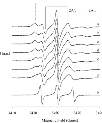
accommodate the aldehyde molecules. Fig. 1 shows the
EPR spectra of 5-DSA-labeled reverse micelles without
(a) and upon octaldehyde addition (b–g) together with
the spectrum of the probe in isooctane (h). Comparison
of spectra (a–g) with spectrum (h) indicate that the spin
probe is located in the reverse micelle membrane, and
therefore, 5-DSA constitutes a good marker to evaluate
changes experimented by the micelle membrane. As
shown in Fig. 1, the EPR spectrum modifications
indi-cate that aldehyde molecules perturb the reverse micelle
membrane. These alterations are analyzed in Fig. 2,
which shows the evolution of the order parameter (
S
)
upon octaldehyde additions to 0.1 M AOT reverse
mi-celle solutions with
W
0¼
26. The inverse linear relations
obtained for the
S
parameter clearly indicates that at
least part of the aldehyde molecules are located within
the reverse micelle membrane increasing its fluidity. On
the contrary, no significant changes were detected for
Fig. 1. EPR spectra of 5-DSA spin labeled 0.1 M AOT reverse micelles with W0¼26. (a) Reverse micelle solution without aldehyde, (b–g)
the polarity parameter, which was found to remain
constant within the experimental error (
a
0¼
14
:
9
0
:
1 G). Similar results were obtained at
W
0¼
20 and for
decylaldehyde at
W
0¼
20 and 26 (data not shown).
Although water-insoluble aldehydes strongly affect
the membrane fluidity, the absence of alterations in
the polarity suggests that the water does not penetrate
into the reverse micelle membrane upon aldehyde
addition.
In contrast, identical studies carried out with
benz-aldehyde resulted in non-detectable fluidity alterations,
indicating that benzaldehyde is not located in the micelle
membrane. Therefore, it should be either in the water
core or close to the membrane–water interface.
The integrity of the 5-DSA-labeled micelle membrane
using either water or imidazole buffer was also checked
for 0.1 M AOT reverse micelles with
W
0¼
20.
Imidazole-containing reverse micelles (
S
¼
0
:
517
0
:
004,
a
0¼
14
:
8
0
:
1 G) are more ordered and less polar than the
water-containing ones (
S
¼
0
:
491
0
:
007,
a
0¼
15
:
3
0
:
1 G). Thus, under the tested conditions, reverse
mi-celles filled with water are less ordered than those filled
with imidazole buffer.
Benzaldehyde catalysis
As observed before [15], the as-purified enzyme
dis-played a simple Michaelis–Menten kinetics for the
benzaldehyde:DCPIP oxidoreductase activity in
aque-ous solution. The parameter
k
catin 50 mM imidazole
buffer at pH 8.0 is given in Table 1. No substrate
inhi-bition was observed for the tested concentration range
(up to
0.3 mM).
Fig. 3A shows the resulting kinetic profiles of
Dg
AOR encapsulated in a 0.1 M AOT solution as a
function of
W
0. As seen in this figure, two bell-shaped
peaks with similar
k
catvalues were obtained at
W
020
and
26. Considering the fact that the protein has
approximately a globular shape [23], peaks of
enzy-matic activity should occur when the size of the reverse
micelle inner core matches the protein size, that is
when
r
mr
p, where
r
mis the inner cavity micelle
radius and
r
pis the mean protein radius. The micelle
Fig. 2. Evolution of the order parameter (S) as a function of octal-dehyde additions to 5-DSA spin labeled 0.1 M AOT reverse micelles withW0¼26.Svalues were obtained from the spectra shown in Fig. 1.
Table 1
Kinetic parameters of Dg AOR in aqueous solution and in AOT reverse micelles with different hydration degrees (W0)
Substrate W0 kcat(s1)
Solution 1.350.04
Benzaldehyde 20 1.30.1
26 1.250.05
Octaldehyde 20 0.80.1
26 1.20.1
Decylaldehyde 20 1.190.07
26 1.220.07
Fig. 3. Profiles of the catalytic constant as a function of the hydration degree (W0¼ ½H2O/[AOT]) forDgAOR encapsulated in 0.1 M AOT
inner cavity radius is given by
r
mð
A
A
Þ ¼
4
þ
1
:
5
W
0[9],
whereas the mean protein radius may be calculated
with the equation:
r
pð
A
A
Þ ¼
0
:
7
ð
M
rÞ1=3
, where
M
rrep-resents the protein molecular weight (g/mol) [24,25].
Both equations predict activity peaks at
W
019 for
the monomer and
W
025 for the dimer, in good
agreement with the experimental data (Fig. 3A). This
indicates that monomeric species are responsible for
the activity peak at
W
020, whereas dimeric species
produce the peak at
W
026. This proves the
hypoth-esis derived from structural data [23] that
Dg
AOR
monomers may act independently.
Interestingly, whereas the kinetic studies confirmed
the presence of
Dg
AOR dimeric form at
W
026, the
sedimentation studies could only detect monomers.
These apparently contradictory results may be
tenta-tively explained considering a monomer–dimer
dy-namical equilibrium. Our current hypothesis is that
this equilibrium would tend towards the dimer in
aqueous solution, as all the previous biochemical and
molecular properties of
Dg
AOR in solution have
shown [13]. On the contrary, in reverse micelles the
predominant form appears to be the monomer, as it
was concluded by our sedimentation analysis. The
detection of an activity peak at
W
026 (the
theoreti-cal catalytic activity maximum for an AOR dimer)
indicates that this equilibrium in reverse micelles is
shifted under turnover conditions (Table 1). Other
substrate-depending effects have been previously
ob-served in reverse micelles. For example, in the case of
lysozyme encapsulated in reverse micelles, the substrate
was found to regulate the equilibrium between
dena-tured and native enzyme towards the native
confor-mation [26].
Octaldehyde and decylaldehyde catalysis
Dg
AOR enzymatic assays carried out in the
aqueous fraction that resulted from partition
experi-ments carried out with octaldehyde and decylaldehyde
in 1:1 isooctane/water mixtures showed no activity
towards the respective aldehyde. However, as it is
shown, their catalysis could be measured in reverse
micelles. As seen in Figs. 3B and C, the
k
catevolution
as a function of
W
0for the water-insoluble aldehydes
also showed two bell-shaped peaks with
k
catvalues
and
W
0positions similar to those observed for the
benzaldehyde assay (Table 1). Further comparison of
Fig. 3A with B and C reveals broader
k
catpeaks for
the water-insoluble aldehydes. As it was concluded
from the partition experiments above, these long chain
aldehydes are found in the micelle membrane. The
consequences of this over the micelle properties may
eventually explain the enzyme stabilization and
con-sequently broader activity peaks over a wider range
of
W
0.
Conclusions
As a system that mimics a cell environment, reverse
micelles offer new possibilities of studying enzymes and
bioconversions of non-polar compounds [27]. The
pres-ent study shows the enzymatic ability of
Dg
AOR
en-capsulated in AOT–water–isooctane reverse micelles.
Taking advantage of this multiphase system, we were
able to demonstrate that
Dg
AOR monomers can be
separated under non-denaturing conditions in a stable
and active form. Considering previous studies that have
shown that
Dg
AOR can catalyze the reactions of a broad
range of aldehydes, we used reverse micelles to show for
the first time the ability of this enzyme to catalyze the
reactions of water-insoluble aldehydes. These aldehydes
are located in the reverse micelle membrane, which
sug-gests that
Dg
AOR, though localized in the cytoplasm,
might catalyze reduction of substrates incorporated into
cell membranes.
Acknowledgments
This work was supported by Fundacß~aao para a Ci^eencia e Tecnologia POCTI/BME/36152/99 (Portugal) and CAI+D-UNL (Argentina). We thank Prof. Natalia L. Klyachko for help with the ultrasedimentation experiments.
References
[1] K. Martinek, A.V. Levashov, N.L. Klyachko, Y.L. Khmelnitski, I.V. Berezin, Micellar enzymology, Eur. J. Biochem. 155 (1986) 453–468.
[2] A. Erjomin, D. Metelitza, Catalysis by hemoproteins and their structural organization in reverse micelles of surfactants in octane, Biochim. Biophys. Acta 732 (1983) 377–386.
[3] R. Bru, A. Sanchez-Ferrer, F. Garcia-Carmona, Kinetic models in reverse micelles, Biochem. J. 310 (1995) 721–739.
[4] Y.L. Khmelnitsky, A.V. Kabanov, N.L. Klyachko, A.V. Leva-shov, K. Martinek, in: M.P. Pileni (Ed.), Structure and Reactivity in Reverse Micelles, Elsevier, Amsterdam, 1989, p. 379. [5] J.D. Nicholson, J.H.R. Clarke, in: K.L. Mittal, B. Lindman
(Eds.), Surfactants in Solution, Plenum Press, New York, 1984, pp. 1663–1674.
[6] A.V. Levashov, R.V. Rarity, K. Martinek, N.L. Klyachko, Artificial glycosylated alpha-chymotrypsin in reverse micelles of aerosol OT in octane—a new approach to elucidation of the role of carbohydrate moieties in glycoproteins, FEBS Lett. 336 (1993) 385–388.
[7] K. Martinek, N.L. Klyachko, A.V. Kabanov, Y.L. Khmelnitsky, A.V. Levashov, Micellar enzymology—its relations to membra-nology, Biochim. Biophys. Acta 981 (1989) 161–172.
[8] A.V. Kabanov, S.N. Nametkin, G.N. Evtushenko, N.N. Chernov, N.L. Klyachko, A.V. Levashov, K. Martinek, A new strategy for the study of oligomeric enzymes—gamma-glutamyltransferase in reverse micelles of surfactants in organic solvents, Biochim. Biophys. Acta 996 (1989) 147–152.
using reversed micelles as matrix microreactors, Protein Eng. 4 (1991) 1009–1017.
[10] A.V. Levashov, Y.L. Khmelnitsky, N.L. Klyachko, V.Y. Chern-yak, K. Martinek, Enzymes entrapped into reverse micelles in organic–solvents. Sedimentation analysis of the protein–aerosol OT-H2O-octane system, J. Colloid Interf. Sci. 88 (1981) 444–457.
[11] R. Hille, The mononuclear molybdenum enzymes, Chem. Rev. 96 (1996) 2757–2816.
[12] M.J. Rom~aao, J. Kn€aablein, R. Huber, J.J.G. Moura, Structure and function of molybdopterin containing enzymes, Prog. Biophys. Molec. Biol. 68 (1997) 121–144.
[13] J.J.G. Moura, A.V. Xavier, M. Brushi, J. LeGall, D.O. Hall, R. Cammack, Molybdenum-containing iron–sulfur protein from
Desulfovibrio gigas, Biochem. Biophys. Res. Commun. 72 (1976) 782–789.
[14] K. Martinek, A.V. Levashov, N.L. Klyachko, I.V. Berezin, Catalysis by water soluble enzymes in organic–solvents. Stabil-ization of enzymes against denaturation (inactivation) when they are included in reverse micelles of surface-active substance, Dokl. Akad. Nauk. 236 (1977) 920–925.
[15] B.A.S. Barata, J. LeGall, J.J.G. Moura, Aldehyde oxidoreductase activity in Desulfovibrio gigas—In vitro reconstitution of an electron transfer chain from aldehydes to the production of molecular hydrogen, Biochemistry 32 (1993) 11559–11568. [16] S.L. Andrade, E.O. Kamenskaya, A.V. Levashov, J.J.G. Moura,
Encapsulation of flavodoxin in reverse micelles, Biochem. Bio-phys. Res. Commun. 234 (1997) 651–654.
[17] A. Cornish-Bowden, Fundamentals of Enzyme Kinetics, Portland Press Ltd, London, 1981.
[18] A.V. Kabanov, A.V. Levashov, N.L. Klyachko, S.N. Namyot-kin, A.V. Pshezhetsky, Enzymes entrapped in reverse micelles of surfactants in organic–solvents. A theoretical treatment of the catalytic activity regulation, J. Theor. Biol. 133 (1988) 327– 343.
[19] Y.L. Khmelnitsky, I.N. Neverova, V.I. Polyakov, V.Y. Grinberg, A.V. Levashov, K. Martinek, Kinetic theory of enzymatic-reactions in reverse micellar systems. Applications of the pseudo-phase approach for partitioning substrates, Eur. J. Biochem. 190 (1990) 155–159.
[20] S. Schreier, C.F. Polnaszek, I.C.P. Smith, Spin labels in mem-branes. Problems in practice, Biochim. Biophys. Acta 515 (1978) 375–436.
[21] J. Harris, T.J. Power, A.L. Bieber, A. Watts, An electron–spin resonance spin-label study of the interaction of purified mojave toxin with synaptosomal membranes from rat-brain, Eur. J. Biochem. 131 (1983) 559–565.
[22] P. Baglioni, E. Ferroni, G. Martini, M.F. Ottaviani, Micellar solutions of sulfate surfactants studied by electron–spin-resonance of nitroxide radicals. 2. Use of C-8, C-12 and C-16 derivatives of piperidinyl-1-oxy, J. Phys. Chem. 88 (1984) 5107–5113.
[23] M.J. Rom~aao, M. Archer, I. Moura, J.J.G. Moura, J. LeGall, R. Engh, M. Schneider, R. Huber, Crystal structure of the xanthine oxidase-related aldehyde oxidoreductase from D. gigas, Science 270 (1995) 1170–1176.
[24] A.V. Levashov, in: I.V. Berezin (Ed.), Chemical and Enzyme Reactions in Surfactants in Solution, VINITI Publishers, Mos-cow, 1987.
[25] Y.E. Shapiro, V.Y. Gorbatyuk, A.V. Levashov, N.L. Klyachko, Segmental mobility of molecules in the reversed micelle of aerosol OT in N-octane containing alpha-chymotrypsin and albumin, Biol. Mem. 7 (1994) 277–290.
[26] B. Steinmann, H. Jakle, P.L. Luisi, A comparative study of lysozyme, conformation in various reverse micellar systems, Biopolymers 25 (1986) 1133–1156.