IntroductIon
Juvenile Sjögren’s Syndrome (JSS) is a rare autoimmune disease that mainly affects children and adolescents sali-vary and lacrimal glands, mostly under diagnosed due to the different initial clinical manifestations and evo-lution along the course of the disease, when compared to adult’s Sjögren’s syndrome (SS). Isolated recurrent parotitis is the most common clinical manifestation during childhood, and may be primary or associated with other autoimmune rheumatic diseases, such as ju-venile systemic lupus erythematosus (SLE), juju-venile idiopa thic arthritis, and juvenile systemic sclerosis1-3.
Exo cri nopathy extended to the skin, the respiratory and urogenital systems may also occur, as well as ex-traglandular and systemic manifestations. With regard to the distribution by gender, there is a predominance of the di sease in female pediatric patients (F: M = 5-7: 1), as well as in the adult population4.
Recent studies in adults show that the salivary glands ultrasound (SGU) detect glandular changes typical of SS, and can be a useful exam in the diagnosis and fol-low-up of patients with positive antibodies, showing 89% sensitivity and 85% specificity5-9. The same ul
-traso nographic findings can be found in JSS, and al-though few studies have been published so far, it is sugges ted that its application may become a new tool to help in the diagnostic aid for children and adoles-cents with recurrent parotitis and classic laboratory abnor malities10.
Between January 2007 and October 2017, 664 pe-diatric patients were admitted to the Rheumatology De-partment of the Pontifícia Universidade Católica of Campinas (PUC-Campinas). Out of these patients, 2 (0.30%) were diagnosed with JSS, exhibiting recurrent parotitis, positive autoantibodies and ultrasound changes of salivary glands, typical of this syndrome. These two cases are described below. This study was approved by the Ethics Committee of the University Hospital.
1. Pediatric Rheumatology Department, Pontíficia Universidade Católica de Campinas.
2. Rheumatology Department, Pontíficia Universidade Católica de Campinas.
Sonographic evaluation of salivary glands
in Juvenile Sjögren’s Syndrome
Guissa VR1, Martinelli EL2, Brandão LMKR2, Garcia LD2, Provenza JR2, Mendonça JA2 ACTA REUMATOL PORT. 2018;43:61-65
AbstrAct
Introduction: Sjögren’s syndrome in childhood is a rare autoimmune disease and mostly under-diagnosed. The aim of this study is to highlight the importance of ul-trasonographic assessment of the salivary glands in chil-dren with recurrent parotitis and positive autoanti-bodies. Two cases of ultrasonographic patterns typical of Sjögren’s syndrome described below.
Case 1: Female, 7-year-old, reporting for 2 years recurrent parotitis, xerophthalmia, xerostomia, poly -arthralgia and fever. Immunological tests were positive for antinuclear antibodies, rheumatoid factor, anti--SSA/Ro and anti-SSB/La. Salivary glands ultrasound was consistent with Grade 4 by the B-mode method and the spectral Doppler with presence of intense Po -wer Doppler signal and decreased vessels internal re-sistance, supporting the diagnosis of juvenile Sjögren’s syndrome.
Case 2: Female, 10 years old, reporting recurrent parotitis for 1 year and polyarthritis for 10 days. The supplementary tests revealed positive antibodies for Sjögren’s syndrome. Salivary glands Ultrasound and Spectral Doppler were consistent with chronic and acti ve inflammatory process of the salivary glands in the juvenile Sjögren’s syndrome.
Discussion: Salivary glands ultrasound can be a use-ful exam in the diagnosis of juvenile Sjögren’s syn-drome.
Keywords: Juvenile Sjögren syndrome; Parotitis; Saliva ry Glands Ultrasound; Autoimmune diseases; Childhood.
cAsE rEports cAsE 1
A 7-year-old white female patient was admitted to the Rheumatology Department of the PUC-Campinas, re-porting recurrent bilateral parotitis, recurrent tooth decay, early loss of deciduous teeth, dry eyes, sym-metrical inflammatory polyarthralgia of small and large joints, intermittent fever and impaired weight gain for 2 years. Diagnosis of idiopathic thrombocytopenic purpura for 2 months was reported; the patient was then treated with an infusion of immu noglobulin and prednisone 5mg/day. No oral ulcers, alopecia, cuta-neous lesions, neurological, cardiac involvement, res-piratory, abdominal or urogenital symptoms were re-ported. The patient’s weight and height were in the 25th
percentile and physical examination found no altera -tions. Laboratory exams revealed hemoglobin 12.4 g/dL, hematocrit 37%, white blood cell count 4.300/ mm3
(48% neutrophils, 46% lymphocytes, 1% eosinophils, 5% monocytes), platelets 198.000/ mm3, C-reactive
protein (CRP) 48 mg/L (normal < 0.5), erythrocyte sedimentation rate (ESR) 42 mm/1st hour (normal < 11), amylase 97 U/L (normal < 80), urea 30 mg/dL (normal 16.6-48.5), crea tinine 0.40 mg/dL (normal 0.53-0.79), aspartate aminotransferase (AST) 32 IU/L (normal 0-32), alani ne aminotransferase (ALT) 9 IU/L (normal 0-33), lacta te dehydrogenase (LDH) 232 U/L (314-333), glucose 79 mg/dL (normal <110), normal urinalysis, negative 24-hour urine protein excretion, C3 121 mg/dL (normal 90-180) and C4 14 mg/dL (normal 10-40). Serologic tests for viral infections were negative (Hepatitis virus B and C, Epstein-Barr virus, Cytomegalovirus, Toxoplasmosis, Parvovirus, Pa ra -myxovirus, Human immunodeficiency virus and
Hu-man T-Lymphotropic Virus). Immunological tests were positive for antinuclear antibodies (ANA): 1/640 (nu-clear fine speckled pattern), rheumatoid factor (RF) 640 IU/mL (normal 1-14), anti-SSA (Ro) 240 U/mL (normal < 10) and anti--SSB (La) 320 U/mL (normal <10) and negative for o ther serum antibodies: double stranded DNA (-dsDNA), Sm, anti-RNP, IgG and IgM anticardiolipin, direct Coombs (DC) and anti-cyclic citrullinated peptide (anti-CCP). The echocardiogram demonstrated normal ventricular function, without pericardial effusion. Chest and ab-domen computed tomo graphy showed no evidence of any lymph node pathology or visceromegaly. The oph-thalmologic exam was consistent with bilateral kera-toconjuctivitis sicca. SGU of parotid, submandibular and sublingual glands was performed with the MyLabTM 50 Esaote Ultrasound (Brazil, São Paulo), equipped with high-frequency linear transducer (12 MHz) for the B-mode and Power Doppler frequency ranging from 6.6 to 8.0 MHz. The B-mode method evidenced irregular contours, hypoechogenic areas > 6mm and multiple calcifications with echo genic bands with de-crease in the size of the glands; the posterior gland bor-der was not visible, consistent with Grade 47,8. In
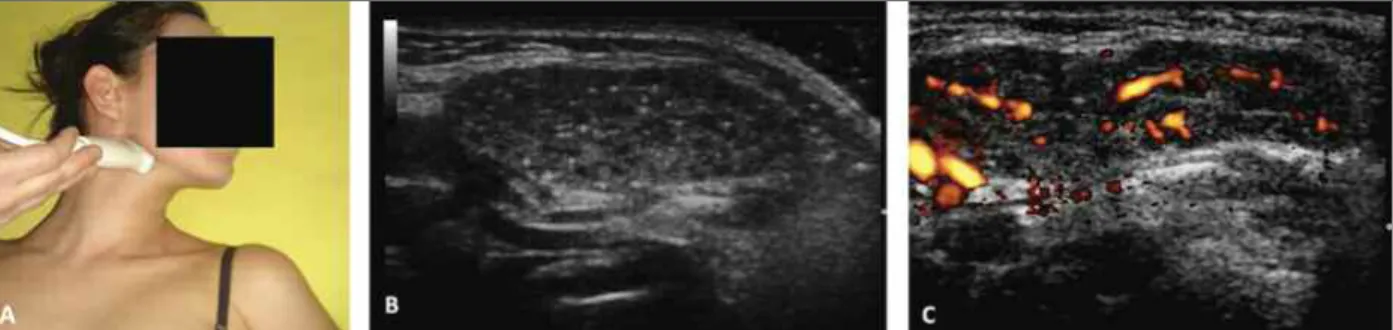
ad-dition, spectral Doppler detected intense signal of Pow-er DopplPow-er (PD) and decreased intPow-ernal resistence (IR) of mean ± Standard Deviation (SD) of 0.42 ± 0.08 (Fig-ure 1). Based on clinical, serolo gical and ultrasono-graphic findings, the diagnosis of primary JSS was es-tablished. Due to the systemic signs and symptoms, treatment with hydroxychloroquine was indicated (5mg/kg/day). The patient remained asymptomatic for only 2 months, with a new relapse of the febrile con-dition and polyarthralgia, maintaining evidence of pos-itive inflammatory activity; a treatment with a
combi-FIGurE 1.Female, 7-year-old, exhibiting salivar glands ultrasound with Grade 4 by the B-mode method, intense power Doppler signal and decreased vessels internal resistance, supporting the diagnosis of juvenile Sjögren's syndrome.
A. Illustrative probe position to evaluate salivary glands. B. Salivary glands classified as Grade 4: irregular contours, several hypoechogenic areas, multiple calcifications with echogenic bands, decrease of the glands size. C. Increased power Doppler signal.
nation of hydroxychloroquine and azathioprine (2 mg/kg/ /day) was then indicated. The case evolved with clinical and laboratorial remission; however, 6 months later the patient presented a thrombotic event, positive antiphospholipid antibodies, hypocomplementemia, fulfilling the new international classification criteria for SLE, according to The Systemic Lupus Interna-tional Collaborating Cli nics (SLIIC)11, supporting the
diagnosis of JSS associated to juvenile SLE.
cAsE 2
A 10-year-old female black patient, with a sickle cell trait, recurrent bilateral parotitis for one year, sym-metric polyarthritis of large joints, and fatigue in the previous 10 days was admitted. Sicca symptoms, fever or other systemic manifestations were not reported. Physical examination showed weight and height in the 75thpercentile and wrists, elbows and knees’ arthritis.
Supplementary investigations revealed: hemoglobin 10.7 g/dL, hematocrit 31%, white blood cell count 4.140/ mm3(40% neutrophils, 49% lymphocytes, 3%
eosinophils, 8 % monocytes), platelets 317.000/ mm3,
CRP 1.0 mg/L, ESR 59 mm 1sthour, amylase 127 U/L,
glucose 72 mg/dL, normal urinalysis, proteinuria 0.07 g/24h, C3 109 mg/dL, C4 18 mg/dL, CH50 163 U/mL (normal 60-265), IgG 2632 mg/dL (normal 570--1320), IgA 300 mg/dL (normal 65-240), IgM 143 mg/ /dL (normal 60-175). Serolo gic tests for viral infections were negative. Immunological tests were positive for ANA: 1/640 (nuclear fine speckled pattern), RF 61 IU/mL, anti-Ro > 240 U/mL, anti-La > 320 U/mL and negative for other serum antibodies: dsDNA, anti--Sm, anti-RNP, IgG and IgM anticardiolipin, DC and anti-CCP. Tomography of the chest and abdomen and echocardiogram were within normality. Schirmer s test and rose bengal unchanged. SGU: parotid and
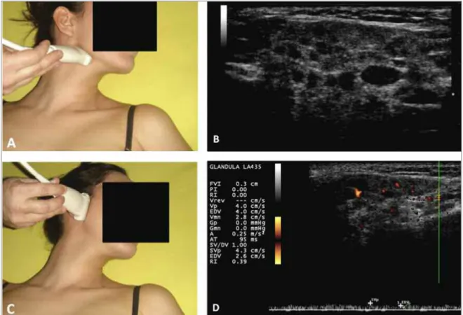
FIGurE 2.Salivary glands Ultrasound and Spectral Doppler of a female, 10-year-old patient, consistent with chronic and active inflammatory process of the salivary glands in the juvenile Sjögren's syndrome.
A. Illustrative probe position to evaluate the submandibular glands. B. Classified as Grade 4: irregular contours, several
hypoechogenic areas, multiple calcifications with echogenic bands, decrease in the size of the glands. C. Illustrative probe position position to evaluate the parotid glands.
subman dibular glands consistent with Grade 4 and sublingual with Grade 17,8(regular edges, small
hy-poechogenic areas and absence of echogenic bands) and presence of intense PD signal and decreased IR, with mean ± SD 0.48 ± 0.07, consistent with active in-flammatory process of the JSS (Figure 2). The patient was submitted to labial salivary gland biopsy (LSGB), with a representative sample of glandular tissue, which demonstrated usual histologi cal analysis, absence of acinar atrophy or lymphocy tic inflammatory infiltrate. After the exclusion of chro nic infections, neoplastic and secondary autoimmune diseases, the clinical, sero-logical and ultrasonographic findings supported the diagnosis of primary JSS and the therapeutic indica-tion. Treatment with a non-hormonal anti-inflamma-tory drug was initiated (napro xen), with remission of joint signs and symptoms within 10 days; however, 2 months later, the patient showed recurrence of bilate -ral parotitis, positive inflammatory activity tests, and initiated hydroxychloroquine (5mg/kg/day). The pa-tient progressed clinically stable until the sixth month of treatment, when she presented with a new episode of parotitis (fifth episode), associated with fever and an-kles and knees arthritis. The indication was then a com-bination of hydroxychloroquine with azathioprine (2 mg/kg/day); the patient remained clini cally asympto -matic and with normal laboratory controls during the last 17 months of the outpatient follow-up.
dIscussIon
In recent years, ultrasound indications in pediatric rheumatologic patients have been widely discussed in the literature, making it a valuable tool for the exten-sion of rheumatologic physical examination, detection of acute and subacute inflammatory activity in organs and tissues, as well as a potential guideline in local therapeutical interventions. In addition to proven diagnos tic sensitivity, ultrasound evidenced advan-tages over other imaging methods because it is safe, non-invasive, dos not require pediatric patient seda-tion and is relatively inexpensive12, 13.
Although ultrasound is not currently included in the diagnostic criteria for SS, ultrasound plays an im-portant role in the evaluation of salivary glands of adult rheumatologic patients and may increase the sensitivi -ty and accuracy of the diagnosis; it is also expected to be an evaluation method of patient’s prognosis and treatment5-7, 9. By means of the B-mode method, it is
possible to semi-quantify the salivary glands in relation to parenchyma homogeneity, echogenicity, gland size and posterior glandular border, classifying the degree of glandular involvement, according to the scoring sys-tem suggested by Cornec and De Vita et al.7, 8, ranging
from Grade 0 (normal salivary glands) to Grade 4 (dif-fuse structural damage of the glandular parenchyma), identifying the presence of glands degeneration, fi-brosis and calcification. In addition, spectral Doppler is a recent method that allows detection of the inflam-matory process through the measurement of internal resistance of vessels, complementing the structural glandular evaluation7, 14.
The diagnosis of JSS is challenging since recurrent parotitis, which is the most common clinical manifesta tion of affected children, normally precedes oral, ocular, extra-glandular and systemic symptoms in years, and may mimic infections, immunodeficiencies, other autoimmune and neoplastic diseases; those con-ditions must be excluded in order to confirm the diagnos tic1,3,15,16. However, biochemical,
hematologi-cal, immunological and histological findings follow the typical patterns of the syndrome in adults, such as the presence of autoantibodies (ANA, anti-Ro, anti-La, RF), ESR increase and lymphocytic infiltration in exo -crine glands1,3,17,18.
The lack of validated diagnostic criteria for the SS in this age group and the syndrome’s initial clinical pecu-liarities mentioned above support the discussion about the applicability of SGU as a new, noninvasive and low risk diagnosis method, provided the patients assessed carry immunological markers suggestive of chronic au-toimmune inflammation of the exocrine glands.
As in the two cases reported above, Nieto-González et al10reported the case of 3 pediatric patients who
were diagnosed with primary JSS, based on the sono-graphic findings of salivary glands in children with re-current parotitis, positive autoantibodies and negative chronic infections and lymphoproliferative diseases. None of those patients underwent salivary gland bio -psy and all of them progressed with clinical improve-ment after starting treatimprove-ment with a non-hormonal anti-inflammatory drug and/or hydroxychloroquine. In a multicenter study with 40 pediatric patients diagno sed with primary JSS, all of them had positive autoantibodies and the most common initial clinical symptom was recurrent parotitis (72.5%). Joint in-volvement was reported in 10-57.7%, fever in 10% and fatigue in 7.5% of children affected. Only two pa-tients were evaluated ultrasonographically, and both
exhibited images consistent with a chronic inflamma-tory process of the salivary glands4. Positive ultraso
-nographic data of salivary glands have also been re-ported in a 2-years and 7-month-old patient diagnosed with primary JSS who presented xerophthalmia, xeros -tomia, arthralgia, recurrent parotitis, positive ANA and RF, and biopsy of minor salivary glands with histologi -cal pattern typi-cal of the disease19. Although few stu
-dies in JSS reported 100% positive findings in the sali-vary gland biopsies of the evaluated patients, there are no comparative studies between the ultrasound fin -dings of the salivary glands and the histological pat-terns found in the biopsies in the JSS18,19. In a recent
study in adults with confirmed SS, out of the 20 pa-tients with positive antibodies and ultrasonographic findings suggestive of the disease, 5 showed LSGB without alterations20. Therefore, in the case #2 repor
ted above, normal biopsy does not invalidate the diag -nosis, since the patient presented with the main clini-cal manifestations of the disease in childhood, posi-tive autoantibodies, exclusion of infectious and neo-plastic diseases, and clinical remission throughout the treatment.
In conclusion, although ultrasound is not validated as a diagnostic criterion for JSS, ultrasonographic pat-terns typical of the disease were found in the patients evaluated above, who exhibited clinical and immuno-logical characteristics suggestive of the syndrome, sup-porting the diagnosis and early treatment, determinants of good prognosis of the disease in that pediatric age group. For the purpose of this study, it is sugges ted that all the children and adolescents affected have their sali-vary glands evaluated through ultrasonography. Besides being a potential tool for diagnosis, ultrasound can screen more accurately those patients who should ac-tually undergo invasive and risky supple mentary ex-aminations for diagnostic classification.
corrEspondEncE to
Vanessa Ramos Guissa
Rua Dr. Sampaio Ferraz, 750, apartamento 22 B E-mail: vanessaguissa@gmail.com
rEFErEncEs
1. Longhi BS, Appenzeller S, Centeville M, Gusmão RJ, Marini R. Primary Sjögren’s syndrome in children: is a family approach indicated? Clinics (São Paulo) 2011; 66: 1991-1993. 2. Baszis K, Toib D, Cooper M, French A, White A. Recurrent
pa-rotitis as a presentation of primary pediatric Sjögren syndrome. Pediatrics 2012; 129: 179-182.
3. Singer NG, Tomanova-Soltys I, Lowe R. Sjögren’s syndrome in childhood. Curr Rheumatol Rep 2008; 10: 147-155. 4. Cimaz R, Casadei A, Rose C et al. Primary Sjögren syndrome
in the paediatric age: a multicentre survey. Eur J Pediatr 2003; 162: 661-665.
5. Saied F, Włodkowska-Korytkowska M, Ma li ska M, et al. The usefulness of ultrasound in the diagnostics of Sjögren’s syn-drome. J Ultrason 2013; 13: 202-211.
6. Hammenfors DS, Brun JG, Jonsson R, Jonsson MV. Diagnostic utility of major salivary gland ultrasonography in primary Sjö-gren’s syndrome. Clin Exp Rheumatol 2015; 33: 56-62. 7. Cornec D, Cornec D, Jousse-Joulin S, Marhadour T. Salivary
gland ultrasonography improves the diagnostic performance of the 2012 American College of Rheumatology classification criteria for Sjögren’s syndrome. Rheumatology (Oxford) 2014; 53: 1604-1607.
8. De Vita S, Lorenzon G, Rossi G, Sabella M, Fossaluzza V. Sali-vary gland echography in primary and secondary Sjögren’s syn-drome. Clin Exp Rheumatol 1992; 10: 351-356.
9. Theander E, Mandl T. Primary Sjögren’s syndrome: diagnostic and prognostic value of salivary gland ultrasonography using a simplified scoring system. Arthritis Care Res (Hoboken) 2014; 66: 1102-1107.
10. Nieto-González JC, Monteagudo I, Bello N, Martínez-Estupi-ñan L, Naredo E, Carreño L. Salivary gland ultrasound in chil-dren: a useful tool in the diagnosis of juvenile Sjögren’s syn-drome. Clin Exp Rheumatol 2014; 32: 578-580.
11. Petri M, Orbai AM, Alarcón GS et al. Derivation and validation of the Systemic Lupus International Collaborating Clinics clas-sification criteria for systemic lupus erythematosus. Arthritis Rheum 2012; 64: 2677-2686
12. Hernández-Díaz C, Ventura-Ríos L, Gutiérrez M, Roth J. Ul-trasonography in pediatric rheumatology in Latin America. Ex-panding the frontiers. Clin Rheumatol 2016; 35: 1077-1080. 13. Laurell L, Court-Payen M, Boesen M, Fasth A. Imaging in ju-venile idiopathic arthritis with a focus on ultrasonography. Clin Exp Rheumatol 2013; 31: 135-148.
14. Mendonça JA. Ultrassonografia em Reumatologia: uma exten-são do exame físico. Rio de Janeiro: Livraria e editora Revinter LTDA, 2014: 55.
15. Houghton K, Malleson P, Cabral D, Petty R, Tucker L. Primary Sjögren’s syndrome in children and adolescents: are proposed diagnostic criteria applicable? J Rheumatol 2005; 32: 2225-2232. 16. Shabana K, Okamoto N, Shindo K, Murata T, Tamai H, Fuji-wara K. Musculoskeletal ultrasound findings of articular ma-nifestations on juvenile primary sjogren’s syndrome. Pediatr Rheumatol Online J 2014 Published Online First: 17 Septem-ber 2014. doi: 10.1186/1546-0096-12-S1-P37.
17. Breda L, Nozzi M, De Sanctis S, Chiarelli F. Laboratory tests in the diagnosis and follow-up of pediatric rheumatic diseases: an update. Semin Arthritis Rheum 2010; 40: 53-72.
18. Saad Magalhães C, de Souza Medeiros PB, Oliveira-Sato J, Cus-tódio-Domingues MA. Clinical presentation and salivary gland histopathology of paediatric primary Sjogren’s syndrome Clin Exp Rheumatol 2011; 29: 589-593.
19. De Oliveira MA, De Rezende NP, Maia CM, Gallottini M. Pri-mary Sjögren syndrome in a 2-year-old patient: role of the den-tist in diagnosis and dental management with a 6-year follow--up. Int J Paediatr Dent 2011; 21: 471-475.
20. Astorri E, Sutcliffe, Richards PS, et al. Ultrasound of the salivary glands is a strong predictor of labial gland biopsy histopatho-logy in patients with sicca symptoms. J Oral Pathol Med 2016; 45: 450-454.