https://doi.org/10.1177/1756283X17722916 https://doi.org/10.1177/1756283X17722916 Ther Adv Gastroenterol
2017, Vol. 10(9) 651 –660 DOI: 10.1177/ 1756283X17722916 © The Author(s), 2017. Reprints and permissions: http://www.sagepub.co.uk/ journalsPermissions.nav
Therapeutic Advances in Gastroenterology
journals.sagepub.com/home/tag 651
Introduction
The introduction of anti-tumour necrosis factor (TNF)α monoclonal antibodies as therapeutic agents in auto-inflammatory disorders has revo-lutionized the medical management strategies of these diseases and the health-related quality of life of patients. In the case of inflammatory bowel diseases [IBDs, which include Crohn’s disease (CD) and ulcerative colitis (UC)], the use of anti-TNFα agents has led to a decrease in
hospitalization rates, risk of surgery and health-related costs.1
However, and despite the anti-TNFα success in the treatment of many IBD patients, some of them do not respond to the drug during the induction phase, whereas others experience a loss of response later during treatment.2 Accumulating evidence from the
literature suggests that the outcomes of CD and UC patients on infliximab (IFX) are strongly related
Clinical performance of an infliximab rapid
quantification assay
Fernando Magro, Joana Afonso, Susana Lopes, Rosa Coelho, Raquel Gonçalves, Paulo Caldeira, Paula Lago, Helena Tavares de Sousa, Jaime Ramos,
Ana Rita Gonçalves, Paula Ministro, Isadora Rosa, Tânia Meira, Patrícia Andrade, João-Bruno Soares, Diana Carvalho, Paula Sousa, Ana Isabel Vieira, Joanne Lopes, Cláudia Camila Dias, Karel Geboes and Fátima Carneiro, on behalf of the Portuguese IBD Study Group (GEDII) Abstract
Background: Therapeutic drug monitoring (TDM)-based algorithms can be used to guide infliximab (IFX) adjustments in inflammatory bowel disease (IBD) patients. This study aimed to explore a rapid IFX-quantification test from a clinical perspective.
Methods: This manuscript describes a prospective cohort study involving 110 ulcerative colitis (UC) patients on the maintenance phase of IFX. IFX trough levels were quantified using a rapid quantification assay and a commonly-used reference kit.
Results: Irrespective of the assay used to measure IFX, its through levels were statistically different between patients with and without endoscopic remission (Mayo endoscopic score = 0), as well as between patients stratified by their faecal calprotectin (FC) levels. Despite the fact that the two methods correlated well with each other [Spearman’s rank correlation coefficient = 0.843, p < 0.001; intraclass correlation coefficients = 0.857, 95% confidence interval (CI): 0.791–0.903], there was a discernible systematic variation; values obtained with the reference kit were on average 2.62 units higher than those obtained with the rapid assay. Notwithstanding, 3 µg/ml was shown to be an acceptable cut-off to assess endoscopic status and inflammatory burden levels using both assays. The percentage of patients that had a positive outcome when the IFX concentration measured by the rapid assay ranked above 3 µg/ ml was 88% both for a Mayo endoscopic score ⩽ 1 and for an FC concentration <250 µg/g. Conclusions: Based on this study, we concluded that using the rapid IFX assessment system with a 3 µg/ml threshold is a reliable alternative to the time-consuming enzyme-linked immunosorbent assays in patients on the maintenance phase of IFX.
Keywords: infliximab, ulcerative colitis, therapeutic window
Received: 29 March 2017; revised manuscript accepted: 2 June 2017.
Correspondence to: Fernando Magro Department of Biomedicine, Unity of Pharmacology and Therapeutics, Faculty of Medicine, University of Porto, Porto, Portugal [email protected] Joana Afonso Department of Biomedicine, Unity of Pharmacology and Therapeutics, Faculty of Medicine, University of Porto, Porto, Portugal MedInUP, Centre for Drug Discovery and Innovative Medicines, University of Porto, Porto, Portugal
Susana Lopes Rosa Coelho Patrícia Andrade
Gastroenterology Department, Centro Hospitalar São João, Porto, Portugal
Raquel Gonçalves João-Bruno Soares
Gastroenterology Department, Hospital de Braga, Braga, Portugal
Paulo Caldeira Gastroenterology Department, Centro Hospitalar do Algarve, Faro, Portugal Paula Lago Gastroenterology Department, Centro Hospitalar do Porto, Porto, Portugal
Helena Tavares de Sousa
Gastroenterology Department, Centro Hospitalar do Algarve, Portimão, Portugal Department of Medical Biosciences and Medicine, University of Algarve, Faro, Portugal
Algarve Biomedical Centre (ABC), University of Algarve, Portugal
with the levels of the drug found in the organism.3–10
From a physician’s perspective, understanding the reasons that lead to unresponsiveness is key to delin-eate future therapeutic strategies, which can include a dose intensification, a switch to another anti-TNFα agent, or adding immunosuppressive drugs or steroids. In this context, the precise and accurate measurement of the circulating drug levels, known as therapeutic drug monitoring (TDM), has a key role. Several TDM-based algorithms and dash-boards are being developed to assist the physician in the therapeutic decision-making process.2,11–13
Moreover, TDM may also be useful to identify cases with supra-therapeutic drug levels (which can be de-escalated to prevent the appearance of adverse effects), and has been proven as a cost-effective strategy when compared with the traditional empir-ical-based adjustment of drug dosage.14,15
Given the importance of TDM in patients on IFX, one can easily find a number of different commer-cial kits that can measure the concentration of this agent from the patient’s serum, most of them rely-ing on an enzyme-linked immunosorbent assay (ELISA) approach. However, these kits have a turn-around time of approximately 8 h, delaying the IFX dose adjustment to the following infusion (usually 6–8 weeks later). A rapid IFX-quantification sys-tem, which allows a fast (15 min) assessment of IFX from a patient’s serum, has been recently launched in the market by the Bühlmann® company
(Schönenbuch, Switzerland). Not only does this system allows an immediate adjustment of the IFX dosage, but it also has the advantage of being a user-friendly desktop device, which can be easily oper-ated by any nurse, technician or physician without the requirement of specific laboratory facilities. We have recently validated the utilisation of the Bühlmann® rapid assay in a laboratorial context,
and concluded that this kit constitutes a reliable and fast alternative to the traditional ELISA kits.16 In this study we aimed to take a step further
and to assess the clinical sensitivity and specificity of this rapid assay, by addressing the existence and interpretability of IFX cut-off values able to guide the therapeutic decision-making process. Material and methods
Cohort
UC patients on the maintenance phase of IFX therapy were prospectively and consecutively
recruited from 10 different university and commu-nity hospitals. Only patients older than 18 years and with at least 14 weeks of IFX treatment were invited to participate. Exclusion criteria included history of malignancy in the previous 5 years, opportunistic infections or demyelinating diseases; existence of adenomatous polyps or known viral infections; and pregnancy and breastfeeding. This study was approved by the ethic committee of all hospitals involved and by the Portuguese Data Protection Authority. All patients enrolled signed an informed written consent.
IFX-quantification assays
A total of 110 samples collected from the same number of patients were assayed to determine their serum trough IFX levels using two different com-mercial kits: Quantum Blue® Infliximab:
Quantitative Lateral Flow Assay (Bühlmann, Schönenbuch, Switzerland), hereafter referred to as QB, and Level Infliximab M2920 kit (Sanquin, Amsterdam, the Netherlands), hereafter referred to as Sanquin. Both kits were used strictly following manufacturers’ instructions. The lower and upper limits of quantification were 0.4 µg/ml and 20 µg/ml for the QB assay, and 0.08 µg/ml (1:200) and 25 µg/ml (1:1500) for the Sanquin assay, respectively. Whenever the results obtained were below or above these limits of quantification, they were considered to be at those same limits. Sanquin was chosen as the reference test as it is a widely used kit in both laboratorial and clinical contexts. All measure-ments were carried out by the same researcher. Assessment of disease outcomes
Disease status, including clinical evaluation, endoscopic and histological activity, and quantifi-cation of faecal calprotectin (FC), was assessed at the same time as the IFX concentration (i.e. immediately before an IFX infusion).
Clinical evaluation. Clinical remission was evalu-ated according to the global Mayo score. Patients were considered to be in clinical remission if their global Mayo score was ⩽2 and no individual sub-score was above 1.
Endoscopic evaluation. Endoscopic activity was evaluated using the Mayo endoscopic subscore:17
mucosal healing was defined as a Mayo endo-scopic subscore equal to 0 or ⩽1.
Jaime Ramos Diana Carvalho Gastroenterology Department, Centro Hospitalar de Lisboa, Lisboa, Portugal
Ana Rita Gonçalves
Gastroenterology Department, Centro Hospitalar Lisboa Norte, Lisboa, Portugal
Paula Ministro Paula Sousa
Gastroenterology Department, Hospital de S. Teotónio, Viseu, Portugal
Isadora Rosa
Gastroenterology Department, Instituto Português de Oncologia de Lisboa, Lisboa, Portugal
Tânia Meira Ana Isabel Vieira
Gastroenterology Department, Hospital Garcia de Orta, Almada, Portugal
Joanne Lopes
Department of Pathology, Centro Hospitalar São João, Porto, Portugal
Cláudia Camila Dias
Department of Community Medicine, Information and Health Decision Sciences, Faculty of Medicine of the University of Porto, Portugal
CINTESIS, Centre for Health Technology and Services Research, Porto, Portugal
Karel Geboes
Department of Pathology, University Hospital of KU Leuven and UZ Gent, Leuven, Belgium
Fátima Carneiro
Department of Pathology, Centro Hospitalar São João, Porto, Portugal Institute of Molecular Pathology and Immunology of the University of Porto [Ipatimup], Porto, Portugal
Histological evaluation. The presence of histologi-cal inflammation was evaluated through the anal-ysis of an average of two biopsy samples from the sigmoid and the rectum. Samples were classified following the Geboes score,18 and histological
remission was defined as a Geboes index ⩽3.0. All samples were the subject of a central reading by two independent pathologists blinded to the patients’ disease status and endoscopic results. Disagreements between pathologists were resolved by a review including a third pathologist (K. Geboes) and using a multiheaded micro-scope, defining the final score.
Quantification of faecal calprotectin. Stool sam-ples were collected and kept at 4°C (for a maxi-mum of 48 h) until shipment to the central laboratory (Department of Pharmacology and Therapeutics, Faculty of Medicine of University of Porto, Portugal). FC was extracted from stools within a maximum of 7 days after collection using the ‘faecal sample preparation kit’ (Roche Diag-nostics, Germany) according to the instructions provided by the manufacturer, and stored at −80°C until quantification. FC concentration in each sample was determined using the QB kit according to the manufacturer’s instructions. Statistical analysis
Categorical variables were described through absolute (n) and relative (%) frequencies and continuous variables were described as mean and standard deviation, median, percentiles, and min-imum/ maximum values when appropriate. All the reported p-values were two-sided, and p-values <0.05 were considered to be statistically signifi-cant. The ability of the measured IFX concentra-tions to assess the various disease outcomes was evaluated by plotting Receiver Operating Characteristic curves and computing the Area Under the Curve. All data were arranged, pro-cessed and analysed with SPSS® v.20.0 data
(Statistical Package for Social Sciences, IBM Corp., Armonk, NY). Graphs were computed with Prism 7 (GraphPad Software, Inc., CA, USA).
Results
Characterization of the cohort and disease outcomes
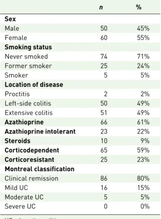
The main baseline characteristics of this study’s cohort are depicted in Table 1. Females
constituted 55% of the entire population, and only 5% of all patients were current smokers. A minority of patients (2%) had a proctitis diagno-sis, whereas 49% of them had left-side colitis and an equal percentage had extensive colitis. Overall 22% of the patients were azathioprine (AZA) intolerant, whereas 59% and 23% were classified as corticodependent and corticoresistant, respec-tively. At the time of study inclusion, 61% and 9% of all patients were on AZA and steroids, respectively.
The disease outcomes addressed during this study are listed in Table 2. Regarding clinical evalua-tion, the majority of patients (72%) had a global Mayo score ⩽2, and 69% of the entire population were considered to be in clinical remission (i.e. had a global Mayo score ⩽2 and no individual subscore >1). Moreover, 58% or 81% of all patients were considered to be in mucosal healing (endoscopic Mayo score = 0 or ⩽1, respectively). Regarding FC levels, 66% of the population were below the threshold of 250 µg/g. Finally, the over-all median [interquartile range (IQR)] of the IFX trough levels was 6.59 µg/ml (3.03–14.66) using the Sanquin kit, and 5.25 µg/ml (1.70–9.58) using the rapid QB assay.
Table 1. Cohort characterization.
n % Sex Male 50 45% Female 60 55% Smoking status Never smoked 74 71% Former smoker 25 24% Smoker 5 5% Location of disease Proctitis 2 2% Left-side colitis 50 49% Extensive colitis 51 49% Azathioprine 66 61% Azathioprine intolerant 23 22% Steroids 10 9% Corticodependent 65 59% Corticoresistant 25 23% Montreal classification Clinical remission 86 80% Mild UC 16 15% Moderate UC 5 5% Severe UC 0 0%
Analytical comparison between the two different IFX-quantification methods
IFX through levels measured by the Sanquin and QB levels were highly correlated [Spearman’s rank correlation coefficient = 0.843, p < 0.001; intraclass correlation coefficient (ICC) = 0.857, 95% CI: 0.791–0.903], as shown in Supplementary Figure 1. However, the mean difference and its CI show that the concentrations obtained with the Sanquin kit were, on average, higher than those obtained with the QB (average difference = 2.62 µg/ml, 95% CI: 1.64–3.60). Finally, the Bland– Altman plot shows that the difference between values measured with both kits increases with the increase in IFX concentrations, but is close to 0 for concentrations below 5 µg/ml (Supplementary Figure 2).
Association between IFX trough levels and outcomes
The medians of serum trough IFX concentra-tions detected with each method for contrasting disease outcomes (concerning clinical remission, endoscopic Mayo score and FC levels) are repre-sented in Figure 1. The results show that IFX trough levels were higher in patients who had positive outcomes irrespective of the assay used, and these results were significant for endoscopic remission (using endoscopic Mayo score = 0 as the criterion for remission) and FC.
We then applied different IFX cut-offs (from 1–10) to the results obtained from each kit, and assessed their ability to predict patient outcomes. The sensitivity, specificity, positive predictive value (PPV), negative predictive value (NPV), accuracy and Kappa for each case are depicted in Supplementary Table 1. A positive test was defined as having an IFX level below the cut-off, whereas the disease status was defined as having a negative outcome (not being in clinical remis-sion, having an endoscopic Mayo score >0 or >1, or having an FC level >250 µg/g). NPV rep-resents the percentage of patients who have a positive outcome (no disease) among those who have an IFX above the defined cut-off (negative test result), whereas PPV represents the percent-age of patients who have a negative outcome (disease) among those that have an IFX below the defined cut-off (positive test result).
Perceptively, the performance values vary widely with the cut-off chosen and the outcome evaluated,
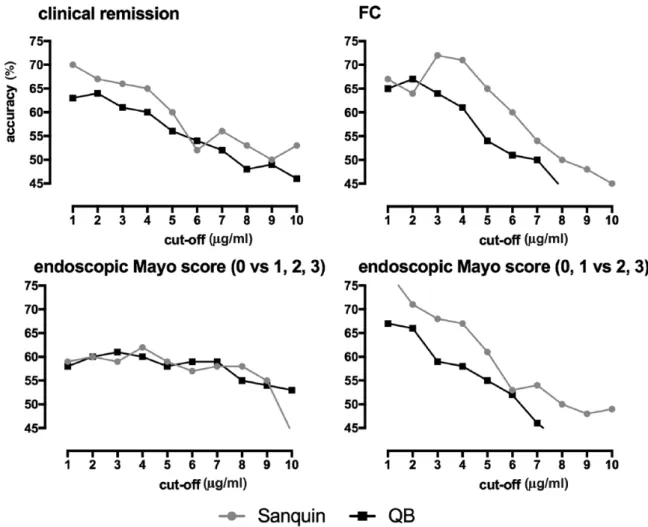
but are considerably similar for both kits when the conditions mentioned are kept stable (i.e. same cut-off and outcome). Figure 2 represents the accu-racy (i.e. the sum of true positives and negatives) of the results obtained with either QB or Sanquin in terms of clinical status, endoscopic score and FC level using different cut-offs. The results show that Sanquin and QB have a very similar variation of the accuracy along the different cut-offs. Overall, a value of 3 µg/ml seems to be an acceptable cut-off for QB, although lower values could be considered in a few situations.
NPV has an important role in this context, as it represents the percentage of patients who have an IFX concentration above the cut-off and would not benefit from a drug adjustment. And in fact, 74, 62, 83 and 86% of patients with an IFX trough level >3 µg/ml measured by the Sanquin
Table 2. Disease outcomes.
n %
Global Mayo score (n, %)
1 60 57% 2 16 15% 3 8 7% 4 3 3% 5 6 6% 6 3 3% 7 4 4% 8 3 3% 9 1 1% 11 2 2% Clinical remission = no 34 31%
Clinical remission = yes 76 69%
Endoscopic Mayo score
(n, %) 0 63 58% ⩾1 45 42% ⩽1 87 81% >1 21 19% FC (µg/g) (n, %) QB <250 59 66% ⩾250 31 34%
IFX, (median, IQR)
Sanquin 6.59 3.03–14.66
Quantum Blue 5.25 1.70–9.58
FC, faecal calprotectin; IFX, infliximab; QB, Quantum Blue® Infliximab: Quantitative Lateral Flow Assay (Bühlmann, Schönenbuch, Switzerland).
kit are in clinical remission, have a Mayo endo-scopic score of 0, have a Mayo endoendo-scopic score ⩽1, and have an FC level <250 µg/g, respectively, whereas these values are 74, 65, 88 and 88% for the QB kit.
When adjusting the IFX cut-off to evaluate clini-cal status to 1 (with Sanquin) or 2 (with QB), the percentage of patients that test above these values and are, indeed, in clinical remission, is 71% and 73%, respectively. This shows that although accuracy can be higher, the NPV is slightly smaller for these lower cut-offs. The same thing occurs when one addresses endoscopic remis-sion (using endoscopic Mayo score ⩽1 as the
remission criterion) using IFX cut-offs <3 μg/ml: the accuracy is higher, but the NPV is lower. On the other hand, the PPV (percentage of patients that are below the IFX cut-off and could benefit from an IFX dose adjustment) are consistently lower than the NPVs, and for a cut-off of 3 µg/ml vary from 23–50% with the Sanquin kit, and from 23–55% with the rapid QB kit.
To test whether the IFX values measured by these kits could also be used to assess deep remission, the Geboes index was considered as a criterion to establish histological remission, and the sensitiv-ity, specificsensitiv-ity, PPV, NPV, accuracy and Kappa for each cut-off concerning the occurrence of
Figure 1. IFX concentrations quantified using the different methods and stratified by disease outcomes. IFX, infliximab; QB, Quantum Blue® Infliximab: Quantitative Lateral Flow Assay (Bühlmann, Schönenbuch, Switzerland); Sanquin, Level Infliximab M2920 kit (Sanquin, Amsterdam, the Netherlands).
deep remission (with or without the histological criterion) are depicted in Supplementary Table 2. Although a cut-off of ⩽3 μg/ml seems to be acceptable to assess histological remission irre-spective of the kit used, the identification of one specific cut-off in what concerns deep remission is hampered by the overall stability of accuracy across the different cut-offs.
Qualitative comparison between the two different IFX-quantification methods
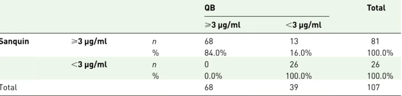
A qualitative comparison of the assays for a cut-off of 3 µg/ml is depicted in Table 3, and shows an accuracy of 88% and a Kappa (standard error of the mean) of 0.718 (0.070). In fact, the distribu-tion of patients according to a 3 µg/ml cut-off is rather similar between both methods, with only 13 patients (12.0%) being placed differently (they
have IFX levels <3 µg/ml when using the rapid QB test, but above that cut-off when using the Sanquin test).
Discussion
Several commercial kits and different protocols have been optimized for an accurate determina-tion of IFX levels from patient serum, but the recent development of a rapid IFX assessment test holds the promise of revolutionizing the TDM-based therapeutic algorithms, by allowing an immediate adjustment of the IFX dosage (as opposed to delaying this intervention to the fol-lowing infusion cycle). This study aimed to assess the clinical sensitivity and specificity of this rapid assay, by using it to measure samples from 110 patients, fully characterized regarding their clini-cal, endoscopic and inflammatory burden status.
Figure 2. Accuracy values of the different cut-off values (only those between 45–75% represented). FC, faecal calprotectin; QB, Quantum Blue® Infliximab: Quantitative Lateral Flow Assay (Bühlmann, Schönenbuch, Switzerland); Sanquin, Level Infliximab M2920 kit (Sanquin, Amsterdam, the Netherlands); vs, versus.
The overall results and the clinical stratification obtained using different cut-offs were compared with those obtained using an already validated and widely used IFX-quantification kit (Sanquin). The results reported here show that although the concentrations obtained by the different methods are strongly correlated, there is a systematic varia-tion: the concentrations measured by the Sanquin kit were, on average, 2.62 units higher than those measured by the rapid QB test, which is consistent with the median IFX values obtained with each method for the entire population. The Sanquin kit’s bias towards measuring higher values when compared with other kits has been noticed before.16,19 Overall, other methodological
com-parisons involving two or more IFX-quantification assays show that, most of the times, the assays compare quite well against each other (even when they are not ELISA-based), but systematic devia-tions are rather common and are likely to result from the fact that different assays use different antibodies with varying IFX affinities.19–25
The association of IFX serum levels with disease outcomes or inflammatory markers such as clinical response, clinical remission, mucosal healing, endo-scopic improvement and C-reactive protein levels have been often reported.3,5,6,8–10,26 Accordingly,
our results show that IFX trough levels were signifi-cantly lower when patients had an endoscopic Mayo score ⩾1 or an FC concentration ⩾250 µg/g. A similar pattern was found for clinical remission and for an endoscopic Mayo score >0 (i.e. patients who have a negative outcome had lower IFX trough lev-els), although in this case the results were not sig-nificant. This might be due to the small size of the cohort, or to the fact that the patients analysed were very stable, most of them (80%) in clinical remis-sion according to the Montreal classification and
with over 14 weeks of IFX therapy (primary nonre-sponders were excluded).
Given the systematic differences encountered in the quantification, one would expect the two dif-ferent methods to have difdif-ferent clinical cut-offs. However, that is not the case: 3 µg/ml is an accept-able threshold for both assays particularly in what concerns assessment of endoscopic status and inflammatory burden (measured by the FC lev-els). Regarding clinical status, although 3 µg/ml may be a satisfactory cut-off, values of 1 and 2 µg/ ml can be considered for the Sanquin and QB assays, respectively. These cut-offs have a margin-ally better accuracy and smaller NPV when com-pared with 3 µg/ml. The same holds true for IFX cut-offs of 1 and 2 µg/ml when addressing endo-scopic activity using an endoendo-scopic Mayo score ⩽1 as criterion for remission: the accuracy raises and the NPV drops when compared with those of a cut-off of 3 µg/ml. Concerning deep remission, however, the different cut-offs seem to behave similarly and it is not easy to choose a single value. This is likely related to the fact that deep remis-sion is a composite endpoint, and therefore reflects the different behaviours of its components. The lack of impact of the systematic bias observed in the optimal clinical cut-off is easily explained by observing the Bland–Altman plot: in fact, this plot shows that the differences encountered in the values measured by both methods are particularly close to 0 for IFX levels <5 µg/ml. In other words, at levels as low as those considered for the clinical threshold, the assays seem to behave in a similar fashion. This is supported by the comparative analysis of the assay’s results, which shows that for a threshold of 3 µg/ml, 88% of the patients fall equally above or below the cut-off irrespective of the method used.
Table 3. Qualitative comparison between Sanquin and QB assays.
QB Total ⩾3 μg/ml <3 μg/ml Sanquin ⩾3 μg/ml n 68 13 81 % 84.0% 16.0% 100.0% <3 μg/ml n 0 26 26 % 0.0% 100.0% 100.0% Total 68 39 107
QB, Quantum Blue® Infliximab: Quantitative Lateral Flow Assay (Bühlmann, Schönenbuch, Switzerland); Sanquin, Level Infliximab M2920 kit (Sanquin, Amsterdam, the Netherlands).
In practical terms, a clinical cut-off should help a physician decide whether a patient may benefit from an IFX dose adjustment. A cut-off of 3 µg/ml has considerably high NPVs, which means that it can exclude patients from benefiting of an IFX dose adjustment with a considerable degree of cer-tainty. Conversely, the PPVs are rather low, which means that having an IFX trough concentration below the defined cut-off does not necessarily imply having clinical activity, endoscopic lesions or a high inflammatory burden. In other words, not all patients with IFX levels below the cut-off will benefit from a dose intensification, and such a decision must be contextualized with other indica-tors (such as symptomatology, presence of anti-bodies to IFX and biomarkers).
The 3 µg/ml (or closer) cut-off has been often referred to in the literature,3,26,27 but so have
lower8,27–29 and higher ones,7,8,10,27 showing that
cut-offs are deeply related to the method used and outcome being assessed, and studies such as these are absolutely necessary to validate thresh-olds and explore their interpretability. One word of caution should be added: our results were derived from a UC patient cohort, and are there-fore only applicable in the context of UC. In fact, the literature shows several instances in which the parallel analysis of CD and UC patients yields different cut-offs or different behaviours of the same cut-off.8,27,28
This study has several strengths that should be noticed, namely its prospective design with a sys-tematic and inclusive evaluation of the therapeu-tic response; and the fact that all quantifications were performed by the same researcher, and therefore the user can be excluded as a source of variation. On the other hand, and as a limitation, one should point out that the sample size was relatively small, although similar to that used in analogous studies;5,6,8,20,28 and that the
occur-rence and amount of anti-IFX in the clinical sam-ples was not taken into consideration.
In conclusion, we have explored the applicability of IFX trough level cut-offs using a recently launched rapid QB test and comparing it with a widely used ELISA kit. Overall, both assays have a good quantitative and qualitative agreement, and a cut-off of 3 µg/ml seems to be appropriate, namely when one is assessing the endoscopic sta-tus (using an endoscopic Mayo score = 0 as the criterion for remission) or the inflammatory
burden. Different cut-offs can be considered for specific situations, and this ultimately depends on whether the user wants to optimize the accuracy or the NPV of the results.
Acknowledgements
The authors thank the Bühlmann company for kindly providing the commercial kits used in this study, all investigators at the hospitals who pro-vided samples and data, GEDII for all the sup-port, Sandra Dias for all the assistance during the data collection and Catarina L. Santos for medi-cal writing assistance.
Author contributions were as follows: FM: Study concept and design; acquisition of data; analysis and interpretation of data; drafting of the manuscript; study supervision; critical revi-sion of the manuscript for important intellec-tual content. JA: IFX and faecal calprotectin assays; analysis and interpretation of data. JL, KG and FC: histological analysis. CCD: statis-tical analysis. All the other authors: recruitment of patients and collection of samples. All authors read and approved the final version of the manuscript.
Funding
This work was supported by the Portuguese IBD Group (GEDII, Grupo de Estudo da Doença Inflamatória Intestinal).
Conflict of interest statement
FM served as speaker and received honoraria from Merck Sharp & Dohme (NJ, USA), Abbvie (IL, USA), Vifor (Glattbrugg, Switzerland), Falk (USA), Laboratorios Vitoria (Amadora, Portugal), Ferring (Saint-Prex, Switzerland), Hospira (IL, USA) and Biogen (MA, USA).
References
1. O’Toole A and Moss AC. Optimizing biologic agents in ulcerative colitis and crohn’s disease.
Curr Gastroenterol Rep 2015; 17: 32.
2. Mould DR and Dubinsky MC. Dashboard systems: pharmacokinetic/pharmacodynamic mediated dose optimization for monoclonal antibodies. J Clin Pharmacol 2015; 55: S51–S59.
3. Adedokun OJ, Sandborn WJ, Feagan BG, et al. Association between serum concentration of infliximab and efficacy in adult patients with
ulcerative colitis. Gastroenterology 2014; 147: 1296–1307, e5.
4. Strik AS, Bots SJA, D’Haens G, et al. Optimization of anti-TNF therapy in patients with Inflammatory Bowel Disease. Expert Rev
Clin Pharmacol 2016; 9: 429–439.
5. Maser EA, Villela R, Silverberg MS, et al. Association of trough serum infliximab to clinical outcome after scheduled maintenance treatment for crohn’s disease. Clin Gastroenterol Hepatol 2006; 4: 1248–1254.
6. Seow CH, Newman A, Irwin SP, et al. Trough serum infliximab: a predictive factor of clinical outcome for infliximab treatment in acute ulcerative colitis. Gut 2010; 59: 49–54.
7. Brandse JF, Mathôt RA, van der Kleij D,
et al. Pharmacokinetic features and presence
of antidrug antibodies associate with response to infliximab induction therapy in patients with moderate to severe ulcerative colitis. Clin
Gastroenterol Hepatol 2016; 14: 251–258.
8. Warman A, Straathof JWA and Derijks LJJ. Therapeutic drug monitoring of infliximab in inflammatory bowel disease patients in a teaching hospital setting: results of a prospective cohort study. Eur J Gastroenterol Hepatol 2015; 27: 242–248.
9. Paul S, Del Tedesco E, Marotte H, et al. Therapeutic drug monitoring of infliximab and mucosal healing in inflammatory bowel disease: a prospective study. Inflamm Bowel Dis 2013; 19: 2568–2576.
10. Ungar B, Levy I, Yavne Y, et al. Optimizing anti-TNF-α therapy: serum levels of infliximab and adalimumab are associated with mucosal healing in patients with inflammatory bowel diseases. Clin Gastroenterol Hepatol 2016; 14: 550–557, e2.
11. Gecse KB, Végh Z and Lakatos PL. Optimizing biological therapy in Crohn’s disease. Expert Rev
Gastroenterol Hepatol 2016; 10: 37–45.
12. Khanna R, Sattin BD, Afif W, et al. Review article: a clinician’s guide for therapeutic drug monitoring of infliximab in inflammatory bowel disease. Aliment Pharmacol Ther 2013; 38: 447–459.
13. Mould DR, D’Haens G and Upton RN. Clinical decision support tools: the evolution of a revolution. Clin Pharmacol Ther 2016; 99: 405–418.
14. Vande Casteele N, Feagan BG, Gils A, et al. Therapeutic drug monitoring in inflammatory
bowel disease: current state and future perspectives. Curr Gastroenterol Rep 2014; 16: 378.
15. Martelli L, Olivera P, Roblin X, et al. Cost-effectiveness of drug monitoring of anti-TNF therapy in inflammatory bowel disease and rheumatoid arthritis: a systematic review.
J Gastroenterol 2017; 52: 19–25.
16. Afonso J, Lopes S, Gonçalves R, et al. Proactive therapeutic drug monitoring of infliximab: a comparative study of a new point-of-care quantitative test with two established ELISA assays. Aliment Pharmacol Ther 2016; 44: 684–692.
17. Kw S, Wj T and Ilstrup Dm. Coated oral 5-aminosalicylic acid therapy for mildly to moderately active ulcerative colitis. A randomized study. N Engl J Med 1987; 317: 1625–1629.
18. Geboes K, Riddell R, Ost A, et al. A reproducible grading scale for histological assessment of inflammation in ulcerative colitis. Gut 2000; 47: 404–409.
19. Ruiz-Argüello B, Del Agua AR, Torres N,
et al. Comparison study of two commercially
available methods for the determination of infliximab, adalimumab, etanercept and anti-drug antibody levels. Clin Chem Lab Med 2013; 51: 287–289.
20. Guiotto C, Daperno M, Frigerio F, et al. Clinical relevance and inter-test reliability of
anti-infliximab antibodies and anti-infliximab trough levels in patients with inflammatory bowel disease. Dig
Liver Dis 2016; 48: 138–143.
21. Schmitz EMH, Van De Kerkhof D, Hamann D,
et al. Therapeutic drug monitoring of infliximab:
Performance evaluation of three commercial ELISA kits. Clin Chem Lab Med 2016; 54: 1211–1219.
22. Steenholdt C, Ainsworth MA, Tovey M, et al. Comparison of techniques for monitoring infliximab and antibodies against infliximab in Crohn’s disease. Ther Drug Monit 2013; 35: 530–538.
23. Hérnandez-Flores D, Valor L, De La Torre I,
et al. Comparison of two ELISA versions for
infliximab serum levels in patients diagnosed with ankylosing spondylitis. Rheumatol Int 2015; 35: 1021–1025.
24. Lee MWM, Connor S, Ng W, et al. Comparison of infliximab drug measurement across three commercially available ELISA kits. Pathology 2016; 48: 608–612.
25. Marini JC, Sendecki J, Cornillie F, et al.
Comparisons of serum infliximab and antibodies-to-infliximab tests used in inflammatory bowel disease clinical trials of Remicade®. AAPS J 2017; 19: 161–171.
26. Vande Casteele N, Ferrante M, Van Assche G,
et al. Trough concentrations of infliximab guide
dosing for patients with inflammatory bowel disease. Gastroenterology 2015; 148: 1320–1329, e3. 27. Silva-Ferreira F, Afonso J, Pinto-Lopes P, et al.
A systematic review on infliximab and adalimumab drug monitoring: levels, clinical
outcomes and assays. Infammatory Bowel Dis 2016; 22: 2289–2301.
28. Steenholdt C, Bendtzen K, Brynskov J, et al. Cut-off levels and diagnostic accuracy of in fl iximab trough levels and anti-in fl iximab antibodies in Crohn’s disease. Scand J Gastroenterol 2011; 46: 310–318.
29. Barlow NL, Mohammed P and Berg JD. Serum trough infliximab and anti-infliximab antibodies in a cohort of gastroenterology and rheumatology patients’ infliximab therapeutic drug monitoring.
Ann Clin Biochem 2016; 53: 477–484.
Visit SAGE journals online journals.sagepub.com/ home/tag