ABSTRACT | Purpose: To compare the use of topical anes thesia and retrobulbar anesthesia during silicone oil removal with a mixed pars plana technique, through evaluating the pain experience of patients. Methods: We selected patients according to their behavior during previous vitreoretinal surgery and ophthalmologic examinations and divided them into two anesthesia groups: topical (n=36) and retrobulbar (n=33). We used a mixed technique for the passive removal of silicone oil in both groups. During each step of the surgery, the patients’ pain experience and the surgeon’s comfort were scored according to a pain scale. Results: The pain experienced during the application of the anesthesia was significantly greater in the retrobulbar group (p<0.001). The topical group experienced greater pain during trocar insertion (p<0.001). There was no significant difference between the groups regarding the overall pain experience or complications. Conclusions: The pain experience of the selected patients during silicone oil removal was comparable between the topical and the retrobulbar anesthesia. Topical anesthesia with the mixed pars plana technique is an effective and safe alternative option for silicone oil removal surgery.
Keywords: Pain; Silicone oils; Anesthetics, local; Administration, topical; Vitreoretinal surgery; Patient satisfaction
RESUMO | Objetivo: Comparar a sensação de dor de pacientes
durante a remoção do óleo de silicone sob anestesia tópica e re trobulbar, usando uma técnica via pars plana combinada.
Métodos: Os pacientes foram selecionados, de acordo com suas atitudes durante cirurgia vitreorretiniana prévia e exames oftalmológicos, e divididos em dois grupos: anestesia tópica e retrobulbar. Para a remoção passiva do óleo de silicone, utili zouse uma técnica combinada em ambos os grupos. A sensação de dor dos pacientes e o conforto do cirurgião foram classificados através de uma escala de dor durante cada etapa da cirurgia. Resultados: Os grupos anestesia tópica e retrobulbar incluíram 36 e 33 pacientes, respectivamente. A sensação de dor durante a aplicação da anestesia foi significativamente maior no grupo retrobulbar (p<0,001). O grupo anestesia tópica sentiu mais dor durante a inserção do trocarte (p<0,001). Não houve diferença significativa entre os grupos em relação à sensação geral de dor e a complicações. Conclusões: A sensação de dor é comparável entre a anestesia tópica e a retrobulbar durante a remoção de óleo de silicone. A combinação de anestesia tópica e uma técnica via pars plana é uma opção alternativa eficaz e segura para a cirurgia de remoção de óleo de silicone.
Descritores: Dor; Óleos de silicone; Anestésicos locais; Admi nistração tópica; Cirurgia vitreorretiniana; Satisfação do paciente
INTRODUCTION
During vitreoretinal surgery for complex cases, surgeons generally use silicone oil tamponade(1). Its ad vantages include quick visual rehabilitation, longterm tamponading of the reattached retina, and relatively stable chemophysical properties(2,3). However, its use is associated with several complications, including ca taracts, keratopathy, corneal endothelial decompen sation, glaucoma, and hypotony. It is therefore recom mended that the silicone oil is removed as soon as possible after achieving retinal stability(36).
Various surgical techniques to remove the silicone oil have been reported(710). Transconjunctival sutureless sys tems result in faster healing, do not require sutures, and result in less discomfort to the patient. However,
Comparison of topical and retrobulbar anesthesia in
terms of pain experience during silicone oil extraction
Comparação da sensação de dor durante a extração de óleo de silicone
sob anestesia tópica e retrobulbar
Alper Mete1, Sabit Kimyon1, Oguzhan Saygılı1, Kıvanç Güngör1, Mithat Temizer2
1. Department of Ophthalmology, School of Medicine, Gaziantep University, Gaziantep, Turkey. 2. Public Health Center, Turkish Public Health Institute, Gaziantep, Turkey.
Submitted for publication: February 6, Accepted: for publication: October 8,
Funding: No specific financial support was available for this study.
Disclosure of potential conflicts of interest: None of the authors have any potential conflict of interest to disclose.
Corresponding author: Alper Mete.
Department of Ophthalmology. Gaziantep University Hospital . 27310 – Gaziantep - Turkey - E-mail: [email protected]
Approved by the following research ethics committee: Gaziantep University Research Ethics Committee (# 2015/173).
the passive removal of silicone oil can prolong the ope ration time during minor sclerotomies, whereas the active vacuum suction needed commonly to extract the silicone oil carries the risk of eyeball collapse or retinal damage(79). A further technique that has been reported for silicone oil removal is the mixed 23gauge infusion and 20gauge pars plana vitrectomy method(10); 20gauge sclerotomy is considered to be an efficient, fast, and inexpensive alternative to the passive removal of sili cone oil of high or low viscosity.
Injection anesthesia methods are commonly used in posterior vitrectomy procedures(11,12). However, these me thods can lead to serious needlerelated complica tions(13,14). Topical anesthesia has been used for coopera tive patients as an alternative to other local anesthesia techniques in vitreoretinal surgery; it has the advantages of being noninvasive, early visual recovery, and ease of administration(1113).
The purpose of this study was to compare retrobulbar anesthesia and topical anesthesia in terms of the pain experience of patients and the comfort of the surgeon during the passive removal of silicone oil using a mixed 23gauge 20gauge technique. There have been only a few previous reports of the use of topical anesthesia for silicone oil removal, and this is the first report to eva luate the pain experience of patients during such use.
METHODS
Study
Thisprospective comparative study included 69 pa tients operated with a mixed pars plana technique for 1000 centistoke (cSt) passive silicone oil removal under topical or retrobulbar anesthesia at the Department of Ophthalmology, Gaziantep University Hospital, Turkey. This study received local ethics committee approval. The procedures were explained to the patients, and they were told they may experience some pain during silicone oil removal and the administration of the retro bulbar block; all patients subsequently provided written informed consent.
Patients
A total of 69 patients who had experienced a comfor table and calm previous vitreoretinal surgery under local anesthesia and preoperative ophthalmologic examina tion were included. All were older than 20 years and had undergone a previous combination of phacoemulsifica tion cataract surgery and posterior vitrectomy with sili
cone oil (1000 cSt) tamponade for retinal detachments (rhegmatogenous retinal detachments, diabetic tractional retinal detachments, or nondiabetic tractional retinal detachments). In addition, the intraocular silicone oil had been in place for at least four months with a stable retina. The exclusion criteria were age younger than 20 years, being phakic or aphakic, redetachments, hypo tony (intraocular pressure (IOP) ≤10 mmHg), extre me an xiety, claustrophobia, speech disorder, ocular in fection, and a known allergy to topical or retrobulbar anesthetic drugs. The participants were matched according to in dications for silicone oil tamponade and an experienced surgeon (A.M.) distributed them randomly between the two anesthesia groups: the topical group (n=36) and the retrobulbar group (n=33).
Surgical procedures and pain–discomfort scoring A single experienced vitreoretinal surgeon (O.S.) per formed all surgeries. For the topical group patients, the surgeon administered topical 0.5% proparacaine hydro chloride drops into the conjunctival sac four times in the 15 minutes preceding surgery, with additional drops administered every 5 minutes throughout the duration of the procedure. During the silicone oil removal, the participants were asked to tell the surgeon if they felt intolerable pain, with additional subtenon anesthesia ready prepared for such an eventuality. For the retrobul bar group patients, a mixture of 3 mL 5% bupivacaine and 3 mL 2% lidocaine was injected into the retrobulbar space using an 0.5inch 26gauge needle. Neither pa tient group received sedation, although we gave an oral analgesic to uncomfortable patients postoperatively. All patients received nasal oxygen (4 L/min) during the procedure.
only two sclerotomies were made. Otherwise, an addi tional sclerotomy was created using a 23gauge trocar cannula system. In all cases, to reduce the patient’s pain and discomfort, the trocars were inserted directly at an angle of approximately 90° to the sclera without displa cing the conjunctiva.
A silicone oilfluid exchange was then performed. In the retrobulbar group, the eyeball was tilted to clear the residual small oil bubbles using colibri forceps. In the topical group, the surgeon instructed the patient to move his or her eyes in the directions needed to clear the residual small silicone oil bubbles. The cannulas were the removed and the IOP was checked. Finally, all the sclerotomies were closed with 80 absorbable sutures. A topical antibiotic drop was instilled in the conjunctival sac. The operative time was recorded.
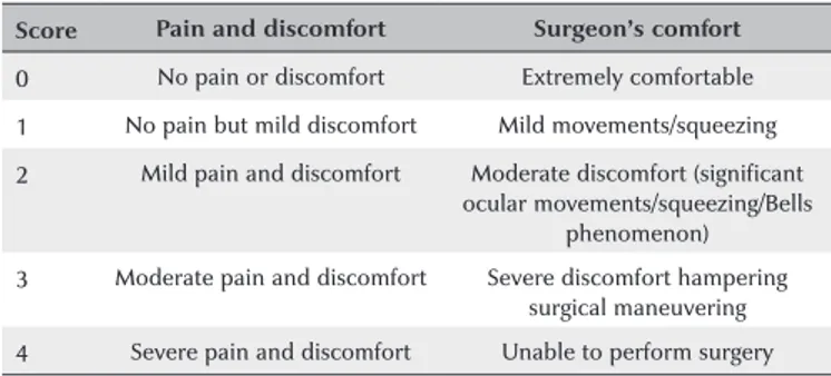
The pain experienced by the patient was scored using a visual analog pain scale (VAPS) at several timepoints: as the anesthesia was administered, during each step of the surgery, and immediately after surgery (Table 1)(11,12). If the patient was unable to see the chart because of low vision, he or she was asked for an oral score. The surgeon’s comfort was also scored using a surgeon’s comfort scale. Each patient underwent followup ocular examinations on the first postoperative day and at the end of the first week and first month.
Data analysis
All statistical analyses were performed using SPSS 16.0 software (SPSS Inc, Chicago, IL). Patient characteristics, clinical characteristics, clinical outcomes, pain scores, and the surgeon’s comfort scores were compared between the two anesthesia groups using MannWhitney U, chisquare, and paired samples t tests. A p value less than 0.05 was considered statistically significant.
RESULTS
Table 2 presents the patients’ characteristics, indica tions for silicone tamponade, duration of silicone tam ponade, operative times, additional procedures during surgery, and preoperative and postoperative bestcor rected visual acuity (BCVA). There was no significant diffe rence between the two groups with regard to age, sex, laterality, distribution of indications for silicone tam ponade, duration of silicone tamponade, operative time, distribution of additional procedures, or preoperative and postoperative BCVA.
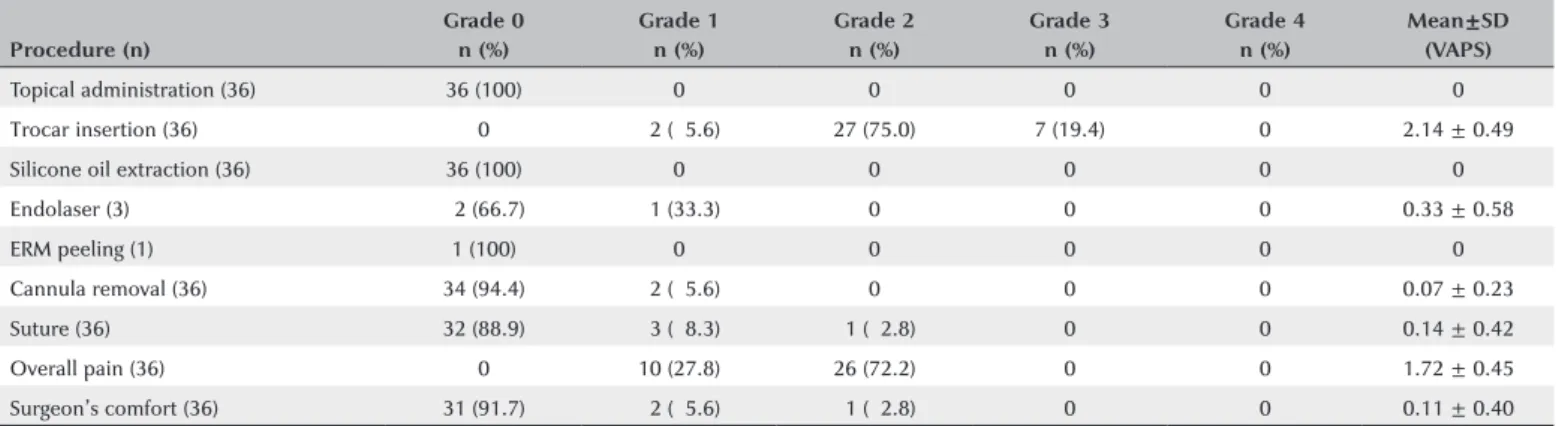
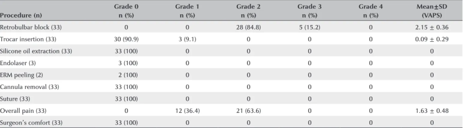
The mean pain scores during the administration of anesthesia differed significantly between the groups (p<0.001). In the retrobulbar group, 28 of the 33 pa tients (85%) scored grade 2 and five (15%) scored grade 3 pain as the retrobulbar block was administered, with a mean pain score of 2.15 ± 0.36. In the topical group, none of the patients reported any pain sensation as the to pical anesthesia was administered. The mean pain scores for all the surgical steps for the topical and retrobulbar groups are shown in tables 3 and 4, respectively.
During trocar insertion, 27 of the 36 patients (75%) in the topical group scored grade 2, seven (19%) grade 3, and two (6%) grade 1 pain; in contrast, only 3 of the 33 patients (9%) in the retrobulbar group scored grade 1 pain. The mean pain scores during trocar entry differed significantly between the groups (p<0.001). However, there was no statistically significant difference between the groups during the passive removal of silicone oil, cannula removal, or the sclerotomy suturing steps of the surgery (p=1.000, p=0.173, and p=0.06, respectively). The mean ( ± SD) comfort score of the surgeon was 0.11 ± 0.40 for the topical group. With the retrobulbar group, the surgeon performed all the procedures with extreme comfort (Grade 0). There was no significant difference in the overall pain and discomfort scores (for anesthesia administration and surgery) or the surgeon’s comfort scores between the groups (p=0.448 and p=0.092; respectively). In both groups, the postoperative mean BCVA increased or remained the same when com pared with the preoperative BCVA. Postoperative BCVA was not changed in seven eyes (19%) in the topical group and in six eyes (18%) in the retrobulbar group.
Additional subtenon anesthesia was not required in any case. None of the patients required oral analgesic or sedation during surgery or postoperatively. Postope ratively, we observed no ocular hypotony or leakage in either group during the followup period. We observed injection anesthesiarelated mild lid edema in three pa Table 1. The visual analog pain scale and the surgeon’s comfort scale
Score Pain and discomfort Surgeon’s comfort
0 No pain or discomfort Extremely comfortable
1 No pain but mild discomfort Mild movements/squeezing
2 Mild pain and discomfort Moderate discomfort (significant ocular movements/squeezing/Bells
phenomenon)
Table 2. Patient characteristics, indications for silicone oil tamponade, duration of silicone oil before its removal, operative time, additional procedures during surgery, and preoperative and postoperative BCVA
Group 1 Group 2
Topical anesthesia n (%) Retrobulbar anesthesia n (%) P value
Age, years 0.670†
Mean±SD 56.2 ± 13.6 54.4 ± 14.9
Range 2381 2478
Sex 0.625*
Male 24 (66.7) 20 (60.6)
Female 12 (33.3) 13 (39.4)
Laterality 0.810*
Right 19 (52.8) 19 (57.6)
Left 17 (47.2) 14 (42.4)
Surgical indications for silicone oil tamponade 0.938*
RRD 19 (52.8) 16 (48.5)
Diabetic TRD 11 (30.6) 11 (33.3)
Nondiabetic TRD 06 (16.7) 06 (18.2)
Duration of silicone oil before its removal, months 0.113†
Mean ± SD 6.3 ± 1.6 06.9 ± 1.7
Range 410 411
Operative time, minutes 0.067†
Mean ± SD 19.5 ± 2.7 18.7 ± 3.2
Range 1628 1526
Additional procedures performed
Endolaser treatment 3 (8.3) 3 (9.1) 1.000*
ERM peeling 1 (2.8) 2 (6.1) 0.603*
BCVA, (logMAR)
Preoperative BCVA (Mean ± SD) 1.16 ± 0.12 1.15 ± 0.13 0.635*
Range (1.521.0) (1.521.0)
Postoperative BCVA (Mean ± SD) 0.76 ± 0.32 0.75 ± 0.35 0.957*
Range (1.300.3) (1.520.3)
n= number of patients; SD= standard deviation; RRD= rhegmatogenous retinal detachment; PDR= proliferative diabetic retinopathy; TRD= tractional retinal detachment; ERM= epiretinal membrane; BCVA= bestcorrected visual acuity.
*= The chisquare test was used; †= The MannWhitney U test was used.
Table 3. Pain experience and surgeon comfort scores for group 1 (topical anesthesia) during the administration of the anesthesia and at each surgical step
Procedure (n)
Grade 0 Grade 1 Grade 2 Grade 3 Grade 4 Mean±SD
n (%) n (%) n (%) n (%) n (%) (VAPS)
Topical administration (36) 36 (100) 0 0 0 0 0
Trocar insertion (36) 0 02 (05.6) 27 (75.0) 7 (19.4) 0 2.14 ± 0.49
Silicone oil extraction (36) 36 (100) 0 0 0 0 0
Endolaser (3) 02 (66.7) 01 (33.3) 0 0 0 0.33 ± 0.58
ERM peeling (1) 1 (100) 0 0 0 0 0
Cannula removal (36) 34 (94.4) 02 (05.6) 0 0 0 0.07 ± 0.23
Suture (36) 32 (88.9) 03 (08.3) 01 (02.8) 0 0 0.14 ± 0.42
Overall pain (36) 0 10 (27.8) 26 (72.2) 0 0 1.72 ± 0.45
Surgeon’s comfort (36) 31 (91.7) 02 (05.6) 01 (02.8) 0 0 0.11 ± 0.40
tients (9%) in the retrobulbar group. Two patients (6%) in the topical group and two (6%) in the retrobulbar group underwent reoperation because of recurrent retinal detachment.
DISCUSSION
Several surgical techniques have been described for silicone oil removal(710,15,16). Surgeons commonly prefer transconjunctival sutureless systems for silicone oil re moval because these systems are less invasive, faster, and more comfortable for the patient than 20gauge sys tems(5,7,9,15,17). Silicone oil removal can be performed by the anterior route in aphakic or pseudophakic patients or along with cataract extraction(1820). Cacciatori et al.(18) reported that the anterior route is more advantageous because it allows the silicone oil to be removed under topical anesthesia, eliminating the potential risks of injection anesthesia and sclerotomyrelated complica tions(18). However, the anterior route has some limita tions in complex posterior vitrectomy procedures, ana tomically limiting surgical maneuvers during these pro cedures(1820). For these cases, the pars plana approach is usually preferred by surgeons because it simplifies the complete evaluation of the peripheral retina during sur gery and the additional procedures(710,1520). In our study, we used the twoport pars plana technique for silicone oil removal. For additional procedures, we crea ted an additional sclerotomy using a 23gauge trocar system. Tan et al.(21) compared twoport and threeport approa ches for silicone oil removal and reported no significant difference between them with regard to the recurrent retinal detachment rate. In contrast, Guo et al.(22) showed that the incidence of recurrent retinal detachment rate
was significantly higher with the twoport than with the threeport technique. The incidence of recurrent retinal redetachment in conventional 20gauge silicone oil re moval surgery has been reported as nearly as 20%(23,24). In our study, the incidence of recurrent retinal detachment with the passive removal of silicone oil using the twoport mixed technique was 6% in both groups. We believe that the low incidence of recurrent detachment in our study was associated with several factors, including perfor ming the silicone oil removal procedures passively in all cases without the use of a powered vacuum system, with its risk of retinal injuries, and suturing all sclerotomies in all patients after the silicone oil had been removed. In this way, we prevented potential ocular hypotony due to leakage. The reported incidence of postoperative ocular hypotony ranges from 0% to 3.8% after 23gauge transconjunctival procedures(2527). However, we did not observe any ocular hypotony in either group during the postoperative period.
Compared with 20gauge systems, 25gauge systems and instruments are more expensive and generally re quire a powered suction device for active silicone oil removal, as well as a prolonged learning curve for the surgeon(15,28). In contrast, 20gauge systems and instru ments are more comfortable and efficient, less expensi ve, and faster when performing additional procedures. Furthermore, 20gauge systems do not generally require powered suction for the silicone oil removal, and they are more effective and faster than transconjunctival su tureless systems for the removal of highdensity silico ne oil(7,10). Active suction for silicone oil removal carries the risk of optic nerve injury or eyeball collapse due to the high vacuum pressure(10). In addition, 23gauge sys tems are widely used for silicone oil removal(7,10,15), and Table 4. Pain experience and surgeon’s comfort scores for group 2 (retrobulbar anesthesia) during the retrobulbar block and at each surgical step
Procedure (n)
Grade 0 Grade 1 Grade 2 Grade 3 Grade 4 Mean±SD
n (%) n (%) n (%) n (%) n (%) (VAPS)
Retrobulbar block (33) 0 0 28 (84.8) 5 (15.2) 0 2.15 ± 0.36
Trocar insertion (33) 30 (90.9) 3 (9.1) 0 0 0 0.09 ± 0.29
Silicone oil extraction (33) 33 (100) 0 0 0 0 0
Endolaser (3) 03 (100) 0 0 0 0 0
ERM peeling (2) 02 (100) 0 0 0 0 0
Cannula removal (33) 33 (100) 0 0 0 0 0
Suture (33) 33 (100) 0 0 0 0 0
Overall pain (33) 0 12 (36.4) 21 (63.6) 0 0 1.63 ± 0.48
Surgeon’s comfort (33) 33 (100) 0 0 0 0 0
some authors have proposed techniques that combine different systems. Lei et al.(10) described a quick and sim ple mixed pars plana technique using a 23gauge entry for infusion combined with a 20gauge entry, which they used to remove 5700cSt silicone oil for 29 patients. They reported no serious complications during surgery and only one recurrent retinal detachment during the followup period. Our outcomes were similar with the findings of that report; we experienced no serious com plications during surgery in either of our groups(10).
Topical anesthesia is a useful alternative option for selected patients in posterior vitrectomy procedures(1113). The wellestablished advantages of topical anesthesia are the absence of needle complications, fast visual recovery, and easy administration(1113). Our experience suggests that the twoport mixed pars plana approach is the most appropriate, suitable, and comfortable te chnique for using with topical anesthesia for silicone oil removal procedures. This mixed technique with topical anesthesia has advantages over other techniques. First, it provides quick and easy removal of silicone oil of various densities through the 20gauge trocar port. Second, the use of 20gauge instrumentation provides costeffective and fast surgery that is comfortable for the surgeon. Third, eye movements during surgery are helpful for the faster removal of silicone oil and air bubbles, and they allow the adequate evaluation of the peripheral retina during surgery without the need for scleral indentation; however, they can result in iatrogenic complications. Sur gical instruments passing through the trocars are useful for the surgeon because they limit sudden eye movements and can provide eye movement in desired directions du ring the procedures(1113). In the present study, we did not experience any complications during surgery.
The assessment of the patients’ pain and discomfort while using topical anesthesia is helpful for following up and for improving the quality of pre (e.g. deciding premedication usage), post, and intraoperative care(29). Pain and discomfort are not objective signs of the quality of anesthetic care; nevertheless, the scored pain values help with evaluating the outcomes from the patient’s perspective(30). We thought that patient selection and surgeon experience played key roles for a comfortable and uncomplicated silicone oil removal surgery under topical anesthesia. The surgeon’s comfort scores did not differ between the retrobulbar and topical groups in our study; however, it was not possible to blind the surgeon to the anesthesia type so there may have been some bias.
The requirement for sedation is another controversial issue associated with silicon oil removal procedures(11). As previously reported, sedation may cause serious complica tions, such as respiratory depression or cardiopulmonary complications(11). We thought that sedation would be re quired for a very small number of patients during silicone oil removal under topical anesthesia. Patient selection considerably reduced the requirement for sedation; in this study, sedation was not used in either group preope ratively or intraoperatively. Nevertheless, we kept addi tional subtenon anesthesia readily available for potential challenges during the surgery.
In conclusion, topical anesthesia provides an alterna tive option for silicone oil removal. We observed com parable pain scores and surgeon’s comfort scores with both topical and retrobulbar anesthesia. Our experience also suggested that the mixed pars plana technique is the most suitable for silicone oil removal under topical anes thesia. Patient selection plays critical role for silicone oil removal under topical anesthesia. To the best of our knowledge, this report is the first on the patients’ pain experience during silicone oil removal using a mixed technique under topical anesthesia.
REFERENCES
1. Krepler K, Mozaffarieh M, Biowski R, Nepp J, Wedrich A. Cataract surgery and silicone oil removal: visual outcome and complica tions in a combined vs. two step surgical approach. Retina. 2003; 23(5):64753.
2. Foulks GN, Harebell DL, Proia AD, Klintworth GK. Histopathology of silicone oil keratopathy in humans. Cornea. 1991;10(1):2937. 3. Casswell AG, Gregor ZJ. Silicone oil removal: operative and posto perative complications. Br J Ophthalmol. 1987;71(12):898902. 4. Kim SW, Oh J, Yang KS, Kim MJ, Rhim JW, Huh K. Risk factors for
the development of transient hypotony after silicone oil removal. Retina. 2010;30(8):122836.
5. Oh HJ, Chang W, Sagong M. Efficacy and safety of active silicone oil removal through a 23gauge transconjunctival cannula using an external vacuum pump. Int J Ophthalmol. 2015;8(2):34752. 6. Gupta S, Chaurasia AK, Chawla R, Kapoor KS, Mahalingam K, Swamy
DR, et al. Longterm outcomes of glaucoma drainage devices for glaucoma postvitreoretinal surgery with silicone oil insertion: a prospective evaluation. Graefes Arch Clin Exp Ophthalmol. 2016; 254(12):244954.
7. Patwardhan SD, Azad R, Shah V, Sharma Y. The safety and efficacy of passive removal of silicone oil with 23gauge transconjunctival sutureless system. Retina. 2010;30(8):123741.
8. Cekiç O, Cakir M, Yilmaz OF. Passive silicone oil removal in 23gauge tranconjunctival vitrectomy. Ophthalmic Surg Lasers Imaging. 2011;42(6):5145.
10. Lei JQ, Xie AM, Shi Q. Clinical presentation of a mixed 23gauge infusion and 20 gauge pars plana technique for active silicone oil removal. Int J Ophthalmol. 2012;5(5):6004.
11. Mete A, Saygılı O, Kimyon S, Pamukcu C, Çeri S, Güngör K, et al. Comparison of pain experience during 23G vitreoretinal surgery under topical and retrobulbar anesthesia. Int Ophthalmol. 2017; 37(2):34956.
12. Mahajan D, Sain S, Azad S, Arora T, Azad R. Comparison of topical anesthesia and peribulbar anesthesia for 23gauge vitrectomy without sedation. Retina. 2013;33(7):14006
13. Bahçecioglu H, Unal M, Artunay O, Rasier R, Sarici A. Posterior
vitrectomy under topical anesthesia. Can J Ophthalmol. 2007;
42(2):2727.
14. Frieman BJ, Friedberg MA. Globe perforation associated with subtenon’s anesthesia. Am J Ophthalmol. 2001;131(4):5201. 15. Kapran Z, Acar N. Active removal of silicone oil with 25gauge su
tureless system. Retina. 2007;27(8):11335.
16. Tsui I, Reddy S, Hubschman JP. An elementary and effective method for silicone oil removal. Retina. 2010;30(3):5246.
17. Abouammoh MA, Yeung J, Gale JS. Sutureless silicone oil removal: a quick and safe technique. Retina. 2012;32(2):3968.
18. Cacciatori M, Nayak H, Arpa P. Silicone oil removal in pseudo phakic and aphakic eyes. Retina. 2007;27(9):13001.
19. Dada VK, Talwar D, Sharma N, Dada T, Sudan R, Azad RV. Pha coemulsification combined with silicone oil removal through a posterior capsulorhexis. J Cataract Refract Surg. 2001;27(8):12437. Comment in: J Cataract Refract Surg. 2002;28(1):5. J Cataract Re fract Surg. 2002;28(10):1719; author reply 1719.
20. El Baha SM, Abouhussein MA, Hemeida TS. Sutureless phacoemulsifi cation with transpupillary removal of silicone oil and intracapsular intraocular lens implantation using illuminated 23gauge infusion system. Retina. 2011;31(2):40812.
21. Tan HS, Dell’omo R, Mura M. Silicone oil removal after rhegma togenous retinal detachment: comparing techniques. Eye (Lond). 2012;26(3):4447. Comment in: Eye (Lond). 2012;26(9):1276; author reply 1277.
22. Guo CM, Wang YS, Hui YN. Analysis of the complications of two kinds of silicone oil removal. Int J Ophthalmol. 2008;11(1):16770. 23. Jiang F, Krause M, Ruprecht KW, Hille K. Risk factors for anatomi
cal success and visual outcome in patients undergoing silicone oil removal. Eur J Ophthalmol. 2002;12(4):2938.
24. Goezinne F, La Heij EC, Berendschot TT, Liem AT, Hendrikse F. Risk factors for redetachment and worse visual outcome after silicone oil removal in eyes with complicated retinal detachment. Eur J Ophthalmol. 2007;17(4):62737.
25. Woo SJ, Park KH, Hwang JM, Kim JH, Yu YS, Chung H. Risk factors associated with sclerotomy leakage and postoperative hypotony after 23gauge transconjunctival sutureless vitrectomy. Retina. 2009;29(4):45663.
26. Parolini B, Prigione G, Romanelli F, Cereda MG, Sartore M, Pertile G. Postoperative complications and intraocular pressure in 943 con secutive cases of 23gauge transconjunctival pars plana vitrectomy with 1year followup. Retina. 2010;30(1):10711.
27. Singh CN, Iezzi R, Mahmoud TH. Intraocular pressure instability after 23gauge vitrectomy. Retina. 2010;30(4):62934.
28. Jusufbegovic D, Gambrell JD, Barak Y, Schaal S. Twentyfivegauge vitrectomy for the removal of 5000 centistokes silicone oil. Retina. 2013;33(9):193842.
29. Fernandes MB, Souza RV, Vasconcelos GC, Ribeiro KG, Andrade BB, Fernandes CR. Assessing patient satisfaction with cataract surgery under topical anesthesia supplemented by intracameral lidocaine combined with sedation. Arq Bras Oftalmol. 2013;76(6):3459. Comment in: Arq Bras Oftalmol. 2014;77(2):132.