FACULDADE DE CIÊNCIAS
DEPARTAMENTO DE BIOLOGIA VEGETAL
DETECTION AND LOCALIZATION OF
LYSA AND GP1 PROTEINS OF THE
MYCOBACTERIOPHAGE Ms6
Alessa Ede Valverde da Silva
MESTRADO EM MICROBIOLOGIA APLICADA
2011
UNIVERSIDADE DE LISBOA
FACULDADE DE CIÊNCIAS
DEPARTAMENTO DE BIOLOGIA VEGETAL
DETECTION AND LOCALIZATION OF
LYSA AND GP1 PROTEINS OF THE
MYCOBACTERIOPHAGE Ms6
Dissertação orientada por Prof.ª Dr.ª Madalena Pimentel (FFUL)
e Prof. Dr. Mário Santos (FCUL)
Alessa Ede Valverde da Silva
MESTRADO EM MICROBIOLOGIA APLICADA
2011
DETECTION AND LOCALIZATION OF
LYSA AND GP1 PROTEINS OF THE
MYCOBACTERIOPHAGE Ms6
Alessa Ede Valverde da Silva
M
ASTER
T
HESIS
2011
This thesis was fully performed at Microbial Genetics Laboratory, Center for
Molecular Pathogenesis, Unit of Retrovirus and Associated Infections
(FFUL) under the direct supervision of Prof.ª Dr.ª Madalena Pimentel.
Prof. Dr. Mário Santos was the internal designated supervisor in the scope
of the Master in Applied Microbiology of the Faculty of Sciences of the
University of Lisbon.
i
A
GRADECIMENTOS/A
CKNOWLEDGMENTSFinalizada uma etapa particularmente importante da minha vida, não poderia deixar de expressar o mais profundo agradecimento a todos aqueles que me apoiaram nesta longa caminhada e contribuíram para a realização deste trabalho. As minhas primeiras palavras são dirigidas à minha orientadora Madalena Pimentel por ter me aceitado no seu grupo de trabalho, pela sua disponibilidade e orientação prestada, pelo seu apoio incondicional e compreensão que sempre manifestou.
Agradeço ao meu orientador interno Mário Santos pela disponibilidade e ajuda prestada.
Ao Centro de Patogénese Molecular pela disponibilização de todo o material e afins necessários para a realização deste trabalho.
À Andreia e Filipa pela amizade e ajuda disponibilizada ao longo deste trabalho.
Por último mas não em último, quero agradecer especialmente à minha mãe pelo apoio incondicional e dedicação para que eu conseguisse chegar a esta etapa da minha vida.
ii
A
BBREVIATIONS aa AG Amp ββββ-gal BCIP bp BRED BS CM DNA ds EDTA FL GFP His HRP IPTG Kan kDa LB MA Met m.o.i. mRNA nt OD OM ORF PAGE PBS PCR PG PhoA p.m.f. PNPP RNA SAR SDS SP ss TCA TMD Tris wt amino acid arabinogalactan ampicillin beta-galactosidase 5-bromo-4-chloro-3-indoxyl phosphate base pairbacteriophage recombineering of electroporated DNA burst size
cytoplasmic membrane deoxyribonucleic acid double-stranded
Ethylenediamine tetraacetic acid free lipids
green fluorescent protein Histidine
horse radish peroxidase
isopropyl β-D-1-thiogalactopyranoside kanamycin kilodalton Luria-Bertani mycolic acids methionine multiplicity of infection messenger ribonucleic acid nucleotide
optical density outer membrane open reading frame
polyacrylamide gel electrophoresis phosphate-buffered saline
polymerase chain reaction peptidoglycan alkaline phosphatase proton-motive force p-nitro-phenyl-phosphate ribonucleic acid signal-arrest-release sodium dodecyl sulphate signal peptide single-stranded trichloroacetic acid transmembrane domain tris(hydroxymethyl)aminomethane wild type
iii
R
ESUMOBacteriófagos, ou fagos, são vírus que infectam bactérias. Estes organismos são dez vezes mais numerosos do que as bactérias no meio ambiente, sendo estes as entidades mais abundantes na Terra. Os fagos desempenham um grande papel no equilíbrio ecológico da vida microbiana. O estudo dos bacteriófagos, teve início em 1915 por Frederick Twort quando este isolou das fezes de um paciente com disenteria um “agente infeccioso e filtrável”. Mais tarde, em 1918, D’Herelle descobriu uma entidade “antagonista” de bactérias, que provocava a sua lise em culturas líquidas e levavam à formação de placas de lise na superfície de meios de cultura com agar semeados com bactérias. D'Herelle criou a designação de bacteriófago para denominar os “vírus que comem bactérias”. Apesar de até a data já terem sito descritos inúmeros fagos, apenas alguns têm sido estudados com algum detalhe. Relativamente à natureza dos ácidos nucleicos, os fagos podem apresentar um genoma quer de ADN de cadeia dupla, ADN de cadeia simples, ARN de cadeia dupla ou ARN de cadeia simples e normalmente o genoma é encapsulado em um invólucro proteico denominado cápside. A maioria dos fagos descritos até à data, apresentam cauda e um genoma de ADN de cadeia dupla (aproximadamente 96%). Existe uma variada gama de diferentes tipos de morfologia nos fagos e a sua taxonomia é baseada nos vários tipos de morfologia, presença ou ausência de envelope, pelo seu tamanho e pelo seu tipo de ácido nucleico.
A classificação actual é derivada de um esquema proposto por Bradley em 1967. Presentemente a classificação inclui uma ordem, 14 famílias oficialmente aceites e 37 géneros.
Os bacteriófagos, como todos os parasitas obrigatórios, requerem células hospedeiras para se manter e se reproduzir, aproveitando-se dos seus mecanismos de biossíntese para sobreviver. Com base no tipo de ciclo de vida, os bacteriófagos podem ser de dois tipos: fagos líticos, que apresentam um ciclo de vida lítico; ou fagos temperados, que apresentam ciclo de vida lisogénico e lítico. No ciclo lítico, o fago redirecciona o metabolismo do hospedeiro para a produção de novos vírus, que são libertados durante a lise celular provocada pelos vírus. No ciclo lisogénico, o genoma do fago é inserido no genoma do hospedeiro e sendo então replicado quando o genoma do hospedeiro se replica até que o ciclo lítico seja induzido, por exemplo por agentes mutagénicos. Para iniciar a infecção, os fagos necessitam adsorver à superfície bacteriana, através do reconhecimento de receptores presentes na parede celular e posteriormente ocorre a entrada do genoma viral na célula hospedeira. Após a injecção do genoma viral, este é transcrito pelas polimerases do hospedeiro eventualmente há produção de novas partículas virais pela maquinaria do hospedeiro. Quando o ciclo lítico sucede, na maioria dos casos é necessário ocorrer lise da célula para que os novos viriões possam encontrar novos hospedeiros. Os fagos de ADN de dupla cadeia desenvolveram um sistema de lise em que utilizam uma estratégia denominada “holina-endolisina” para realizar uma lise celular eficiente no hospedeiro.
O fago Ms6 é um micobacteriófago temperado, com genoma constituído por ADN de cadeia dupla, que infecta a bactéria Mycobacterium smegmatis. Os micobacteriófagos representam excelentes sistemas modelo para estudar micobactérias hospedeiras. Estes bacteriófagos têm vindo a desempenhar um papel fundamental no desenvolvimento de sistemas genéticos para o estudo da patogénese
iv
molecular das micobactérias patogénicas para o homem, sendo as espécies mais reconhecidas Mycobacterium tuberculosis e Mycobacterium leprae, os agentes causadores da tuberculose e lepra, respectivamente. Mycobacterium smegmatis é considerada uma micobactéria não-patogénica representando assim um bom modelo de estudo.O micobacteriófago Ms6, sendo de cadeia dupla, utiliza a estratégia “holina-endolisina” para induzir uma lise efectiva do hospedeiro. As endolisinas são enzimas muralíticas com actividade hidrolítica das ligações específicas que se estabelecem na camada de peptidoglicano e as holinas são proteínas membranares de pequenas dimensões, que conduzem a uma alteração do potencial de membrana como consequência da abertura de poros na membrana, actuando assim como controladora da activação da endolisina ou permitindo o seu acesso à mureína.
Na maioria dos casos descritos até à data, o acesso da endolisina ao seu alvo é feito pela passagem através dos poros formados pela holina. Actualmente têm sido descritos fagos que produzem endolisinas cujo transporte para o meio extracitoplasmático é independente da holina, utilizando os sistemas de transporte bacterianos. Estas endolisinas apresentam na sua estrutura uma sequência sinal, que permite a translocação da proteína, através da membrana citoplasmática, utilizando o sistema sec (translocase) do hospedeiro, até ao espaço periplásmico. Foram já descritas endolisinas que apresentam na região N-terminal um péptido sinal, como é o caso da Lys44 produzida pelo fago fOg44 de Oenococcus oeni. Recentemente foram descobertas outras endolisinas, de fagos que infectam bactérias Gram-negativas, que apresentam na sua extremidade N-terminal uma região de carácter hidrofóbico, designada por Signal-Arrest-Release (SAR), que permite igualmente, o transporte da endolisina através da membrana citoplasmática com o auxílio do sistema sec,, com a particularidade de não ser clivada. Apesar de nestes casos a holina não participar no transporte da endolisina, pensa-se que esta possa desempenhar um papel na activação de lisinas que se encontram no espaço periplásmico e na determinação do tempo de lise.
A cassete lítica do fago Ms6 é constituída por 5 genes. Para além dos genes da endolisina (lysA ou gp2) e holina (hol ou gp4), a cassete lítica do Ms6 inclui mais 3 genes, gp1, gp3 e gp5.
Catalão e colegas (2010) mostraram que a endolisina LysA é exportada com a ajuda do produto do gene gp1, de uma forma independente da holina. É demonstrado que o gene gp1 codifica uma proteína “chaperone-like” que se liga à endolisina, auxiliando a sua exportação para o ambiente extra-citoplasmático, o que é necessário para uma eficiente lise do fago Ms6. Actualmente não se sabe como o endolisisna permanece inactiva até a holina determinar o tempo de lise. Assim, o objectivo principal deste trabalho foi investigar o papel do produto do gene gp1 no transporte de LysA e outras proteínas sem péptido sinal, para o periplasma. Para alcançar este objectivo, construiu-se um fago Ms6 mutante onde o gene gp1 contém na sua extremidade 3 ' uma sequência que codifica para uma cauda C-Myc permitindo a produção de uma proteína recombinante Gp1-C-Myc. Esta proteína foi detectada com o anticorpo anti C-Myc na fracção solúvel e membranar quando células de M. smegmatis foram infectadas com o fago mutante Ms6Gp1-CMyc.
v
O mesmo resultado foi obtido em E.coli confirmando que Gp1 se distribui entre os compartimentos solúveis e membranares. Para apoiar este resultados foram utilizados duas proteínas repórter, a fosfatase alcalina bacteriana (PhoA) como repórter para a localização periplasmática e a proteína verde fluorescente (GFP) como repórter para a localização citoplasmática. A capacidade de Gp1 translocar proteínas foi também apoiada pelo resultado obtido com fusões com a PhoA e GFP. Os resultados da fusão da PhoA revelaram actividade da fosfatase alcalina, enquanto a fusão Gp1-GFP resultou em perda de emissão de fluorescência.Na experiência realizada com a proteinase K obteve-se um perfil alterado de proteínas quando LysA foi sujeita a um tratamento com proteinase K, sugerindo que a LysA pode estar localizada no periplasma na presença de Gp1.
Os resultados apresentados reforçam o papel da Gp1 em auxiliar outras proteínas, sem um péptido sinal, para atravessar a membrana para o periplasma.
Os resultados obtidos neste trabalho experimental abrem novas perspectivas no sentido de oportunidades para novos estudos em vias de secreção de micobactérias. Também uma melhor caracterização do genoma dos bacteriófagos poderá levar a contribuição para a identificação de novos alvos terapêuticos na parede das micobactérias.
vi
A
BSTRACTBacteriophages are viruses that infect bacteria. The majority described to date (≈96%) are endowed with a tail and contains a double-stranded DNA (dsDNA) genome. The phage Ms6 is a temperate phage that infects Mycobacterium smegmatis. Like dsDNA phages, Ms6 uses the holin-endolysin mechanism to accomplish lysis of the host. In addition to holin-endolysin (lysA) and holin (hol) genes, Ms6 genome encodes three accessory lysis proteins, Gp1, LysB and Gp5. Endolysins are proteins that degrade the rigid murein layer, and holins are small membrane proteins that control the activation of the endolysin or its access to the murein. Recently it has been shown that the endolysin LysA is exported with the help of the gp1 gene product, which is involved in its delivery to the peptidoglycan, in a holin-independent manner. Thus, the purpose of this project is to investigate the role of gp1 gene product in the transportation of LysA and other proteins without a peptide signal, to the periplasm. To achieve this we constructed a mutant Ms6 phage where the gp1 gene carries, at its 3’ end, a sequence coding for a C-Myc tag allowing the production of a recombinant protein that was detected with an antibody anti C-C-Myc in the soluble and membrane fraction of M. smegmatis infected cells. The same result was obtained in E.coli confirming that Gp1 distributes between the soluble and membrane compartments. The ability of Gp1 to translocate proteins is suggested by the obtained result with alkaline phosphatise (PhoA) and green fluorescence protein (GFP) fusions. A Gp1-PhoA fusion results in alkaline phosphatase activity while a Gp1-GFP fusion results in loss of fluorescence emission. In addition, an altered profile of the proteins when LysA is subject to a proteinase K treatment suggests that LysA may be periplasm localized in the presence of Gp1. The results presented here strengthen the role of Gp1 in helping other proteins, without a peptide signal, to cross the membrane to the periplasm.
vii
T
ABLE OF CONTENTS Agradecimentos/Acknowledgments ... i Abbreviations ... ii Resumo ... iii Abstract ... vi Introduction ... 1 Classification... 1The phage life cycle ... 2
Bacteriophage-Induced host cell lysis ... 4
The sec-Mediated Lysis ... 7
Mycobacteriophages ... 9
Mycobacteriophage Ms6 and its Lysis Operon... 9
Objectives ... 12
Materials and Methods ... 13
Bacteriophages, bacterial strains, plasmids and growth conditions ... 13
DNA extraction from Ms6 wt ... 14
One-step growth experiments ... 14
Gp1 expression in M. smegmatis-infected cells ... 15
Cellular fractionation of M. smegmatis ... 15
Protease accessibility experiments ... 16
Plasmid construction ... 16
Detection and localization of LysA and Gp1 proteins of the mycobacteriophage Ms6 Table of Contents
viii
Expression and determination of solubility from Gp1 and Gp1-GFP in E. coli cells ... 17
PCR reactions ... 18
Results ... 19
Construction of the Ms6Gp1-CMyc mutant ... 19
Evaluation of the Ms6Gp1-CMyc growth parameters ... 21
Gp1 expression in M. smegmatis-infected cells ... 22
Subcellular localization of Gp1 in M. smegmatis-infected cells ... 23
Expression and localization of Ms6 Gp1 in E. coli ... 24
Analysis of a Ms6 Gp1 fusion to Alkaline Phosphatase ... 25
Analysis of a Ms6 Gp1 fusion to Green-fluorescent protein ... 28
Protease accessibility experiments of LysA in E. coli ... 31
Discussion/ Conclusion ... 33
1
I
NTRODUCTIONBacteriophages (phages) are viruses that have the ability to infect bacteria. They are among the most common biological entities in the biosphere, which play a major role in the ecological balance of microbial life (Pedulla 2003; Deschavanne, 2010). They also play a profound role in the evolution of their host (bacteria) and represent one of the three major mobile genetic elements that contribute significantly to horizontal gene transfer in bacterial genomes by transduction (Boyer et al., 2008). Whole-genome sequencing studiesin bacteria have shown that phage elements contribute significantly to sequence diversity and can potentially influence bacterial pathogenicity.
Phages are ten times more abundant than bacteria, with an estimated 1032 bacteriophages on earth (Hanlon, 2007). On a global scale, it is estimated that ~ 1025 phages initiate an infection every second (Pedulla et al., 2003; Brüssow et al., 2004; Hendrix, 2005; Hatfull, 2010) and in each of those infections the phage encounters DNA (bacterial or prophage DNA), and can potentially recombine to generate new genomic arrangements (Canchaya et al., 2003; Hatfull et al., 2006).
Bacteriophages were first discovered by Frederick Twort in 1915, when he described the glassy transformation of “Micrococcus” colonies by a transmissible agent, and by Felix d'Herelle in 1917
(Weinbauer, 2003; Kutter et al., 2004) when he observed and described the lysis of Shigella (Ackermann, 2003). D’Herelle created the term bacteriophages for these infectious agents lysing the bacteria, which literally means “eaters of bacteria”. He also developed several techniques that are still in use, postulated the intracellular multiplication of viruses, and was the pioneer to the phage therapy of infectious diseases (Ackerman, 1997).
Classification
There exists a range of different morphological types of phages and taxonomy is based on their capsid shape, the presence or absence of an envelope, their size and also by their nucleic acid type.
The actual phage classification is derived from a scheme proposed by Bradley in 1967. Currently the classification includes one order, 14 families officially accepted and at least five other are awaiting for the classification, and 37 genera (Ackermann, 2009).
Phage genome consists either of double-stranded DNA (dsDNA), single-stranded DNA (ssDNA) or very rarely of single-stranded RNA (ssRNA) and normally the phage’s genome is encapsulated in a protein coat (capsid). Bacteriophages can be tailed or tailless. Phages that have a tail in its structure, belong to the order Caudovirales and represents around 96% of all the phages described so far. Each phage of the Caudovirales order possesses a double-stranded DNA structure and is grouped in one of three families: Myoviridae (with long contracted tails), Siphoviridae (with long non contracted tails) and Podoviridae (with short tails). The remaining phages can be either filamentous, polyhedral or pleiomorphic and are grouped in the families Microviridae (ssDNA), Leviridae (ssRNA), Cystoviridae
Detection and localization of LysA and Gp1 proteins of the mycobacteriophage Ms6 Introduction
2
(dsRNA with envelope) or Inoviridae (ssDNA with long filaments) (Ackermann, 2003; Ackermann, 2009; Hanlon, 2007).The phage life cycle
Viruses are obligate pathogens that require host cells in order to replicate (Short et al., 1999). Each phage multiplies and produces new viral particles inside bacteria using the host biosynthetic machinery to survive (Young, 1992; Young et al., 2002).
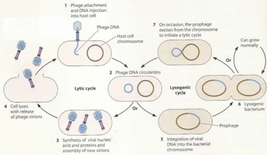
Upon infection of the bacterial host, bacteriophages can follow several life cycles: lytic, lysogenic, pseudolysogenic and chronic infections (Weinbauer, 2004). In the lytic cycle, the phageredirects the host metabolism towards the production of new phages, which are released during the lysis of the host cell. In the lysogenic cycle, the phage’s genome remains in the host in a dormant stage (as prophage or as plasmids) and replicates along with the host, until the lytic cycles is induced (Weinbauer, 2004; Hanlon, 2007; Ackerman 2009). The life cycle of a temperate phage is exemplified in Figure 1.
In chronic infections, the cell is infected and phage progeny is continuously released from the host cell by budding or extrusion without lysing the cell. In persistent infections (pseudolysogeny) phages multiply in a fraction of the population (Weinbauer, 2004).
Infection of cells by viruses requires direct contact between a virus particle and its host (Short et al., 1999). To initiate an infection, a phage virion has to first adsorb to the surface of a susceptible host cell (Shao et al., 2008).Bacterial virus do not randomly attach to the surface of a host cell, they fasten to specific receptor sites that may be any one of a wide variety of cell surface components, including proteins, oligosaccharides, teichoic acids, peptidoglycan or lipopolysaccharides. (Kutter et al., 2004; Guttman et al., 2004; Hanlon, 2007;). In some cases, the attachment site might be present on the cell capsule, flagella or even conjugative pili. At first, the attachment is reversible but then it becomes irreversible and is followed by transfer of phage genetic material into the host cell. The entrance of the phage genome into the host cell can occur by a variety of mechanisms, depending on the physiology of the virus (Weinbauer, 2004; Hanlon, 2007). In some phages, the bases present on the phage DNA are chemically modified to confer protection against attacks by cellular restriction and nuclease enzymes. After DNA injection, the viral genome is transcribed by the host cell RNA polymerase, which starts to synthesize early mRNAs to take over the metabolic machinery of the bacterium, redirecting its metabolic processes to the manufacture of new virus particles(Hanlon, 2007). Early mRNA synthesis is followed by the phage DNA replication and later mRNA synthesis. Late mRNAs direct the synthesis of three kinds of proteins: phage structural proteins, proteins that help with the phage assembly without becoming part of the virion structure, and proteins involved in the cell lysis and phage release (Guttman et al., 2004). Afterwards, the construction and assembly of new phage particles within the host cell takes place, and in the majority of the phages, the release of the new virions implies host cell lysis (Hanlon, 2007). In some phages, the cell lysis is mediated by the production of late proteins, which attack the bacterial peptidoglycan (Young et al., 2006; Hanlon, 2007).
3
In the lysogenic cycle, temperate bacteriophages do not automatically enter a lytic cycle, but alternately, integrate their DNA into the host cell DNA after infection (Figure 1). The bacterial cells are then designated lysogenic cells. When the bacterial DNA replicates, the phage DNA replicates simultaneously and so each daughter cell will withhold the viral DNA (prophage). Each prophage produces a repressor protein that blocks the transcription of its own genes and also those of similarly related bacteriophages (Weinbauer, 2004). The existence of a prophage can therefore grant, upon a bacterial cell, some sort of immunity to infection by the same or close related bacterial virus. Lysogenic cells may undergo several rounds of division, but sporadically, one cell can lyse spontaneously and release the phage’s progeny. Nerveless, a population of lysogenic cells may be induced to enter the lytic cycle through environmental stress, such as treatment with mutagenic agents or exposure to ultraviolet light (Hanlon, 2007).Figure 1. Two lifestyles of a temperate bacteriophage: lysogenic and lytic cycle (Shaechter et al., 2006).
When a prophage escapes regulation by the repressor, its DNA is cut free allowing it to follow a lytic cycle. The excision of prophage DNA is frequently imprecise and bacterial genes adjacent to the prophage DNA may be incorporated into the infectious phage DNA and then transferred to subsequent host cells (Hanlon, 2007). This mechanism is known as restricted transduction, and is responsible for the horizontal transfer of genes, from one bacterial cell to another (Canchaya et al., 2003; Hanlon, 2007). The acquisition of prophages would be an irrelevant process for the evolution of pathogenic bacteria if phages wouldn’t transfer useful genes to the lysogen cell, which are known to increase the survival fitness of lysogens (Brüssow et al., 2004).
Detection and localization of LysA and Gp1 proteins of the mycobacteriophage Ms6 Introduction
4
Bacteriophage-Induced host cell lysisMost bacteriophages must lyse their host cell to release the newly assembled progeny virions to the extracellular environment (Young et al., 2006) Lysis is a programmed event of major importance regarding the phage survival and ecological fitness (Wang, 2006). A sharply time-defined and efficient release of phage progeny is crucial to maximize both burst-size (number of phage progeny released) and the opportunity to infect new hosts (Wang, 2006; São-José et al., 2007; Shao et al., 2008).
If lysis occurs too early, prior to the assembly of a significant number of progeny, the infectious cycle would not be profitable. On the other hand, if lysis happens too late it might preclude the phage’s progeny from taking advantage of other potential host cells in the environment of the infected cell. It is likely that these opposing evolutionary pressures are balanced, giving a characteristic optimal lysis time for each bacteriophage (Bernhardt et al., 2002).
The major barrier to lysis is the continuous meshwork of peptidoglycan, which is a stable structure that allows the bacterial envelope to resist internal osmotic pressure (Young et al., 2000; Wang, 2006). Filamentous phages have a unique morphology and morphogenesis and can extrude from the cytoplasm without lethal consequences for the host. The rest of the phages must either degrade or compromise the peptidoglycan to provoke lysis (Young et al., 2000; São-José et al., 2007).
There are at least two distinct strategies for bacteriophages lysis: one employed by phages with small single-stranded nucleic acids and the other by phages with double-stranded genome. Single-stranded DNA and ssRNA phages accomplish lysis with a single lysis protein that does not encode a muralytic enzyme activity. This protein causes lysis through acting as a specific inhibitor of an enzyme of the multi-step pathway of murein biosynthesis, thus inhibiting the cell wall synthesis (Young et al., 2000; Bernhardt et al., 2002).
In double-stranded DNA (dsDNA) phages, two complementary functions have independently evolved to accomplish both rapid progeny release and optimal time lysis (São-José et al., 2007). Double-stranded DNA phages evolved a lytic system that uses a “holin-endolysin” strategy to accomplish an efficient host cell lysis. These phages produce, during the lytic cycle’s late phase of gene expression, a soluble muralytic enzyme called endolysin or lysin. Endolysin is a rather generic term used to describe a range of bacteriophage-encoded peptidoglycan hydrolases. These enzymes are also known as phage lysozymes, lysins, or muralytic enzymes. They are characterized by their ability to target bonds in the peptidoglycan (PG) layer of the host cell wall. Therefore, endolysins degrade the rigid murein layer and release the newly assembled virions by lysis (Loessner, 2005). Generally, endolysins have muralytic activity against one of the three different types of covalent bonds (glycosidic, amide, and peptide) of the peptidoglycan polymer of the cell wall. However, a small number of endolysins with more than one type of muralytic activity have been described, such as, endolysins encoded by Streptococcus agalactiae bacteriophage B30 (muramidase and peptidase) (Pritchard et al., 20004), Staphylococcus aureus phage
5
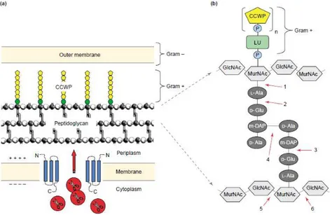
Φ11 (endopeptidase and amidase) (Navarre et al., 1999) and S.agalactiae phage NCTC 11261 (endopeptidase and muramidase) (Cheng et al., 2005).Depending on the enzymatic specificity, endolysins can be divided into five major functional types: (i) N-acetylmuramidases (lysozymes), (ii) endo-b-N-acetylglucosaminidases, and (iii) lytic transglycosylases, which all cleave the sugar moiety of peptidoglycan; (iv) endopeptidases, which cleave the peptide moiety; and (v) N-acetylmuramoyl- L-alanine amidases, which cut the amide bond between both moieties (Figure 2) (Borysowski et al., 2006)
Figure 2. Bacterial cell wall structure and endolysin targets. (a) Schematic representation of the bacterial cell wall, and of how phage endolysins,like λR, gain access to their substrate. Holin proteins (blue) insert themselves into the cytoplasmic membrane and can oligomerize, thereby forming membrane lesions. The endolysins (red) pass through these pores to access the peptidoglycan. (b) The bonds potentially attacked by endolysins of different enzymatic specificities are indicated by numbers: 1) N-acetylmuramoyl-L-alanine amidase; 2) L-alanoyl-D-glutamate endopeptidase; 3) D-glutamyl-m-DAP endopeptidase (this activity has not yet been identified in a phage endolysin); 4) interpeptide bridge-specific endopeptidases; 5) N-acetyl-b-D-glucosaminidase; and 6) N-acetyl-b-D-muramidase (also known as muramoylhydrolase and ‘lysozyme’) and lytic transglycosylase (Figure from Loessner, 2005).
Abbreviations: CCWP, carbohydrate cell wall polymer; GlcNAc, acetyl glucosamine; LU, linkage unit; m-DAP, meso-diaminopimelic acid; MurNAc, N-acetyl muramic acid; P, phosphate group
The endolysins structure, from phages infecting Gram positive host, is composed by two domains, positioned at the N and C-terminus connected by a short linker (L) (Fig. 3). Generally the N-terminal domain contains the catalytic activity of the enzyme, and the C-N-terminal domain binds to a specific substrate (usually carbohydrates) found in the cell wall of the host bacterium (Fischetti, 2005; Lossner, 2005). Cleavage requires interaction of the binding domain with its cell wall substrate. This offers some degree of specificity to the lysins because these substrates appear to be found only in
Detection and localization of LysA and Gp1 proteins of the mycobacteriophage Ms6 Introduction
6
enzyme-sensitive bacteria. Sequence comparisons of enzymes with the same activity indicate that the catalytic region is highly conserved whereas the C-terminal region is variable (Fischetti, 2005).Figure 3. Basic structure of phage lytic enzymes (Fischetti, 2005).
Most endolysins described so far lack a secretory signal sequence and are thus unable to access the murein layer. During a phage infection these endolysins accumulate in the cytoplasm, until another lysis protein, the holin, forms pores in the cytoplasmic membrane that allow the endolysin to reach its target (Figure 2) (Young et al., 2000). Holins are small hydrophobic proteins thatform a hole in the cell membrane and serves to release or activate the endolysin at a programmed time, (Young, 1992; São-José et al., 2003; Loessener, 2005) regulating the timing of the lysis. Holins are thus, subject to intense evolutionary pressure to achieve lysis at an optimal time (Young, 2005; Wang et al., 2006). Unlike endolysins, holins are more clustered and frequently unique with respect to their primary structure. Holins are small proteins, with a hydrophilic and highly charged C terminal sequence (São-José et al., 2003). They are currently grouped into three classes, based on their membrane topology. Class I, with three transmembrane domains (TMDs) (N side out, C side in), and class II, which have two TMDs (N side in, C side in). Both classes I and II have multiple, unrelated gene families, but only one gene family, the T4 holin gpt and its relatives in T-even phages, defines a class III, which have one TMD (N side in and Cside out (Young, 2005).
During late gene expression, holins progressively accumulate in the membrane and when the proton motive force (PMF) is sufficiently depleted, the holins undergo some massive rearrangement, resulting in oligomers and form holes in the cytoplasmic membrane through which some endolysins can then pass through (Loessener, 2005; Young, 2005; São-José et al., 2007). The structure alterations introduced in the holin may either prevent lysis or lead to premature or delayed lysis (São-José et al., 2007). Holins can be prematurely triggered by membrane depolarization with energy poisons such as cyanide and dinitrophenol (Wang et al., 2000; Young, 2005).
In addition to holins, phages can also synthesize other proteins, such as antiholin, an inhibitor of the holin activity.If present, the inhibitor is usually transcribed from the same reading frame as the holin (dual start motif), but contains a functionally defective transmembrane domain (Bernhardt, 2002; Loessener, 2005). In the case of the λ phage, the S107 (antiholin) differs from the holin gene product, S105, by two residues, Methionine and Lysine (Young, 2005). Antiholins can be either soluble or membrane-bound (São-José et al., 2007). In others cases, the antiholin, can be coded by an independent gene. In 20 out of 46 cases of phages infecting Gram-positive hosts in which a typical holin-lysin cassette was reported, a second holin-like gene was located immediately upstream of the hol-lys pair (São-José et al., 2003). In most cases, the endolysin and holin genes are adjacent and frequently clustered with other
7
genes providing supplementary lysis functions in a ‘‘lysis cassette’’, encoding up to five proteins (Young, 2002).The best-characterized example of holin-endolysin systems is the λ phage, which is composed by 5 genes (São José et al., 2007). The first genes of the late operon are the phage’s lysis genes: S, R, Rz and Rz1. The S gene encodes the holin (S105) and the antiholin (S107) and R encodes the endolysin with a
transglycosylase activity (Wang et al., 2003; Young, 2005).The DNA sequence in the lysis cassette contains two additional genes Rz and Rz1, that encode an inner membrane and a lipoprotein, respectively. Together, they interact and form a complex allowing the fusion of inner and outer membrane (Young et al., 2006).
The lysis cassette of the P2 phage contains the genes Y, K, and lysABC, with Y encoding a class I holin, K encoding the endolysin, and the lysBC genes resemble the Rz and Rz1 genes of λ phage and lysA seems to be an antiholin (Young, 2005).
Genes encoding similar gene products were identified in several other phages infecting Gram-negative hosts (Summer et al., 2007). Therefore, the mechanism of bacteriophage lysis suggests that the complexity of the phage’s lytic cassettes depends on their hosts. Hosts with a more simpler envelope require the phage to possess a simple lytic cassette, while hosts with a complex envelope require the phage to have a more complex lytic cassette (Gil et al., 2010).
The sec-Mediated Lysis
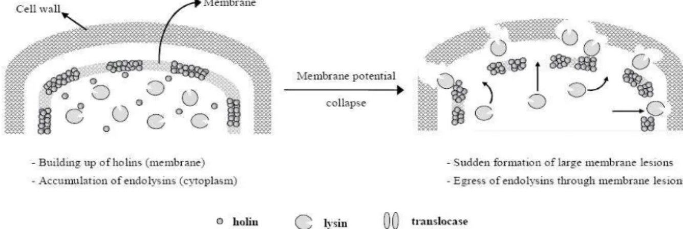
One of the characteristics of lysis regulation is that the phage endolysins would not reach the cell wall before the formation of holin lesions (see Figure 4).
Figure 4. Model representation of host-lysis strategies of phages producing non-secreted endolysins (São-José et
al., 2003).
This was long thought to be universal, but recently it was shown that some phages produce endolysins whose transport across the cytoplasmic membrane is not dependent on holin function. These endolysins have a signal sequence in its structure that allows for translocation of the protein through the host cytoplasmic membrane to the periplasmic space involving the host sec system (Xu et al., 2004). This
Detection and localization of LysA and Gp1 proteins of the mycobacteriophage Ms6 Introduction
8
paradigm began with studies by Santos and colleagues on the phage fOg44, which grows on the Gram-positive bacterium Oenococcus oeni. This phage encodes an endolysin (Lys44), which possesses a cleavable N-terminal signal sequence that also functions in Eschericha coli. Lys44 is continuously exported during assembly to the extracytoplasmic environment by sec machinery and its signal sequence is proteolytically removed by the leader peptidase, generating an active enzyme (Xu et al., 2004; Young, 2005; São-José et al., 2007; Catalão et al., 2010). In spite of the presence of a secretory enzyme, fOg44 has a holin gene. Moreover, fOg44 infections, showed that the mature endolysin was already detectable long before lysis was achieved, demonstrating that the sec-mediated export of the endolysin is not sufficient to provoke lysis.These remarks give raise to a number of questions about the role and mode of action of holins and how lysis timing is regulated in phages where the endolysin is sec-exported (Xu et al., 2004; São-José et al., 2007). The authors proposed that if endolysin can use host endogenous pathways to reach their substrate, the holin function would seem expendable. Their model suggests that, in phages producing secreted endolysins, the activity of the target endolysins would be inhibited in the cell wall until dissipation of the membrane potential by the holins (Figure 5), therefore, holins would activate the exported endolysins by collapsing the membrane potential, instead of releasing them (São-José et al., 2007).
Figure 5. Model representation of host-lysis strategies of phages producing peptide-bearing endolysins (São-José et
al., 2003).
More recently, it was demonstrated that a number of endolysins from phages that infect Gram- hosts, have intrinsic export signals, named signal-arrest-release (SAR) domains (Xu et al., 2004; Xu et al,. 2005). A particular interesting case is that of the endolysin of enterobacteriophage P1 (Lyz) and the lambdoid coliphage 21 (R21): they do not require the holin for export, but differ from the fOg44 secretory endolysin in that export is mediated by a N-terminal transmembrane domain (TMD), which is not proteolytically cleaved.. The N-terminal domain of P1Lyz phage is both necessary and sufficient, not only for the export of this endolysin through the membrane, but also for its release into the periplasm. This class of endolysins also requires the host sec translocon to cross the cytoplasmic membrane. The
9
unusual N-terminal domain, rich in residues that are weakly hydrophobic, functions as a SAR sequence, which acts as a normal signal-arrest domain to direct the endolysin to the periplasm in a membrane tethered form where it remains enzymatically inactive. This functional regulation is essential to avoid premature lysis of the infected host (Sun et al., 2009). After that, SAR allows P1’s Lyz to be released from the membrane, as a soluble active enzyme in the periplasm (Xu et al., 2004; Xu et al., 2005; Young, 2005).A model for triggering of lysis with SAR endolysins was proposed: firstly the SAR endolysin is tethered in an inactive form to the energized membrane and the holin protein is accumulated without affecting the proton-motive force (pmf). At the programmed lysis time, the holin triggers, disrupting the membrane sufficiently to abolish the pmf, and possibly assists the release of the endolysin from the membrane, which results the activation of the endolysin (Xu et al., 2004).
Interestingly the class represented by the lambdoid bacteriophage 21, utilizes endolysins having N-terminal SAR domains and pinholins, as opposed to the large-hole-forming holins (small hole size) (Park et al., 2006; Park et al., 2007). The pinholins are not large enough to allow endolysin to cross through them. The term ‘‘pinholin’’ has been proposed to differentiate the small-hole (pinhole) characteristic of the phage’s 21 holin from the canonical holins that form large, non-specific holes (Pang et al., 2009).
Mycobacteriophages
Mycobacteriophages are virus that infect mycobacteria hosts. The interest in these phages derives, in part, from the medical significance of their hosts (Hatfull, 2000; Hatfull, 2006). Mycobacteria are acid-fast staining bacteria with characteristic waxy cell walls, containing a peptidoglycan-arabinogalactan polymer linked to a long chain of mycolic acids (MA). The cell wall provides an extraordinarily efficient permeability barrier which contributes to the high intrinsic resistance to many drugs (Hoffmann, 2008). Mycobacteria are divided into two groups based on their growth rate: slow-growers such Mycobacterium tuberculosis and fast-slow-growers such Mycobacterium smegmatis. Many mycobacterial species are human and animal pathogens, the most recognized being M. tuberculosis and M. leprae, the causative agents of tuberculosis and leprosy, respectively (Hatfull et al., 1994; Hatfull, 2006; Zuber, 2008). Even though M.smegmatis is considered non-pathogenic, it provides a popular model for studying the virulence mechanisms of the pathogenic mycobacteria (Arora et al., 2008).
Mycobacteriophage Ms6 and its Lysis Operon
Mycobacteriophage Ms6 is a temperate phage that infects Mycobacterium smegmatis.
Electronic microscopy studies revealed that these phage particles are composed by an isometric polyhedral head with 80 nm in diameter, hexagonal form and an extensive non-contractile tail 210 nm long. The Ms6 genome is constituted by a molecule of dsDNA with a length over 50 kbp (unpublished
Detection and localization of LysA and Gp1 proteins of the mycobacteriophage Ms6 Introduction
10
data) and GC content of 62%. The morphological characteristics of the mycobacteriophage allowed for its classification in the Siphoviridae group (Portugal et al., 1989).Freitas-Vieira and colleagues (1998) have characterized the genetic elements involved in the site-specific integration events between phage Ms6’s DNA and the mycobacterial genome recombination mechanism required for chromosomal integration. This recombination is catalysed by a phage-encoded recombinase and involves a common core sequence present in both phage (attP) and bacterial (attB) genomes (Freitas-Vieira et al., 1998).
In 2002, the genetic organization and some transcriptional control elements of the mycobacteriophage Ms6 lysis functions were described. Garcia and colleagues identified and isolated, from Ms6, a strong promoter region (Plys) by using transcriptional fusions with lacZ reporter gene. Two
tandem σ70-like promoter sequences (P1 and P2), which are recognized by the host’s RNA polymerase, were found in this region by genetic analysis. Transcription of the lysis genes is dependent on the Plys
promoter located about 6kb away from the integration locus (Figure 6A), which is positioned in the middle of the Ms6 genome. Furthermore, an intrinsic transcription termination signal was detect in the leader sequence upstream of the first open reading frame (ORF). These data suggest that an anti-termination mechanism may be involved in the regulation of Ms6 lysis genes transcription (Garcia et al., 2002).
Figure 6. (A) Schematic representation of Plys on the 57-kb Ms6 DNA. (B) Genetic organization of the Ms6 lysis locus. Figure Adapted from Garcia et al., 2002.
The lytic cassette of Ms6 consists of five genes. Apart from the endolysin (lysA) and holin (hol) genes, the Ms6 lytic cassette includes three extra genes (gp1, gp5 and gp3) (Fig.6B). The gp1 gene, 231 bp long, is separated from the +1 position by the leader sequence and has the potential to encode a 77-amino acid (aa) protein that was recently identified has a chaperon-like protein that specifically interacts with the N-terminal region of LysA and is involved in its delivery to the peptidoglycan in a holin-independent manner (Catalão, et al., 2010).
11
gp2 or lysA, with 1152 bp, starts at a GTG codon that overlaps the gp1 TGA stop codon, which
is in a different reading frame. It encodes the Ms6 endolysin with 384 aa (Garcia et al., 2002). Analysis of the deduced amino acid sequence have identified an amidase domain and and its hydrolase activity was
already demonstrated; the protein was shown to cleave the bond between L-Ala and D-muramic acid
(Piechota et al., unpublished). Recently it was shown that during an Ms6 infection, lysA synthesizes two proteins: Lysin384 translated from the start codon at position 1 and Lysin241 translated from a second start codon at position 430 of the nucleotide sequence (Catalão et al., 2011a). It was demonstrated that both proteins are necessary for an efficient lysis, although it’s not known the reasons why Ms6 produces two endolysins.Downstream of lysA is gp3 (lysB), with 996 bp and starts at an ATG codon that overlaps, in a different reading frame, the TGA stop codon of gp2 and encodes a 332 aa protein (Garcia et al., 2002). The LysB enzyme was shown to have lipolytic activity that targets the mycobacteria’s outer membrane (Gil et al., 2008; Gil et al., 2010).
Mycobacteria, apart of having a mycobacterial outer membrane composed of mycolic acids and free lipids, have a second barrier, an arabinogalactan-peptidoglycan layer (Hoffmann et al., 2008). Recently, Gil et al., 2010 have shown that Ms6’s LysB cleaves the ester bond between mycolic acids and arabinogalactan. In addition, LysB also hydrolyzes other lipids containing mycolic acids present in the envelope of mycobacteria, such as the trealose dymicolate, a lipid involved in pathogenesis of slow growing mycobacteria (Gil et al., 2010). Payne et al., (2009) also reported that mycobacteriophage Giles Lysin B is a novel mycolarabinogalactan esterase, which cleaves the mycolylarabinogalactan bond to release free mycolic acids. The authors proposed that LysB acts at a late stage in the lysis, severing the connection of the mycobacterial outer membrane to the cell wall, providing a faster and more complete lysis of the host cell (Figure 7).
The gp4 (hol) gene begins with ATG codon located 10 nucleotides downstream of the lysB stop codon and encodes a 77 aa protein with holin-like activity. Ms6 hol shares some structural characteristics with class II holins (Garcia et al., 2002). The Ms6 Gp4 possesses two TMDs, TMD2 and TMD1, which TMD1 has characteristics of a SAR domain. Unlike some holins, such as λ S, Gp4 lacks a dual-start motif.
Figure 7. A model for mycobacteriophage lysis of mycobacteria. Mycobacterial cell walls are unusual in that the cytoplasmic membrane (CM) is surrounded by a peptidoglycan layer (PG) to which a network of arabinogalactan (AG) is covalently attached. A mycobacterial outer membrane consisting of mycolic acids (MA) and free lipids (FL) is covalently attached via an ester linkage of mycolic acids to arabinogalactan. LysA — assisted by holins encoded by at least some of mycobacteriophages — perform an essential step in lysis involving degradation of the peptidoglycan layer, and the lysis is completed through LysB-mediated cleavage of the outer membrane from arabinogalactan (Payne et al., 2009).
Detection and localization of LysA and Gp1 proteins of the mycobacteriophage Ms6 Introduction
12
Catalão and colleagues suggests that Gp4 is a pinholin because the present of a SAR domain is followed by a typical TMD, which is analogous to other pinholins already characterized, such as the holin of phage 21 (Catalão et al.,2011b).At last, gp5 with 372 bp starts at an ATG codon that overlaps the gp4 TGA stop codon in a different reading frame. It encodes a 124 aa protein (Garcia et al., 2002) with a predicted single TMD at the N-terminal region. Catalão and their colleges hypothesized that Gp5 might function as a holin-like protein because they demonstrated that the overexpression of Gp5 in E. coli results in a drastic inhibition of the cell growth (Catalão et al., 2011b).
As mentioned above, Catalão and colleagues showed that endolysin LysA is exported with the help of the gp1 gene product, in a holin-independent manner. gp1 is shown to encode a chaperon-like protein that binds the endolysin, assisting its export to the extra-cytoplasmic environment and which is required for an efficient lysis in Ms6. Currently is not known how the endolysins remain inactive until holin determines the time of lysis. Thus, the purpose of this project is to investigate the role of the gp1 gene product in the export of LysA and other proteins, without signal peptide, from the cytoplasm to the periplasm.
Objectives
The main goal of this study is to investigate the gp1 gene product localization in M. semgmatis and evaluate its role in the transportation of LysA and other proteins without a peptide signal, from the cytoplasm to the periplasm.
To achieve this, the specific objectives are:
- Construct a Ms6 mutant phage where the gp1 gene carries, at its 3’ end, a sequence coding for a C-Myc tag allowing the production of a recombinant protein that can be detected with an antibody anti C-Myc in M. smegmatis infected cells.
- Evaluate the capacity of Gp1 to translocate proteins across the cytoplasmic membrane, by fusing Gp1 with reporter proteins.
13
M
ETHODS/E
XPERIMENTALP
ROCEDURESBacteriophages, bacterial strains, plasmids and growth conditions
Phages, bacterial strains, plasmids and oligonucleotides used in this study are listed in Table 1. Ms6 phage was isolated from Mycobacterium smegmatis strain HB5688 (Portugal et al., 1989; Snapper et al., 1990). M. smegmatis was grown in Middlebrook 7H9 (DifcoTM ) supplemented with 0.5% glucose and 1 mM CaCl2 at 37 °C overnight under vigorous shaking, or on M iddlebrook 7H10 also supplemented.
The phage stocks were obtained from confluent lysis in supplemented 7H10 after infection of M. smegmatis with the appropriate phage dilution, and then eluted for at least 4h at 4⁰C with SM buffer (50 mM Tris.HCl pH 7.5, 10 mM MgSO4.7H2O, 100 mM NaCl) supplemented with 1 mM CaCl2. E. coli strains
were grown at 37⁰C with shaking, in Luria-Bertani (LB) broth or agar supplemented with 100 µg/mL ampicilin or 30 µg/mL kanamycin for plasmid selection.
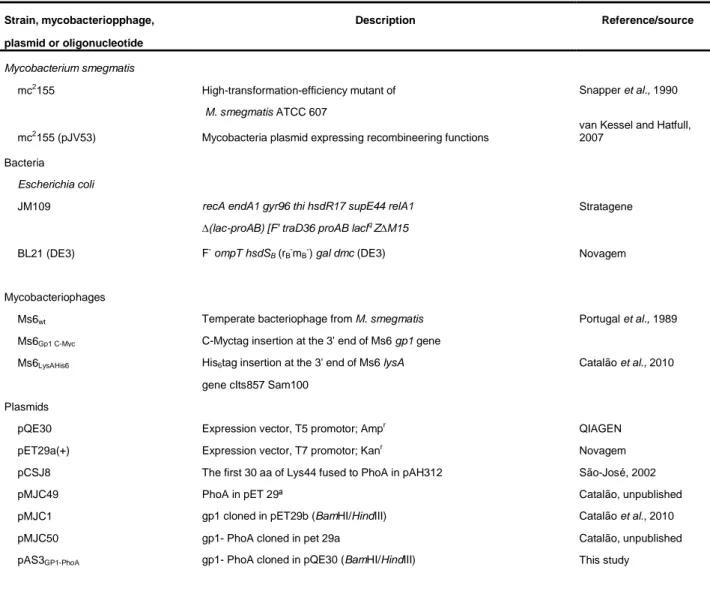
Table 1. Strains, mycobacteriophages, plasmids and oligonucleotides used in this study.
Strain, mycobacteriopphage, Description Reference/source
plasmid or oligonucleotide
Mycobacterium smegmatis
mc2155 High-transformation-efficiency mutant of Snapper et al., 1990
M. smegmatis ATCC 607
mc2155 (pJV53) Mycobacteria plasmid expressing recombineering functions van Kessel and Hatfull, 2007
Bacteria Escherichia coli
JM109 recA endA1 gyr96 thi hsdR17 supE44 relA1 Stratagene
∆(lac-proAB) [F' traD36 proAB laclq Z∆M15
BL21 (DE3) F- ompT hsdSB (rB-mB-) gal dmc (DE3) Novagem
Mycobacteriophages
Ms6wt Temperate bacteriophage from M. smegmatis Portugal et al., 1989
Ms6Gp1 C-Myc C-Myctag insertion at the 3' end of Ms6 gp1 gene
Ms6LysAHis6 His6tag insertion at the 3' end of Ms6 lysA Catalão et al., 2010
gene cIts857 Sam100 Plasmids
pQE30 Expression vector, T5 promotor; Ampr QIAGEN
pET29a(+) Expression vector, T7 promotor; Kanr Novagem
pCSJ8 The first 30 aa of Lys44 fused to PhoA in pAH312 São-José, 2002
pMJC49 PhoA in pET 29ª Catalão, unpublished
pMJC1 gp1 cloned in pET29b (BamHI/HindIII) Catalão et al., 2010
pMJC50 gp1- PhoA cloned in pet 29a Catalão, unpublished
Detection and localization of LysA and Gp1 proteins of the mycobacteriophage Ms6 Methods/Experimental Procedures
14
pAS1SP-PhoA SPLys44-PhoA cloned in pQE30 (BamHI/HindIII) This study
pAS2SP-PhoA SPLys44-PhoA cloned in pET29a(+) (BamHI/HindIII) This study
pGLO Plasmid containg the GFPuv gene Biorad
pAS4GFP gfp cloned in pQE30 This study
pAS5Gp1-GFP gp1-gfp in pQE30 This study
pMJC3 gp1 and lysA cloned in pET29b Catalão et al., 2010
pMJC4 lysA cloned in pET29b Catalão et al., 2010
Oligonucleotides
Pr Gp1 C-Myc CTCCATCCCCGTCCTCGGCGGAATCCTCGGGAGCAAACGGGAACAG This study
AAACTGATCAGCGAAGAGGATCTGTGACGGGAGCAAACGGTGA CCACGAAAGATCAAGTCGCCC
Pr Gp1 C-Myc Extended fwd CTGACCAACCTTCCAGCGCAAGTCATGGACATCATCGACAGCGCGCT This study
GCGCTCCAGACAGCGCGCTGCGCTCCATCCCCGTCCTCGGCGGAATC Pr Gp1 C-Myc Extended 3' rv GCATTCGCTGCGGGTGTAGCCGCGCGCCTTGGCTTCGGCGATGGTGA This study
TTTGGGCGACTTGATCTTTCGTGGTCAC
Pr Screening Myc fwd CTCGGGAGCAAACGGGAACAGAAACTG This study
Pr Gp1A fwd CGGGATCCATGGACCGCTTAGGCATCGTCC Catalão et al., 2010
Pr LysA60aa rv GTCGAAGCGGTGTGGGTAGGAGCCG
Pr SPLys44 fwd GGCGGATCCACACGTAAAAAGTTAAA
Pr phoA3' rv CCAAGCTTTTTCAGCCCCAGAGC
PrGp1 rv, EcoRI TCGTGGGGAATTCCGTTTGCTCCC This study
PrGFP fwd, EcoRI GAAAAAATGAATTCCAAAGGAGAAGAAC This study
PrGFP rv, PstI GGAATTCACTGCAGTTATTTGTAGAGCTC This study
PrGFP fwd, BamHI CCAGGATCCGCTAGCAAAGGAG This study
DNA extraction from Ms6 wt
The extraction of Ms6 genomic DNA was adapted by a protocol from Sambrook and Russel (2003). 600 µl of phage stock were incubated with proteinase K (50 µg/mL) and 0,5% SDS for 1 hour at 56⁰C. The mixture was treated three times with an equal volume of phenol-chloroform-isoamyl alcohol (25:24:1) and one time with chloroform. Finally, the DNA was precipitated with 10% 3M sodium acetate and an equal volume of cold isopropanol. After a maximum centrifugation step of 45 min at 4⁰, the DNA was washed with 200 µL of 70% ethanol, centrifuged for 15 min, and then the pellet was dried at 37⁰C and resuspended in water. The samples were stored at -20⁰C and were quantified in Spectrophotometer ND-1000 (Nanodrop).
One-step growth experiments
One-step growth experiments were performed as described in Catalão et al., 2010. Mycobacterium smegmatis cells were grown to an OD600 0,5-0,6 and then diluted to an OD600 between
15
0,2-0,3. Then 10 mL were centrifuged and resuspended in 1 mL of a phage dilution, using a multiplicity of infection (m.o.i.) of 1. The mixture was incubated for 50 min at 37⁰C to allow adsorption of the phages.100 µL of 0,4% H2SO4 was added and incubated 5 min at 37⁰C, to inactivate the non-adsorbed phages.
The suspension was neutralized with 100 µL of 0,4% NaOH and diluted to 1:100. 1 mL of the suspension were withdrawn every 30 min until reaching 210 min and 100 µL of serial dilutions of each sample were plated with 200 µL of M. smegmatis cells as top agar lawns on 7H10 medium. Phage titer for each sample was determined after 24 h at 37°C.
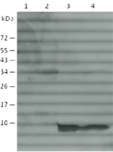
Gp1 expression in M. smegmatis-infected cells
Examination of Gp1 synthesis in M. smegmatis was performed as previously described (São-José et al., 2000). An exponentially-growing culture of M. smegmatis was infected with Ms6LysAHis6 or Ms6Gp1 C-Myc at a multiplicity of infection (m.o.i.) of 10 and incubated at 37⁰C for 30 minutes. Ten milliliter samples were withdrawn at 30-min intervals, centrifuged (8000rpm 13’ at 4ºC) and the pellet was immediately frozen at -20⁰C. After thawing, cells were resuspended in 100 µL of 10mM Tris-HCl buffer supplemented with 20 mg/mL of lysozyme and 1 µL of a protease inhibitor cocktail (Calbiochen) (Sambrook et al., 2001). After an incubation period at 37°C for 3 hours, 25 µL of 5x SDS-PAGE sample buffer were added followed by incubation at 100°C f or 5 min to complete cell lysis. The proteins were analyzed by 15% SDS–PAGE followed by Western-blotting, using a horse-radish-peroxidase (HRP) conjugated anti-C Myc antibody (Roche).
Cellular fractionation of M. smegmatis
A 200 mL culture of M. smegmatis infect with Ms6Gp1C-Myc was centrifuged and washed twice in a large volume of PBS and resuspended in 5 mL of ice cold Phosphate Buffered Saline (PBS) with 50 µL of a cocktail of proteases inhibitor (Calbiochem).The cell suspension was sonicated with a microtip probe at 50% output for 3 min, incubated in ice for 10 min and spun in centrifuge 30 min maximum speed at 4ºC. The supernatant, which has the cytosol-enriched fraction, was filtered through a 0.22 mm filter to remove membrane fragments that did not pellet, the volume was measured, brought up to 0.8 mL in ice cold PBS and 0.2 mL of 10% Triton X-100 was added (final concentration of 2.0% Triton X-100). The pellet, having the membrane and cell wall-enriched fraction, was resuspended in 0.8 mL of ice cold PBS and 0.2 mL of 10% Triton X-100 (final concentration 2.0% Triton X-100). The fractions were incubated on ice for 1h with intermittent vortexing (approximately every 15 min) and spun in an Eppendorf centrifuge 30 min maximum speed at 4ºC. The supernatant from the cytosol-enriched fraction was transferred to a new tube (thereby having the membrane enriched fraction) and the pellet (cell wall-enriched fraction) was resuspended in 1 mL of ice cold PBS. All fractions were then precipitated with 9 mL of cold acetone at – 70ºC for 2h and were afterwards analysed in western-blot (Hatfull, personal communication).
Detection and localization of LysA and Gp1 proteins of the mycobacteriophage Ms6 Methods/Experimental Procedures
16
Protease accessibility experimentsThe procedure was performed using a protocol from Chou and Kendal (1990). Expression of Gp1 and LysA in E.coli was induced from plasmids pMJC3 and pMJC4. E. coli cells were grown to an OD600 of
0,5 and a 1 mL aliquot was collected, meant for a negative control. The E.coli cultures were induced for 1h and then collected and immediately chilled on ice. An equal volume of 10% trichloroacetic acid (TCA) was added to the uninduced sample and one of the induced samples, and then the precipitate was collected by centrifugation (15 000 x g, 30 min at 4⁰C), precipitated with ice cold acetone, air dried and resuspended in 100 µL of 2x SDS-PAGE sample buffer. The two remaining aliquots were washed in 30 mM Tris-HCl pH 8 and then resuspended in 0.5 M sucrose, 30 mM Tris-HCl pH 8, containing 20 µg/mL lysozyme and 1 mM EDTA to digest the cell wall and to expose the plasma membrane. Following 20-min incubation on ice, proteinase K (25 g/mL) was added in one of the aliquots while the same volume of enzyme storage buffer was added to the other aliquot. After 20 min of incubation on ice, followed by 15 min room temperature and subsequently precipitation with TCA the samples were washed with ice cold acetone and resuspended in 2xSDS-PAGE sample buffer. All samples were boiled at 100⁰C for 8 min and analysed by Western-bolt.
Plasmid construction
To construct plasmids pASSP-PhoA and pAS2SP-PhoA, a DNA fragment containing the 5’ end,
corresponding the first 30 amino acids of the fOg44 endolysin gene (lys44) fused with the mature form of phosphate alkaline gene was amplified by Polymerase chain reaction (PCR) from plasmid pCSJ8 (São-José C., 2002). The reaction was performed with Pr SPLys44 fwd and Pr phoA3' rv designed to contain the recognition sequence of BamHI and HindIII (Fermentas), respectively. The PCR product was restricted with BamHI and HindIII and inserted into vector pQE30 (QIAGEN) and pEt29a (Novagen) restricted with the same enzymes. The ligation reaction was performed at 25⁰C for 2h with T4 DNA ligase (Biolabs) according to the manufacter instructions. Transformations were performed in competent in E. coli cells previously treated with 50 mMCaCl2. Recombined cells were selected in LB agar supplemented
with the w appropriate antibiotic. The recombinant plasmids were then extracted using FastPlasmid Mini Kit (Eppendorf) and insertion of the DNA fragment of interest was checked by PCR with the same pair of primers used for cloning. To construct plasmid pAS3GP1-PhoA, a DNA fragment containing the Ms6 gp1
fused with the mature form of phosphate alkaline gene was amplified by PCR from pMJC50 (Catalão, unpublished). The reaction was performed with Pr Gp1A fwd and Pr phoA3' rv designed to contain the recognition sequence of BamHI and HindIII (Fermentas) respectively and inserted into pQE30 previously restricted with the same enzymes. After purification, the product was cloned into pQE30, restricted with the same restriction enzymes, and the ligated as above. Plasmid pAS4GFP was constructed by insertion of
17
fwd, BamHI and PrGFP rv, PstI. After purification the product was cloned into pQE30 restricted with BamHI and PstI and then ligated as previously described. Transformations were performed in competent E. coli JM109 cells (for pQE30 derivatives) or BL21 (DE3) (for pET29 derivatives), previously treated with 50 mMCaCl2 (Samborook et al., 2003). Recombinant cells were selected in LB agar supplemented withthe appropriate antibiotic. The recombinant plasmids were then extracted using FastPlasmid Mini Kit (Eppendorf) and insertion of the DNA fragment of interest was checked by PCR with the same pair of primers used for cloning.
Plasmid pAS5Gp1-GFP was constructed by ligation of three DNA fragments generated as follows: 1) gp1 gene was amplified from Ms6 DNA with primers PrGp1 fwd, BamHI and PrGp1 rv, EcoRI and restricted with BamHI and EcoRI; 2) GFPuv gene was amplified from plasmid pGLO with primers PrGFP fwd, EcoRI and PrGFP rv, PstI and restricted with EcoRI and PstI; 3) plasmid pQE30 was restricted with BamHI and PstI. The ligation of the three fragments was performed at 25⁰C for 2h followed by incubation at 16⁰C overnight. The reaction mixture was inserted into electrocompetent E.coli JM109 cells by electroporation (Hanahan, 1983; Sambrook et al., 2001). Recombinant cells were selected in LB agar with ampicillin and plasmid extraction was performed as above. The presence of the inserted fragment was checked by enzymatic restriction.
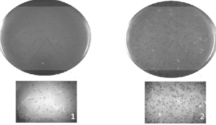
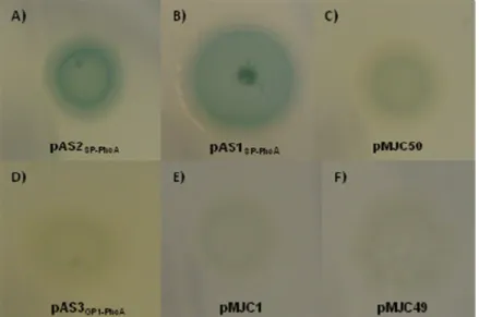
Detection and quantification of Alkaline Phosphate activity
For the PhoA screening: E. coli recombinant strains were grown in LB agar supplemented with 1mM IPTG and 40 µg/mL BCIP (5-Bromo-4-chloro-3-indolyl phosphate) and the appropriate antibiotic. For the quantification assay: E coli cells were grown in LB broth to an OD600 of 0.4 - 0.5 at 37ºC with
shaking (~ 250rpm) and expression was induced with 1 mM IPTG. 1 mL of culture was collected at times 0’, 30’ and 90’ following induction, and centrifuged at 12 000x g for 1 min at 4ºC. Supernatant was discarded and the cells were resuspended in 1 mL of MOPS-buffer. Cell density was read at 600nm either
directly or after dilution with MOPS-buffer. 100µL of 40 mg/mL p-nitro-phenyl-phosphate (PNPP) was added to the assay tubes and the tubes were incubated at 37⁰C until a significant yellow color was observed. To stop the reaction, 100 µL of K2HPO4 was added. Then the OD was read at 420nm and
550nm. Units of phosphatase activity were calculated by the following formula (Brickman et al., 1975; Belin
2010):
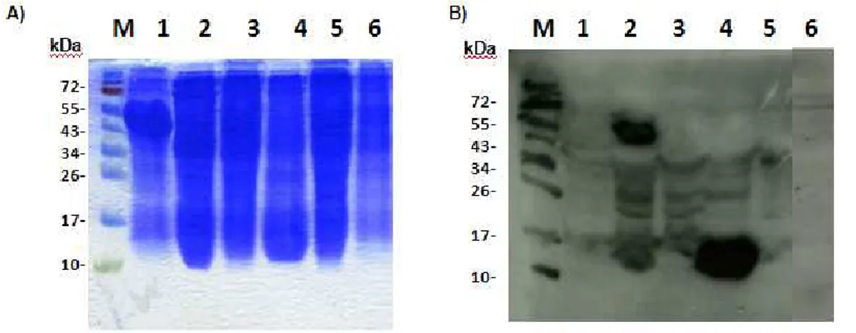
Expression and determination of solubility from Gp1 and Gp1-GFP in E. coli cells
E. coli recombinant cells carrying plasmids PQE30, pAS4GFP or pAS5Gp1-GFP were grown in LB medium supplemented with ampicillin, at 37°C wit h shaking until OD600 equals 0.6. At this time an
Detection and localization of LysA and Gp1 proteins of the mycobacteriophage Ms6 Methods/Experimental Procedures
18
aliquot of 1 mL was collected (noninduced control) and the expression was induced with 1 mM IPTG for 1h and 3h. 1 mL samples were collected, centrifuged at 4000 x g for 10 min at 4°C and the pellet was resuspended in 100 µL of phosphate buffer (50 mM NaHPO4 and 30 mM CaCl2) and incubated withlysozyme (1mg/mL) and 1 µL of a protease inhibitors cocktail at 4°C for 30 m in. 4 cycles of freezing (with liquid nitrogen) and thawing (at 42°C) were then ca rried out and the lysates were centrifuged at 15 000 x g for 30 min at 4°C. The supernatant corresponds th e soluble fraction (cytoplasmic proteins), while the pellet corresponds to the insoluble fraction (membrane proteins). The pellet was resuspended in 100 µL SDS-PAGE buffer and the supernatant was added by the same volume of 2 X SDS-PAGE buffers. The samples were boiled for 10 min and analysed by SDS-PAGE followed by Coomassie-Blue staining.
PCR reactions
The reaction mixture for PCR reactions, whose composition is summarized in Table 2, was subject to the following conditions: 1 min at 95 ° C, 30 cycles of 1 min at 95 ° C, 1min at 56 ° C and 1min at 72 ° C, and 10min at 72 °C and then stored at 4° C. The reactions were performed in the BioRad thermocycler MyCycler.
Table 2.
Reaction mixture Final concentration
polymerase buffer 1x MgCl2 2,5mM DNA 2 ng/µL dNTP 0,25mM Primer fwd 1pmol/µL Primer rv 1pmol/µL
19
RESULTS AND DISCUSSION
Construction of the Ms6Gp1-CMyc mutant
To determine the localization of Ms6 Gp1 during infection of Mycobacterium smegmatis, a Ms6 derivative mutant was constructed, where the gp1 gene carries a sequence at its 3’ end that encodes a C-Myc tag. Construction of Ms6 mutant phages was performed using Bacteriophage Recombineering of Electroporated DNA (BRED) as described previously by Marinelli and colleagues (2008). 10-20 ng of a 110 bp oligonucleotide with 40 bp of upstream and downstream homology to the insertion region, was extended by PCR using two 75-bp extender primers, which have 25 bp of homology at each end of the 100-mer (see Table 1 and Figure 8A). The PCR reaction was performed as follows: 1 cycle of 7 min at 95 ° C, 30 cycles of 1 min at 95 ° C, 1 min at 56 ° C and 1 min at 72 ° C, and 10 min at 72 °C and then stored at 4°C. The final 220-bp dsDNA product was p urified using MinElute PCR Purification Kit (Qiagen) and then co-electroporated with 70 ng of Ms6 wt genomic DNA into electrocompetent M. smegmatis (pJV53). The cells were resuspended in supplemented 7H9 medium, incubated at 37⁰C for 2 hours with shaking and plated as a lawn in 7H9 molten agar with 200 µL of wild-type M. smegmatis. After 24h of incubation at 37⁰C, 40 phage plaques were picked and eluted into an eppendorf with 100 µL phage buffer (SM) with 1 mM CaCl2, for 2 hours at room temperature. To detected plaques containing the phage
mutant, 21 of them were screened by Polymerase chain reaction (PCR) with primers Pr Screening Myc fwd, which hybridizes with the C-Myc coding sequence, and Pr LysA60aa rv, which hybridizes with the LysA DNA at position 48 and 73 to detect the C-Myc tag insertion (Table 1).