Photodynamic Therapy and possible action against SARS- CoV-2
Terapia fotodinâmica e possível ação contra SARS-CoV-2
DOI:10.34117/bjdv6n7-761
Recebimento dos originais: 27/06/2020 Aceitação para publicação: 28/07/2020
Gabriella Brandimarte Queiroz
Graduate student in dentistry, State University of Northern Paraná – UENP
Health Sciences Center - Dentistry, State University of Northern Paraná – UENP, Jacarezinho, PR, Brasil
Prolongamento da Av. Pedro Coelho Miranda, S/N. Jardim Panorama; CEP 86400-000- Jacarezinho - Paraná - Brasil
E-mail: gabsbrandimarte@gmail.com
Augusto Alberto Foggiato
Dentist, Specialist in Radiology, Orthodontics and Facial Orthopedics, Ms and PhD Orthodontics Health Sciences Center - Dentistry, State University of Northern Paraná – UENP, Jacarezinho, PR,
Brasil
Prolongamento da Av. Pedro Coelho Miranda, S/N. Jardim Panorama; CEP 86400-000- Jacarezinho - Paraná - Brasil
E-mail: augusto.foggiato@uenp.edu.br
João Lopes Toledo Neto
Dentist and PhD of oral-biology by the Faculty of Dentistry of Piracicaba Unicamp
Health Sciences Center - Dentistry, State University of Northern Paraná – UENP, Jacarezinho, PR, Brasil
Prolongamento da Av. Pedro Coelho Miranda, S/N. Jardim Panorama; CEP 86400-000- Jacarezinho - Paraná - Brasil
E-mail: joaoneto@uenp.edu.br
Douglas Fernandes da Silva
Biologist, biotechnological engineer and PhD in applied microbiology - Paulista State University – UNESP
Health Sciences Center - Dentistry, State University of Northern Paraná – UENP, Jacarezinho, PR, Brasil
Prolongamento da Av. Pedro Coelho Miranda, S/N. Jardim Panorama; CEP 86400-000- Jacarezinho - Paraná – Brasil
ABSTRACT
Photodynamic therapy is based on the selective photosensitization of a target tissue using a topically or systemically administered agent, which is activated by light, promoting an oxygen-dependent cytotoxic reaction. This study consisted of a literature review that selects articles that show the use of Photodynamic Therapy in health areas, contrasting its use as antimicrobial therapy and the use of action against SARS-CoV-2. In this bibliographic research, the use of photodynamic therapy used in microbial control has demonstrated high effectiveness because it does not cause negative effects or drug interactions. In addition to acting not only on the host's tissues, but also preventively, since the results obtained in disinfection processes are seen. Through this study, it is possible to use a series of benefits of using photodynamic therapy to combat the new Coronavirus (COVID-19), in addition to being an effective method of disinfecting contaminated instruments or suggesting as a method of action against SARS-CoV-2.
Keywords: Microbial control, Photodinamic therapy (PDT), Betacoronavirus, Health; RESUMO
A terapia fotodinâmica é baseada na fotossensibilização seletiva de um tecido-alvo usando um agente administrado por via tópica ou sistêmica, que é ativado pela luz, promovendo uma reação citotóxica dependente de oxigênio. Este estudo consistiu em uma revisão de literatura que seleciona artigos que mostram o uso da Terapia Fotodinâmica nas áreas da saúde, contrastando seu uso como terapia antimicrobiana e o uso de ação contra SARS-CoV-2. Nesta pesquisa bibliográfica, o uso da terapia fotodinâmica utilizada no controle microbiano demonstrou alta eficácia, pois não causa efeitos negativos ou interações medicamentosas. Além de atuar não apenas nos tecidos do hospedeiro, mas também de forma preventiva, uma vez que são observados os resultados obtidos nos processos de desinfecção. Através deste estudo, é possível usar uma série de benefícios do uso da terapia fotodinâmica para combater o novo Coronavírus (COVID-19), além de ser um método eficaz de desinfetar instrumentos contaminados ou sugerir como método de ação contra o SARS-CoV-2.
Palavras-chave: Controle microbiano, Terapia fotodinâmica (TFD), Betacoronavírus, Saúde;
1 INTRODUCTION
Light is used to treat diseases of antiquity, but it was after scientific discoveries from pioneers like Finsen, Raab and Von Tappeiner, a combination of light administration and drugs used for photochemotherapy surgery as a therapeutic method (ACKROYD e colab., 2001).
Photodynamic Therapy (PDT) has application of in different areas of health, whose principle is the selective photosensitization of a target tissue through a topically or systemically administered agent that is then activated by light promoting an oxygen-dependent cytotoxic reaction (KURWA e BARLOW, 1999).
PDT is an inherently complex technology that depends on several variables, including the chemical and photochemical properties or Photosensitizer (PS). The PS dosing and delivery vehicle,
the drug's time interval - light, wavelength, energy dose, the power density and pulse structure of light, and the state of tissue oxygenation (CASTANO e colab., 2004).
Therefore, the action of the PDT mechanisms results from a sequence of photochemical and photobiological processes that will cause damage to the target cell, such as cell death through the three main types of cell death, being: apoptotic, necrotic cell death and associated with autophagy (ABRAHAMSE e HAMBLIN, 2016; TENNERT e colab., 2014). In addition, this process can also activate the immune system if it compromises the supply of oxygen and essential nutrients (SANTOS e colab., 2019).
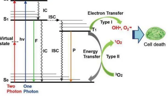
In the presence of oxygen found in cells, the activated photosensitizer (PS) can react with neighboring molecules through two reactions, by transferring electrons or hydrogen, leading to the production of free radicals, which is the type I reaction, and by transferring energy to oxygen, leading to the production of singlet oxygen, which is the type II reaction (PERUSSI, 2007). This mechanism is explained in the jablonski diagram shown in figure 1 (LAN e colab., 2019), the excitation and relaxation of a chromophore is then shown by the jablonski diagram, where the PS absorbs a photon with the appropriate wavelength and can move from the state (S0) to the first excited state, which it
forms singlet oxygen (S1) or that for the second excited state of singlet oxygen (S2), which will cause S2
to be rapidly deteriorated to S1 through internal conversion (IC). Thus, where in the presence of oxygen
the excited PS can react with the substrates, forming radicals or radical ions (BASKARAN e colab., 2018).
In addition to using light, photosensitizer and oxygen to cause cell death, which consists of a biological process essential for growth and physiological development, maintaining homeostasis and related to manifestations and control of various diseases (ALMEIDA e colab., 2004; QUEIROZ e colab., 2020). The process of aPDT foment apoptosis or necrosis, depending on the sensitizer, the dose of PDT and the cell genotype (CASTANO e colab., 2005). In the case of apoptosis, which is through the most active cell death of the PDT, the PS will be placed without tissues and be illuminated, promoting an acute stress response, causing changes in lipid and lipid metabolism in the production of stress cytokines and proteins (CASTANO e colab., 2005). Thus, enzymes, proteins and kinases, are activated and transcription factors are expressed, usually occurring in the mitochondria (CASTANO e colab., 2004; QUEIROZ e colab., 2020). Therefore, these effects promote the induction of approval by the mitochondrial pathway, which is detected with the release of cytochrome c or by the pathways involving ceramide or death receptors, being a great area of research of treatments, mainly in tumor (ALMEIDA e colab., 2004; CASTANO e colab., 2004; QUEIROZ e colab., 2020).
PDT has low toxicity, apparent antitumor activity, having the potential to be used together with chemotherapy and mediating an immune response, causing the death of tumor cells in places distant (BANERJEE e colab., 2017). However, photodynamic therapy (PDT) also depends on the capture of a photosensitizing dye, usually a macrocycle related to porphyrin, visible light of adequate wavelength corresponding to the absorption spectrum of the dye and molecular oxygen, to generate intermediates of reactive oxygen that initially leads to damage to the organelles to which the dye is attached to thereby arrive at cell death (OLEINICK e EVANS, 1998).
The light sources needed to activate the photosensitizer used in the PDT process need to be visible, low power, pecific wavelength and most photosensitizers are activated by red light between
630 and 700 nm. This valor is corresponding to a light penetration depth of 0.5 cm to 1.5 cm (RAJESH e colab., 2011). According to the same authors, the total light dose, dose rates and depth of destruction may vary with each treated tissue and also with the photosensitizer used.
The most used PS for medical purposes belong to the classes of tricyclic dyes with different mesoatoms, such as: Acridine orange, proflavin, riboflavin, methylene blue, fluorescein, eosin, erythrosine, cane rose, Tetrapyrroles, Porphyrins and derivatives, chlorophyll, phyllererythrin, phthalocyanine, Furocoumarins, Psoralen, methoxy xanthotoxin and bergapten (RAJESH e colab., 2011). The 5-aminolevulinic acid (5-ALA) and mainly its derivative, methyl aminolevulinate (MAL), are the types of topical photosensitizing prodrugs most used in PDT, being applied exogenously ignoring the step of limiting the intracellular rate along the way of heme synthesis to produce real photosensitizers, and porphyrin (HAMBLIN e HASAN, 2004; WANG e colab., 2017).
Based on the clinical and literary understanding that has evolved over the years, it is noted that photodynamic therapy is in extensive expansion, being extremely important to understand this functions and applications, which with correct use, can bring health benefits and treatments. Especially in microbial control, since the increase in antibiotic resistance has led to the development and research of new antimicrobial strategies, and in the case of PDT, are improbable to develop resistance (RKEIN e OZOG, 2010).
In addition, the use of the PDT method is currently being applied to the treatment of diseases caused by yeasts, viruses and parasites, as well as the disinfection of blood and other products, preventing cross-infections (JORI e BROWN, 2004), therefore, it can be applied to combat the transmission of the new Coronavirus (SARS-CoV-2). SARS-CoV-2 or COVID-19 is a virus that emerged in China in december 2019 and has spread throughout the world, being known to be transmitted from bats to humans through intermediate host palm civets, and have a high pathogenic manifestation (LIU e colab., 2020). According to the World Health Organization (KHEDKAR e PATZAK, 2020), this virus was characterized the situation as a pandemic, there were 118,000 cases in 114 countries with 4291 deaths on March 11, 2020. The COVD-19 is being associated to severe acute respiratory syndrome (SARS) (JORI e BROWN, 2004; KHEDKAR e PATZAK, 2020). According to the same authors, there is no effective treatment against the virus, and the only proven form of control is to avoid contact with the active virus.
Therefore, this work purpose to raise a possible use of the PDT process in the deactivation of Covid-19 and, consequently, in its control. Because, although at first it is not a process that causes viral deactivation capable of fighting the virus, it can be applied as an efficient therapy to reduce viral load in the respiratory tract or even on different surfaces, thus PDT is a safe treatment option to the patient and harmful to the vírus (DIAS e colab., 2020).
1.1 PDT IN MICROBIAL CONTROL
Photodynamic therapy with bactericidal action is denominade antimicrobial photodynamic therapy (aPDT), which is a method of coloring an object with a photosensitizing dye and then sterilizing by radiating the dye at the excitation wavelength (SHIOTSU-OGURA e colab., 2019).
Several studies have addressed that it is possible to kill bacteria with a light source from a low-powered laser after the microorganisms have been sensitized to low concentrations of photosensitizers (HUANG e colab., 2019). Phenothiazine, sucstoluidine blue and methylene blue (MB) are among the most studied PS for antimicrobial photodynamic therapy (aPDT), which if combined with light in the appropriate parameters, the cytotoxic photodynamic dose required for microbial inactivation is generally less than necessary to cause damage to host cells, such as keratinocytes and fibroblasts
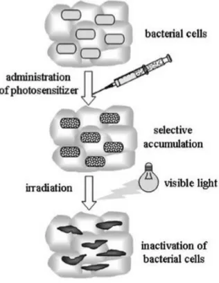
(SOUKOS e colab., 1996). Thus, this therapy can be used to treat various conditions, including infectious diseases caused by microorganisms, including Gram-positive and Gram-negative bacteria, viruses, protozoa and fungi (FOGGIATO e colab., 2018; FOGGIATO e SILVA, 2019; LYON e colab., 2011). Microbial photodynamic inactivation, is based on the administration of a non-toxic photosensitizer, which is preferentially accumulated in microbial cells, causing subsequent irradiation with visible light, in the presence of oxygen, specifically produces cellular damage that inactivates microorganisms, such as shown by Durantini (DURANTINI, 2006) (Figure 2). In addition, low-intensity light, lasers and light-emitting diode (LED), can also be associated with the administration of non-toxic PS to locally promote photochemical reactions that can induce microbial control by cell death (FOGGIATO e colab., 2018; SILVA e colab., 2019).
The actions of the PS can reach the cell when the PS is only close to a bacterial cell without effective binding, which can limit oxidative damage to external structures, such as the cell wall or cytoplasmic membrane or intracellular components, such as cytoplasmic proteins or DNA (CIEPLIK e colab., 2018). PDT is also an option in the fight against different oral diseases and associated infections, having an advantage under and antibiotic therapy, which can have multiple intracellular targets, causing opportunistic infections, hypersensitivity reactions and microbial resistance (SILVA e colab., 2019).
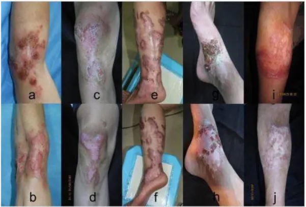
In the case of fungi, most studies are focused on fungal pathogens caused by the greater occurrence of nosocomial infections or opportunistic mycoses in immunocompromised patients, mainly in the use of PDI for pathogenic or potentially pathogenic fungi (PERUSSI, 2007). The antifungal action of PDT appears to be voltage dependent, and the type of biological medium has been shown to affect the effectiveness of PDT in vivo (LIANG e colab., 2016). In view of this, and with the emergence of resistance between species of fungi, PDT has been used as a combined therapy with antifungal drugs, as shown in figure 3 the clinical aspect of chromoblastomycosis lesions in
patients before and after treatment with photodynamic therapy with 5-aminolevulinic acid (ALA- PDT) combined with oral antifungals (HU e colab., 2019).
Figure 3: Clinical aspect of chromoblastomycosis lesions in patients before and after treatment (HU e colab., 2019).
In the case of viruses, most research is focused on the disinfection of blood and blood products in vitro, as well as applications for localized infections that target viral lesions, and lesions that can progress to malignancy (CASTANO e colab., 2004; JORI e BROWN, 2004). The recent study of Inomata et al. (INOMATA e colab., 2019), it was demonstrated that PDT is a promising alternative technique for decontamination of materials, suggesting its future use in hospitals. The study of SILVA
et al. (SILVA e colab., 2019), which, through an Ultrasonic Photodynamic Inactivation device,
proposes PDT as a low cost and non-toxic alternative to the disinfection of biomedical devices, non-critical instruments and also for use in the food industry. In Figure 4 is observed the positive
clinical results of photodynamic therapy, used as the first choice treatment for herpes zoster infection (TEITELBAUM e colab., 2020).
Figure 4: (A) Clinical appearance after 3 days of mild irradiation in herpes zoster infection. (B) Eight months of follow- up after the last application of light irradiation treatment, with no evidence of recurrence (TEITELBAUM e colab., 2020).
1.2 PDT APPLIED TO VIRAL CONTROL
In viral control, the effectiveness of PDT depends on the characteristics of the damaged tissue and cells, as well as the generation of individual oxygen, which remains within the cells for a short period of time (NAMVAR e colab., 2019). Being a unique method compared to vaccines or antiviral drug approaches because, independent of the antigen virus or replication processes (LIM e colab., 2012). Also, according to the author, PDT works by directly inactivating viruses without depending on the host's responses, being a potential antiviral therapy to be applied inside or outside biological systems (LIM e colab., 2012). Second Jori et al. (JORI e BROWN, 2004), the design and mode of action of the sensitizer are essential for obtaining efficient results in PDT and must be focused on the structure and function of the specific viral target. Thus, if the photosensitizer binds to target cells, it can selectively destroy damaged cells without injuring adjacent cells (NAMVAR e colab., 2019). According Zverev et al. (ZVEREV e colab., 2016), studies have shown that membrane viruses lose their infectiousness after treatment with a photosensitizer under red light irradiation. In addition, PDT has demonstrated the potential to treat viral infections such as hepatitis C, herpes simplex and acquired immunodeficiency syndrome, as well as being suggested for the treatment of cold sores, as well as infections, injuries and cancers associated with viruses (LIM e colab., 2012; LOTUFO e colab., 2020; ZVEREV e colab., 2016).
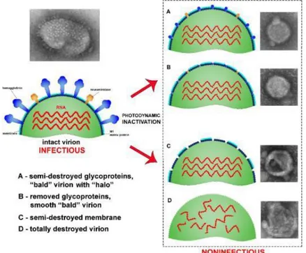
Therefore, the photodynamic inactivation (PDI) of viruses proved to be a promising alternative antiviral strategy, as shown in the study of Korneev et al. (KORNEEV e colab., 2019) that investigated the ultrastructural properties of the photodynamic inactivation of the influenza virus H5N8, inducing three distinct forms of damage to the virion by photodynamic inactivation. Korneev et al. (KORNEEV e colab., 2019) demonstrated the effects of photodynamic inactivation on the influenza vírus (Figure 5). The study showed that the PDI treatment of the virus induces the removal of glycoproteins from the viral surface and create virions non-infectious, even without damage to the membrane.
In the decontamination of products infected by microorganisms and blood, a photodynamic technology is quite advanced, mainly in the disinfection of instruments, it was demonstrated in the study of Silva et al. (SILVA e colab., 2019). These results can be directed to surfaces contaminated by viruses, including against SARS-CoV-2.
Figure 5: Illustration of the effects of photodynamic inactivation on the influenza virus; A) Semi-destroyed glycoproteins; B) Glycoproteins removed; C) Partial destruction of the membrane; E and D) Total virion destruction (KORNEEV e colab., 2019).
1.3 PDT AND POSSIBLE ACTION AGAINST SARS-COV-2
SARS-CoV-2 can be transmitted in three main ways: personal contact; aerosol transmission or contact transmission; and transmission by respiratory droplets during coughing or sneezing (YANG e colab., 2020). The studies of Zheng et al. (ZHENG, 2020) demonstrated that typical symptoms of patients infected with SARS-CoV-2 are: fever, cough, pneumonia, symptoms of acute respiratory infection, with some who quickly developed acute respiratory failure and other serious complications.
Yang et al. (YANG e colab., 2020) states that there is no clear, unified and effective treatment plan for COVID-19. Most guidelines emphasize early identification, early isolation, early diagnosis and early treatment (YANG e colab., 2020). Furthermore, according to the same authors, there are currently no evidence-based drugs to support the effectiveness of antiviral drugs for COVID-19. In the case of vaccines, they are still being developed, but with difficulty in obtaining the desired results, which prevents their production and the regression of the pandemic (AMANAT e KRAMMER, 2020).
To reduce the risk of contracting SARS-CoV-2, the world health organization (WHO) recommended several hygiene and personal practices to be adopted, such as regular hand washing with soap and water, alcohol-based smears that consist of in ethyl alcohol (ethanol); isopropyl alcohol (isopropanol, propan-2-ol) and n-propanol; respiratory hygiene (covering your mouth when coughing or sneezing, wearing a mask, maintaining an adequate social distance of at least 3 feet from those around you and particularly avoiding any form of physical contact or activity that may encourage the projection and transfer of small droplets) (YAN e colab., 2020). All these precautions are necessary to contain the transmission.
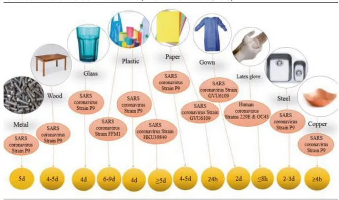
In studies of Pagliano et al. (PAGLIANO e KAFIL, 2020) demonstrated that SARS-CoV-2 can persist on a variety of surfaces from hours to days, (Figure 6). This increases the need for disinfection and environmental cleaning procedures in a correct and consistent manner, in addition to a higher frequency between disinfection intervals.
Thus, efforts to contain the virus are underway however, given the many uncertainties regarding the transmissibility and virulence of pathogens, the effectiveness of these efforts is unknown (LI e colab., 2020). Actually, is not yet known about the appropriate disinfectants to combat SARS-CoV-2, nor about the safety of blocking and histological processing (HENWOOD, 2020).
Therefore, as PDT has obtained positive results in disinfection processes, its use in disinfecting surfaces and instruments infected with SARS-CoV-2 is suggested. In addition, in fact this virus can cause aggressive inflammation and severe acute respiratory syndrome, oxygenation and faster rehabilitation of damaged tissue, antiviral effects and the reduction of inflammatory agents are one of the most important methods of managing the SARS-CoV-2 (FEKRAZAD, 2020). Therefore, photodynamic therapy can be used as adjunctive therapy or even as an alternative therapy in all of these mechanisms, without side effects and drug interactions.
2 CONCLUSION
In view of the discussion presented, photodynamic therapy has demonstrated several applications in the health field. In addition, its action as an antimicrobial therapy is effective against a diversity of microbial, including viruses, without side effects and drug interactions. Understanding the applications of photodynamic therapy with anti-viral allows the development of new therapeutic targets and future studies and, possibly, a promising option in combating the new Coronavirus (SARS- CoV-2) and also in prevention, considering its attributes for disinfection.
ACKNOWLEDGMENTS
REFERENCES
ABRAHAMSE, Heidi e HAMBLIN, Michael R. New photosensitizers for photodynamic therapy. Biochemical Journal, v. 473, n. 4, p. 347–364, 2016.
ACKROYD, Roger e colab. The History of Photodetection and Photodynamic Therapy Photochemistry and Photobiology, v. 74, n. 5, p. 656, 2001.
AGOSTINIS P, BERG K, CENGEL K., Et al. Republic of Yemen Ministry of Public Health and
Population Nutrition and Mortality Survey Report Ibb Governorate , Yemen. Ca Cancer J Clin, v.
61, n. April, p. 250–281, 2017.
ALMEIDA, Ramiro D. e colab. Intracellular signaling mechanisms in photodynamic therapy. Biochimica et Biophysica Acta - Reviews on Cancer, v. 1704, n. 2, p. 59–86, 2004.
AMANAT, Fatima e KRAMMER, Florian. SARS-CoV-2 Vaccines: Status Report. Immunity, v. 52, n. 4, p. 583–589, 2020. Disponível em: <https://doi.org/10.1016/j.immuni.2020.03.007>.
BANERJEE, S. M. e colab. Photodynamic therapy: Inception to application in breast cancer. Breast, v. 31, p. 105–113, 2017. Disponível em: <http://dx.doi.org/10.1016/j.breast.2016.09.016>. BASKARAN, Rengarajan e LEE, Junghan e YANG, Su-Geun. Clinical development of
photodynamic agents and therapeutic applications. Biomaterials Research, v. 22, n. 1, p. 1–8, 2018.
CASTANO, Ana P. e DEMIDOVA, Tatiana N. e HAMBLIN, Michael R. Mechanisms in
photodynamic therapy: Part one - Photosensitizers, photochemistry and cellular localization.
Photodiagnosis and Photodynamic Therapy, v. 1, n. 4, p. 279–293, 2004.
CASTANO, Ana P. e DEMIDOVA, Tatiana N. e HAMBLIN, Michael R. Mechanisms in
photodynamic therapy: Part two - Cellular signaling, cell metabolism and modes of cell death.
Photodiagnosis and Photodynamic Therapy, v. 2, n. 1 SPEC. ISS., p. 1–23, 2005.
CIEPLIK, Fabian e colab. Antimicrobial photodynamic therapy–what we know and what we
don’t. Critical Reviews in Microbiology, v. 44, n. 5, p. 571–589, 2018. Disponível em:
<https://doi.org/10.1080/1040841X.2018.1467876>.
DIAS, Lucas D. e BLANCO, Kate C. e VANDERLEI S. BAGNATO 1, 2. COVID-19: Beyond the
virus. The use of Photodynamic Therapy for the Treatment of Infections in the Respiratory Tract. Photodiagnosis and Photodynamic Therapy, v. 1000, n. 20, p. 101804, 2020. Disponível em:
<https://doi.org/10.1016/j.pdpdt.2020.101804>.
DURANTINI, Edgardo. Photodynamic Inactivation of Bacteria. Current Bioactive Compounds, v. 2, n. 2, p. 127–142, 2006.
FEKRAZAD, Reza. Photobiomodulation and Antiviral Photodynamic Therapy as a Possible
Novel Approach in COVID-19 Management. Photobiomodulation, photomedicine, and laser
surgery, v. 38, n. 5, p. 1–3, 2020.
FOGGIATO, Augusto Alberto e SILVA, Douglas. Terapia fotodinâmica. 488 OrtodontiaSPO, v. 52, n. 5, p. 0–2, 2019.
FOGGIATO, Augusto Alberto e SILVA, Douglas Fernandes e CASTRO, Renata Cristina Faria Ribeiro. Effect of photodynamic therapy on surface decontamination in clinical orthodontic
Disponível em: <https://doi.org/10.1016/j.pdpdt.2018.09.003>.
HAMBLIN, Michael R. Antimicrobial photodynamic inactivation: a bright new technique to kill
resistant microbes. Current Opinion in Microbiology, v. 33, p. 67–73, 2016. Disponível em:
<http://dx.doi.org/10.1016/j.mib.2016.06.008>.
HAMBLIN, Michael R. e HASAN, Tayyaba. Photodynamic therapy: A new antimicrobial
approach to infectious disease? Photochemical and Photobiological Sciences, v. 3, n. 5, p. 436–
450, 2004.
HENWOOD, Anthony F. Coronavirus disinfection in histopathology. Journal of Histotechnology, v. 00, n. 00, p. 1–3, 2020. Disponível em: <https://doi.org/10.1080/01478885.2020.1734718>.
HU, Yongxuan e colab. Photodynamic therapy combined with antifungal drugs against
chromoblastomycosis and the effect of ALA-PDT on Fonsecaea in vitro. PLoS Neglected Tropical
Diseases, v. 13, n. 10, p. 1–14, 2019.
HUANG, Tsun Chin e colab. Antimicrobial efficacy of methylene blue-mediated photodynamic
therapy on titanium alloy surfaces in vitro. Photodiagnosis and Photodynamic Therapy, v. 25, p. 7–
16, 2019. Disponível em: <https://doi.org/10.1016/j.pdpdt.2018.11.008>.
INOMATA, Katia Cilene Ayako e colab. Effects of photodynamic therapy on materials used in
hospitals. 2019 SBFoton International Optics and Photonics Conference, SBFoton IOPC 2019, p. 1– 5,
2019.
JORI, Giulio e BROWN, Stanley B. Photosensitized inactivation of microorganisms.
Photochemical and Photobiological Sciences, v. 3, n. 5, p. 403–405, 2004.
KHEDKAR, Pratik Hemant e PATZAK, Andreas. SARS-CoV-2: What do we know so far? Acta physiologica (Oxford, England), n. March, p. e13470, 2020.
KORNEEV, Denis e colab. Ultrastructural aspects of photodynamic inactivation of highly
pathogenic avian H5N8 influenza virus. Viruses, v. 11, n. 10, p. 1–11, 2019.
KURWA, H. A. e BARLOW, R. J. The role of photodynamic therapy in dermatology. Clinical and Experimental Dermatology, v. 24, n. 3, p. 143–148, 1999.
LAN, Minhuan e colab. Photosensitizers for Photodynamic Therapy. Advanced Healthcare Materials, v. 8, n. 13, p. 1–37, 2019.
LI, Ruiyun e colab. Substantial undocumented infection facilitates the rapid dissemination of
novel coronavirus (SARS-CoV2). Science (New York, N.Y.), v. 493, n. May, p. 489–493, 2020.
LIANG, Yi e colab. Photodynamic therapy as an antifungal treatment (Review). Experimental and Therapeutic Medicine, v. 12, n. 1, p. 23–27, 2016.
LIM, Meng Earn e colab. Photodynamic inactivation of viruses using upconversion nanoparticles. Biomaterials, v. 33, n. 6, p. 1912–1920, 2012. Disponível em: <http://dx.doi.org/10.1016/j.biomaterials.2011.11.033>.
LIU, Zhixin e colab. Composition and divergence of coronavirus spike proteins and host ACE2
receptors predict potential intermediate hosts of SARS-CoV-2. Journal of Medical Virology, n.
LOTUFO, Monica Andrade e colab. “Efficacy of photodynamic therapy on the treatment of
herpes labialis: A systematic review”. Photodiagnosis and Photodynamic Therapy, v. 29, n. August,
2020.
LYON, Juliana Pereira e colab. Photodynamic therapy for pathogenic fungi. Mycoses, v. 54, n. 5, p. 265–271, 2011.
NAMVAR, Mahsa Alavi e colab. Effect of photodynamic therapy by 810 and 940 nm diode laser
on Herpes Simplex Virus 1: An in vitro study. Photodiagnosis and Photodynamic Therapy, v. 25, p.
87–91, 2019. Disponível em: <https://doi.org/10.1016/j.pdpdt.2018.11.011>.
OLEINICK, Nancy L. e EVANS, Helen H. The Photobiology of Photodynamic Therapy: Cellular
Targets and Mechanisms. Radiation Research, v. 150, n. 5, p. S146, 1998.
PAGLIANO, Pasquale e KAFIL, Hossein Samadi. Protection and disinfection policies. Le Inferziono in Medicina, v. 2, n. April, p. 185–191, 2020.
PERUSSI, Janice Rodrigues. Inativação fotodinâmica de microrganismos. Quimica Nova, v. 30, n. 4, p. 988–994, 2007.
QUEIROZ, Gabriella Brandimarte e colab. CELL DEATH AND ITS CONCEPT APPLIED IN
GENERAL HEALTH AND MICROBIOLOGICAL ACTION: LITERATURE REVIEW.
CPAQV, v. 12, p. 1–24, 2020.
RAJESH, S. e colab. Antimicrobial photodynamic therapy: An overview. Journal of Indian Society of Periodontology, v. 15, n. 4, p. 323–327, 2011.
RKEIN, Ali M e OZOG, David M. Photodynamic Therapy, Methods in Molecular Biology. Chapter 12. Antimicrobial Photodynamic Inactivation and Photodynamic Therapy for Infections, p. 155–173, 2010.
SANTOS, Anc��ly Ferreira e colab. Photodynamic therapy in cancer treatment - an update
review. Journal of Cancer Metastasis and Treatment, v. 2019, 2019.
SHIOTSU-OGURA, Yukako e colab. Antimicrobial photodynamic therapy using a plaque
disclosing solution on Streptococcus mutans. Photodiagnosis and Photodynamic Therapy, v. 26, n.
January, p. 252–257, 2019. Disponível em: <https://doi.org/10.1016/j.pdpdt.2019.04.003>.
SILVA, Douglas Fernandes e colab. Effect of photodynamic therapy potentiated by ultrasonic
chamber on decontamination of acrylic and titanium surfaces. Photodiagnosis and Photodynamic
Therapy, v. 27, n. March, p. 345–353, 2019.
SOUKOS, Nikolaos S. e colab. Photodynamic effects of toluidine blue on human oral
keratinocytes and fibroblasts and Streptococcus sanguis evaluated in vitro. Lasers in Surgery
and Medicine, v. 18, n. 3, p. 253–259, 1996.
TANG, Wei e colab. Red blood cell-facilitated photodynamic therapy for cancer treatment. Advanced Functional Materials, v. 26, n. 11, p. 1757–1768, 2016.
TEITELBAUM, Susana e AZEVEDO, Luciane Hiramatsu e BERNAOLA-PAREDES, Wilber Edison.
Antimicrobial Photodynamic Therapy Used as First Choice to Treat Herpes Zoster Virus Infection in Younger Patient: A Case Report. Photobiomodulation, Photomedicine, and Laser
TENNERT, Christian e colab. Effect of photodynamic therapy (PDT) on Enterococcus faecalis
biofilm in experimental primary and secondary endodontic infections. BMC Oral Health, v. 14, n.
1, p. 1–8, 2014.
WANG, B. e colab. Gain with no pain? Pain management in dermatological photodynamic
therapy. British Journal of Dermatology, v. 177, n. 3, p. 656–665, 2017.
YAN, Yuxin e colab. The first 75 days of novel coronavirus (SARS-CoV-2) outbreak: Recent
advances, prevention, and treatment. International Journal of Environmental Research and Public
Health, v. 17, n. 7, 2020.
YANG, Yongshi e colab. The deadly coronaviruses: The 2003 SARS pandemic and the 2020
novel coronavirus epidemic in China. Journal of Autoimmunity, v. 109, n. February, p. 102434,
2020. Disponível em: <https://doi.org/10.1016/j.jaut.2020.102434>.
ZHENG, Jun. SARS-coV-2: An emerging coronavirus that causes a global threat. International Journal of Biological Sciences, v. 16, n. 10, p. 1678–1685, 2020.
ZVEREV, V. V. e colab. In vitro studies of the antiherpetic effect of photodynamic therapy. Lasers in Medical Science, v. 31, n. 5, p. 849–855, 2016.