F
ACULDADE DEM
EDICINA DEL
ISBOAA
UTOIMUNIDADE E CÉLULAS REGULADORAS
T
CD4
+CD25
HIGHNA
I
MUNODEFICIÊNCIA
C
OMUM
V
ARIÁVEL
Susana Clara Barão Lopes da Silva dos Anjos
MESTRADO EM IMUNOLOGIA MÉDICA
2007
F
ACULDADE DEM
EDICINA DEL
ISBOAA
UTOIMUNIDADE E CÉLULAS REGULADORAS
T
CD4
+CD25
HIGHNA
I
MUNODEFICIÊNCIA
C
OMUM
V
ARIÁVEL
Susana Clara Barão Lopes da Silva dos Anjos
Mestrado em Imunologia Médica
Dissertação orientada pelo Professor Doutor Antero G. Palma-Carlos
Todas as afirmações efectuadas no presente documento são da exclusiva responsabilidade do seu autor, não cabendo qualquer responsabilidade à Faculdade de
A impressão esta dissertação foi aprovada em Comissão Coordenadora
do Conselho Científico da Faculdade de Medicina de Lisboa, em
Good research brings you more questions than answers.
R
ESUMOIntrodução: Vários mecanismos têm sido sugeridos para explicar a elevada prevalência de
doenças autoimunes (DAIs) na Imunodeficiência Comum Variável (ICV). Procurámos avaliar a prevalência de DAIs numa população com IDCV, caracterizar estes doentes e verificar se um defeito quantitativo na população T CD4+CD25high poderia estar associado à maior prevalência de autoimunidade na ICV.
Métodos: Foram incluídos 47 doentes com ICV sob terapêutica substitutiva com
imunoglobulina endovenosa (IGEV). Através de revisão dos processos clínicos e entrevista individual foram recolhidos dados clínicos e laboratoriais relativamente às manifestações de apresentação e evolução clínica, incluindo DAIs e níveis séricos de imunoglobulinas no diagnóstico de ICV. Em estudo transversal, foi quantificada IgG sérica e populações T, B e NK e células T CD4CD25 por citometria de fluxo em sangue total.
Resultados: Foram diagnosticadas DAIs em 19 doentes (40,4%), sendo as citopénias
autoimunes as mais frequentes. As DAIs foram diagnosticadas antes da ICV em 8 doentes, nenhum deles sob terapêutica imunossupressora no ano anterior ao diagnóstico de ICV. A idade média dos doentes com DAI era superior no momento do estudo, diagnóstico de ICV e no início da terapêutica com IGEV. Também apresentavam uma prevalência mais elevada de diarreia crónica não infecciosa e hiperplasia linfoide e IgG sérica mais elevada no diagnóstico. O estudo transversal não evidenciou diferenças significativas na IgG sérica pré-infusional ou populações linfocitárias entre doentes com e sem DAI. As frequências de CD4+CD25high foram significativamente mais baixas em doentes com DAI comparados com doentes sem DAI e com controlos saudáveis e no conjunto dos doentes com ICV comparados
Conclusões: Estes resultados sugerem que a deficiência quantitativa de CD4+CD25high poderá contribuir para a elevada prevalência de DAIs na ICV. Uma avaliação longitudinal e mais detalhada da população T CD4+CD25high, incluindo marcadores fenotípicos adicionais e estudo funcional, contribuirão para clarificar esta questão.
P
ALAVRASC
HAVEA
BSTRACTBackground: Several mechanisms have been proposed to explain the high incidence of
autoimmune diseases (AID) in Common Variable Immunodeficiency (CVID). We aimed to evaluate AID frequency within a CVID population and to characterize patients with AID. We also investigated whether a quantitative defect in the immunoregulatory population CD4+CD25high could be associated with increased prevalence of autoimmunity in CVID.
Methods: 47 patients with CVID on regular intravenous immunoglobulin substitution therapy
were enrolled. Chart review and questionnaire-guided interview were used to collect clinical and laboratory data concerning presentation symptoms and clinical evolution, including AID. Serum immunoglobulins were quantified at diagnosis. A cross-sectional evaluation was performed before IVIG infusion, including serum IgG level, T, B and NK cell quantification by flow-cytometry in freshly whole blood. CD4+CD25+ cells were simultaneously quantified in whole blood by flow-cytometry and compared with age-matched healthy volunteers.
Results: AIDs were diagnosed in 19 patients (40.4%) and autoimmune cytopenias were the
most frequent. AID was diagnosed before CVID in eight patients, none on immunosuppressors in the year before CVID diagnosis. Patients with AID were older at the time of the present evaluation, at CVID diagnosis and at beginning of IVIG. They also exhibited higher prevalence of chronic non-infectious diarrhea and lymphoid hyperplasia and higher serum IgG at diagnosis. There were no significant differences in IgG pre-infusional levels and lymphocyte subpopulations between patients with and without AID. CD4+CD25high frequencies were significantly lower in patients with AID compared to those without AID and controls and in the whole group of CVID compared to controls.
Conclusions: Our results suggest that CD4+CD25high deficiency may possibly contribute to the high incidence of AID in CVID. More detailed and longitudinal evaluation of CD4+CD25high T cells in larger cohorts, including the use of additional markers and suppressor cells function assessment, will help to clarify this issue.
K
EY-W
ORDSÍ
NDICE Resumo... I Abstract... III Índice... V Preâmbulo... 1 Agradecimentos... 3 Lista de abreviaturas... 5 Resumo extenso... 7 Referências bibliográficas... 20 Artigo científico... 31 Abstract... 33 Introduction... 35Patients and methods... 38
Patients... 38
Clinical and laboratory data collection... 39
Cross-sectional laboratory evaluation - CD4+CD25high quantification….. 40
Statistical Analysis... 41
Results ... 42
Autoimmune diseases in patients with CVID... 42
Other clinical features of patients with AID ... 43
Immunological features of patients with AID ...
Comparison between patients with and without AIDst...
44 44
CD4+CD25high ... 45
Discussion... 47
Acknowledgements ... 67
References... 69
Tables and Figures... 91
Table 1... 93 Table 2... 95 Table 3... 97 Table 4... 99 Table 5... 101 Figure 1... 103 Figure 2... 105 Figure 3... 107 Figure 4... 109
A decisão de fazer o Mestrado em Imunologia Médica foi motivada essencialmente pela vontade de aprofundar o conhecimento na especialidade que escolhi para a prática clínica. Sendo parte integrante da formação e actividade em Imunoalergologia, as Imunodeficiências Primárias (IDPs) são, desde logo por motivos epidemiológicos, uma área com a qual contactamos com menor frequência, apesar de a prevalência global destas doenças se situar em 1: 20 000 nascimentos, se englobarmos todos os grupos de IDPs. A Imunodeficiência Comum Variável é a IDP sintomática mais frequente, encontrando-se actualmente em seguimento no Serviço de Imunoalergologia do HSM cerca de 35 doentes com ICV. São frequentemente casos complexos e absorventes, que se caracterizam por uma diversidade de patologias, com espectro de gravidade alargado.
Do ponto de vista conceptual, as IDPs são extremamente instrutivas, constituindo verdadeiros modelos vivos que nos permitem compreender melhor o sistema imunitário. Para além do desafio que a sua complexidade de diagnóstico e as dificuldades na evolução clínica e terapêutica oferecem, o seguimento de doentes com IDPs torna-se hoje cada vez mais gratificante. O investimento da investigação nesta área tem contribuído para uma significativa melhoria da qualidade de vida dos doentes.
Todos estes motivos em conjunto contribuíram para aumentar o meu interesse pelas IDPs e reforçam a pertinência da escolha deste tema, em particular da Imunodeficiência Comum Variável, para área de trabalho prático no Mestrado em Imunologia Médica.
No decurso do Internato Complementar, estagiei durante o primeiro trimestre de 2004 no Hospital Vall d`Hebron, em Barcelona, centro de referência para crianças e adultos com IDPs da Catalunha. O dinamismo da orientação da Dra Teresa Español e a excelente recepção por
No Hospital Vall d´Hebron encontram-se em seguimento cerca de 15 crianças e 70 adultos com ICV, a maioria dos quais recorre àquela instituição para terapêutica substitutiva com IGEV. A possibilidade de acesso a uma população alargada e a disponibilidade do Laboratório de Imunologia, permitiram realizar o presente trabalho. Tendo colocado a hipótese original de um defeito quantitativo das células reguladoras CD4+CD25high poder estar associado à elevada incidência de doenças autoimunes na ICV, sublinho e agradeço a disponibilidade e a coragem de toda a equipa para colaborar sem reservas com esta ideia. A realização do trabalho prático durante o tempo do estágio foi um objectivo extremamente ambicioso / exigente para este período, que no entanto contribuiu de forma decisiva para o seu sucesso. A avaliação clínica, baseada na revisão dos processos e entrevista clínica, foi obviamente afectada pelos condicionalismos da metodologia retrospectiva. O protocolo laboratorial foi desenhado em conjunto com a equipa do Laboratório de Imunologia, o qual suportou todos os encargos financeiros e num período record o integrou na sua rotina e nos demais trabalhos em curso.
Obtiveram-se resultados significativos relativamente à hipótese colocada, original e integrada em linhas actuais de investigação em autoimunidade e imunodeficiências primárias humorais. Estes resultados, aliados ao facto de a autoimunidade constituir um problema simultaneamente frequente e intrigante para aqueles que na prática clínica e no laboratório lidam com doentes com ICV, motivaram a elaboração em inglês do artigo CD4+
CD25high and Autoimmunity in Common Variable Immunodeficiency: searching new answers for an old question.
A
GRADECIMENTOSA todos aqueles que tanto me ajudaram na concretização deste trabalho.
À Dra Teresa Español, anfitriã de excepção, pelo exemplo inspirador de dinamismo e empenho na concretização deste projecto. À Dra Drahomira Detková, pela amizade, pela análise das células CD4+CD25high e discussão dos resultados, em conjunto com o Dr Manuel Hernandez. Ao Dr Javier de Gracia e Dr José Maria Bertran, responsáveis pelos Hospitais de Dia de Pneumologia e Imunologia Infantil de Vall d´Hebron, pelo seu apoio desinteressado. À Dra Emília Faria, amiga e colega das IDPs, também em Barcelona, pelo apoio e carinho. Ao Dr José Gonçalo Marques, pelo primeiro desafio de trabalhar as IDPs, pelo apoio constante e por um trabalho conjunto cada vez mais estimulante.
Aos colegas do Serviço de Imunoalergologia, meus apoiantes na longa e difícil fase de escrita desta tese, já submergida em plena actividade assistencial, em particular à Dra Amélia Spínola Santos, Dra Anabela Pregal, Professor Manuel Branco Ferreira, Dra Elisa Pedro e ao Professor Doutor Manuel Barbosa, impulsionador do estágio em Barcelona.
À Unidade de Imunologia Clínica do Instituto de Medicina Molecular, pelo estímulo permanente, em particular à Dra Adriana Albuquerque, Dra Rita Cavaleiro, Professora Doutora Ana Espada de Sousa, Professora Doutora Maria Conceição Santos e Professor Doutor Rui Victorino, pelas leituras críticas e construtivas desta tese em preparação.
Ao Professor Doutor A. G. Palma-Carlos, meu orientador, pelo exemplo e incentivo para início do mestrado e pela orientação deste projecto.
Aos meus pais e irmãs. Ao Rui.
L
ISTA DEA
BREVIATURASAID Autoimmune disease
AIHA Autoimmune hemolytic anemia
AZT Azathioprine
BAFF-R B-cell activating factor receptor (BAFF-R) CD Crohn´s disease
CT Computed tomography
CTLA-4 Cytotoxic T Lymphocyte associated-Antigen 4
CVID Common Variable Immunodeficiency
Cy Cyclosporine
DAI Doença autoimune
EDTA EthyleneDiamineTetrAcetic acid
ELISA Enzyme-Linked Immunosorbent Assay
ESID European Society for Primary Immunodeficiencies
F Female
FITC Fluorescein isothiocyanate
FoxP3 Forkhead transcription factor
GITR Glucocorticoid-Induced TNF-Receptor family-related gene
HCV Hepatitis C Virus
HIV Human Immunodeficiency Virus
ICOS Inducible Costimulator -
ICV Imunodeficiência Comum Variável
Ig Immunoglobulin / Imunoglobulina
ITP Immune thrombocytopenia
IUIS International Union of Immunological Societies
IVIG Intravenous immunoglobulin
M Male
NK Natural Killer
NSAIDs Non-steroidal anti-inflammatory drugs OS Oral steroids
PBMC Peripheral Blood Mononuclear Cell
PCR Polymerase Chain Reaction
PE Phycoerythrin
PerCP Peridinin chlorophyll protein
PID Primary immunodeficiency
RA Rheumatoid arthritis
SD Standard deviation
SLE Systemic Lupus Erythematosus
TACI Transmembrane activator and calcium-modulator and cyclophilin-ligand Interactor
TRECs T-cell receptor-rearrangement excision circles
T reg T regulatory / T reguladora
R
ESUMOE
XTENSOA Imunodeficiência Comum Variável (ICV) é a imunodeficiência primária (IDP) sintomática mais frequente, tendo uma prevalência estimada em 1 / 25 0001,2 entre a população ocidental. O diagnóstico definitivo de ICV baseia-se na diminuição de IgG, IgA e/ou IgM, pelo menos 2 desvio-padrões em relação ao normal para a idade, associada à deficiência de produção de isohemaglutininas e/ou de anticorpos específicos e após exclusão de outras causas primárias ou secundárias de hipogamaglobulinémia3,4. O espectro clínico da ICV é extremamente amplo no tipo de manifestações clínicas e sua gravidade. Para além das infecções recorrentes, mais frequentemente respiratórias e digestivas, as doenças autoimunes5, a hiperplasia linfoide6, em alguns casos com padrão granulomatoso, e a incidência aumentada de neoplasias hematológicas7 são alguns dos problemas clínicos mais frequentes na ICV.
A prevalência de doenças autoimunes (DAIs) na ICV tem sido estimada entre 21%8 a 50%9,10,11, contrastando com os 5-7% calculados para a população geral12 e sugerindo a existência de defeitos imunológicos favorecedores da autoimunidade na ICV.
Este intrigante aumento da incidência de DAIs mediadas por células e/ou por anticorpos, numa IDP predominantemente atribuída a défice de anticorpos constitui um paradoxo aparente e tem originado várias hipóteses visando a sua explicação13.
A existência de uma predisposição genética para a autoimunidade em doentes com ICV é sugerida por estudos de linkage e tipagem HLA que têm demonstrado associações entre genes de susceptibilidade major para ICV e/ou défice de IgA e outros para DAIs9,14.
Na ICV, como em outras IDPs, a infecção poderá constituir o elo entre a imunodeficiência e a autoimunidade15. A incapacidade de lidar com super-antigénios16 e de eliminação de antigénios externos, secundária aos múltiplos defeitos da imunidade inata e adquirida
infecciosos ou por uma resposta inflamatória exacerbada aos mesmos, a reactividade cruzada entre tecidos do doente e antigénios estranhos ou à deposição de complexos imunes.
Entre os múltiplos defeitos identificados na diferenciação / função dos linfócitos B, salientam--se defeitos na maturação de células de memória CD19+CD27+ 10,17,18,19,20. Warnatz et al e Ko
et al demonstraram que doentes com maior deficiência de células B de memória
class-switched têm maior prevalência de DAIs18,20. Contrariamente, Piqueras et al não confirmaram
esta associação, mas verificaram maior prevalência de esplenomegália, proliferação linfoide e doença granulomatosa no grupo de doentes com maior deficiência na maturação de linfócitos B19.
Múltiplos defeitos de imunidade celular estão também descritos na ICV, nomeadamente linfopénia T21, sobretudo CD4 naive22,23, para a qual podem contribuir a redução de progenitores mononucleares na medula óssea24, deficiência de timopoiese2,24,, deficiência de IL225 e IL726 e aumento da apoptose27,28,29. Outros defeitos funcionais na imunidade celular incluem ainda alterações na activação e proliferação T8,30 e na produção de citocinas, estando descrito neste contexto um desvio Th12 e diminuição de citocinas Th2, nomeadamente IL4, IL5 e IL1031, 32, 33, 34, 35.
A falência de mecanismos de indução e/ou manutenção de tolerância central ou periférica pode também contribuir para o aumento da incidência de DAIs na ICV.
As células T reguladoras, entre as quais as CD4+CD25high, estão envolvidas na manutenção de tolerância ao self, através da supressão activa da activação e expansão de células T auto-reactivas existentes à periferia de todos os indivíduos saudáveis36. As células T reguladoras estão também envolvidas não só na supressão de reacções alérgicas e reacção enxerto vs hospedeiro após transplante, mas também da resposta a infecções e tumores37,38.
Com este estudo pretendemos verificar se uma deficiência quantitativa de CD4+CD25high se poderia associar ao aumento da prevalência de DAI descrito na ICV, podendo este ser um
defeito universal ou definir um perfil particular de um subgrupo de doentes com expressão clínica / laboratorial de autoimunidade. Embora tivesse já sido anteriormente sugerida a existência de um compromisso funcional das células T supressoras na ICV e sua associação com o aumento de prevalência de DAIs14, a hipótese de um defeito quantitativo de CD4+CD25high nunca antes tinha sido testada.
Sakagushi et al demonstraram que a depleção de células reguladoras CD4+CD25+ resulta no desenvolvimento de DAIs em ratinhos39. O mesmo grupo demonstrou em animais que uma população minor de células T CD4+CD25+ é crucial para o controlo de células T autoimunes
in vitro40,41. Diversas AID foram induzidas em estirpes susceptíveis de ratinhos, em
protocolos envolvendo remoção completa ou alteração do desenvolvimento das células T
CD4+CD25+, nos quais a co-transferência de células T CD4+CD25+ evitava o
desenvolvimento de DAIs38,41,42. Adicionalmente, os ratinhos com deficiência primária de CD25 demonstraram ser susceptíveis a autoimunidade grave que podia ser evitada pela inoculação de células T CD4+CD25+ de ratinhos singénicos43. No seu conjunto, estes dados sugerem que as células T CD4+ que expressam primariamente a cadeia α do receptor da IL2 (CD25) desempenham um papel importante na patogénese das DAIs.
Uma população com propriedades in vitro fenotípicas e funcionais idênticas foi posteriormente definida nos humanos, no sangue periférico, timo e sangue venoso umbilical de recém-nascidos saudáveis44,45,46,47,48. A capacidade supressora destas células foi preferencialmente associada às células T CD4 com maior intensidade de expressão de CD25 (CD4+CD25high)44 e num estadio final de diferenciação, sendo maioritariamente CD4+CD25+CD45RA-CD45RO+ 44. A população total CD4+CD25+ contém uma proporção relativamente elevada de células T activadas, já que o CD25 é expresso transitoriamente à superfície de células T CD4+ não reguladoras após activação, não conferindo actividade
Muitos trabalhos têm procurado clarificar o papel das células T CD4+CD25+ na patogénese das DAIs no ser humano. Defeitos quantitativos e / ou funcionais têm sido descritos em diversas DAIs, embora os resultados sejam escassos e discrepantes. Diferentes autores encontraram uma diminuição do número de células T CD4+CD25+ circulantes na diabetes insulino dependente49, hepatite autoimune50 e lúpus eritematoso sistémico51,52 e defeito funcional, mas não quantitativo, no síndrome poliglandular tipo II53, esclerose múltipla54 e diabetes autoimune55,56. Outros estudos não detectaram qualquer deficiência de CD4+CD25high na miastenia gravis57, esclerose múltipla58, diabetes insulino-dependente59 e síndrome de Sjögren60. Na artrite reumatoide foi encontrada maior quantidade de células T reguladoras, com actividade supressora mais intensa, no líquido sinovial de articulações inflamadas, em comparação com o sangue periférico dos mesmos doentes61,62, um fenómeno com fundamento possivelmente equiparável ao aumento de células reguladoras CD4+CD25+ verificado na mucosa intestinal de doentes com doença inflamatória intestinal61,62.
No presente trabalho, foi estudada uma população de 47 doentes com ICV seguidos no Hospital Vall d´Hebron, em Barcelona, com os objectivos de avaliar a frequência de DAI nesta população, caracterizar os doentes com ICV e DAI e comparar o seu perfil clínico e imunológico com o de doentes sem DAI. Pretendemos ainda avaliar a frequência de CD4+CD25high em doentes com ICV e DAI em comparação com controlos saudáveis.
A caracterização clínica foi realizada através da revisão do processo clínico hospitalar e entrevista guiada com cada doente. Foram colhidos dados relativos à ICV, nomeadamente idade e tipo de apresentação, evolução clínica, idade de diagnóstico e de início de terapêutica substitutiva com imunoglobulina endovenosa (IGEV), para além da evolução de eventuais DAIs, incluindo tipo de DAI, idade de diagnóstico e respectivo tratamento. Foram ainda recolhidos dados laboratoriais, nomeadamente doseamento de IgG, IgA, IgM, subclasses de IgG e produção de anticorpos específicos na altura do diagnóstico de ICV. Em paralelo,
realizou-se uma avaliação laboratorial transversal desta população, incluindo IgG pré– infusional, hemograma e imunofenotipagem com quantificação por citometria de fluxo das populações B, NK, T CD4+, T CD8+ e expressão de HLA-DR nas duas últimas subpopulações T. Em paralelo, foi feita a avaliação quantitativa da percentagem de CD25high entre as células T CD4+, por citometria de fluxo em amostras de sangue total com tripla marcação CD4 / CD25 / CD45-RO, cujos resultados foram comparados com os obtidos numa população controlo de 29 saudáveis.
Foram identificados 19 doentes (40,4%) com manifestações de DAI ao longo da sua evolução clínica. As DAIs detectadas foram Síndrome de Evans, trombocitopénia autoimune, anemia perniciosa, eritroblastopenia, artrite reumatoide, vitiligo, alopecia areata, psoríase, Síndrome de Sjögren, hepatite autoimune, doença de Crohn e hipotiroidismo primário. Cunninghan-Rundles et al descreveram DAI em 52 / 248 doentes8, embora prevalências ainda mais altas tenham sido reportadas na literatura (28%63 a 50%10). Tal como em outras séries, as citopénias autoimunes foram as DAIs mais frequentemente diagnosticadas (6 / 47)10,64, seguidas da artrite reumatoide e anemia perniciosa (6,4%).
Verificámos um predomínio não significativo do sexo feminino, tanto no conjunto de toda a população com ICV, como entre os doentes com DAIs. A idade de início dos sintomas foi muito variável, mas em média ligeiramente mais precoce que em outras séries (15,6 ± 14,7 anos)8,9.
As primeiras manifestações atribuíveis à imunodeficiência no grupo de doentes com DAI foram as infecções respiratórias recorrentes, seguidas da autoimunidade em oito doentes, nenhum deles sob terapêutica imunossupressora durante o ano que precedeu o diagnóstico de ICV. Apenas dois doentes estavam sob terapêutica imunossupressora (ciclosporina) aquando da realização da avaliação laboratorial transversal.
A idade média dos doentes com ICV e DAI era significativamente superior à dos doentes sem DAI, não só no momento da realização do estudo, como no aparecimento dos primeiros sintomas atribuíveis à ICV e no início da terapêutica com IGEV. Os doentes com DAI apresentavam demora média desde os primeiros sintomas até ao diagnóstico de ICV significativamente mais longa.
Não se verificaram diferenças na idade de apresentação ou de diagnóstico de ICV quando comparados doentes que tiveram manifestações de DAI como primeiros sintomas com os restantes, com outros tipos de apresentação. No entanto, os doentes com DAI, mas em que esta não foi a primeira manifestação de ICV tiveram demora média significativamente mais longa desde a apresentação até ao diagnóstico de ICV do que doentes sem DAI.
Verificámos que os doentes com DAI tinham mais frequentemente diarreia crónica não infecciosa e hiperplasia linfoide, sendo esta diferença significativa. As infecções respiratórias recorrentes e bronquiectasias, associadas frequentemente a tosse crónica e sinusite, e as gastrenterites infecciosas foram frequentes no conjunto de todos os doentes, no entanto sem diferença significativa entre os doentes com e sem DAI.
Atendendo aos níveis de referência de imunoglobulinas séricas para cada idade, a IgA e IgG no diagnóstico estavam diminuídas em todos os doentes com DAI, encontrando-se a IgM dentro dos valores de referência em cinco destes doentes. A IgG sérica no diagnóstico era significativamente superior em doentes com DAI, particularmente nos que tinham esta forma de apresentação inicial, quando comparados com doentes sem DAI.
Aquando da avaliação transversal todos os doentes estavam sob terapêutica com IGEV com doses e periodicidade muito variáveis (de 373 a 1360 mg/Kg/mês). A IgG pré-infusional, hemograma e as populações B, NK, T CD4+, T CD8+ não revelaram diferenças significativas entre os doentes com e sem DAI.
Os doentes com ICV e DAI apresentaram médias de frequências de células T CD4+CD25+ e de CD4+CD25high significativamente inferiores às dos doentes sem DAI e controlos saudáveis. O conjunto total de doentes com ICV apresentou também frequência média CD4+CD25high inferior quando comparada com a dos controlos, sendo esta diferença mais significativa no grupo dos doentes com DAI e mantendo significado estatístico após exclusão dos dois doentes sob terapêutica com ciclosporina.
A percentagem de células CD25high entre as células T CD4+ foi extremamente variável nos doentes e nos controlos, salientando-se o facto de a média das frequências de CD4+CD25high obtida no grupo dos controlos saudáveis (1,25 ± 0,26 %) ter sido muito semelhante à obtida por Baecher-Allan et al no trabalho utilizado como referência metodológica para definição da população CD25high no presente estudo44. Naquele trabalho, as células CD4+CD25high foram estimadas em 1-2% da população T CD4+, sendo definidas por citometria de fluxo, com dupla marcação CD4 e CD25, como uma subpopulação que se destaca da população contendo
CD4+CD25low e CD4+CD25- 44.
Mais recentemente, diversos autores têm descrito outras formas de definir a mesma população52,55,56,59,60, alguns deles com maior objectividade, sendo outros omissos em relação à metodologia, incluindo critérios de definição de elevada expressão de CD2551,53,57, o que dificulta a comparação entre resultados. No nosso trabalho, tentámos minimizar a subjectividade do método escolhido através da quantificação de CD4+CD25high por um único investigador, sem acesso aos dados clínicos e aplicando os mesmos critérios em todos os doentes.
Na nossa população, as células CD4+CD25high eram maioritariamente de memória CD45RO+, tanto nos doentes com ICV como nos controlos. Foi avaliada a expressão de HLA-DR nas células T CD4+ e T CD8+, não tendo sido encontradas diferenças nos valores absolutos ou
com e sem DAI. Estes resultados e a ausência de correlação entre CD4+CD25highCD45RO+ e essas subpopulações sugerem que as diferenças encontradas nas frequências de CD4+CD25high não seriam apenas directamente dependentes de uma maior activação imunológica.
Outros marcadores têm sido associados às células T reguladoras CD4+CD25+ T, incluindo CD152 (Cytotoxic T Lymphocyte associated-Antigen 4 - CTLA-4), GITR (Glucocorticoid-Induced TNF-Receptor family-related gene), CD62L e o factor de transcrição FoxP3. Este último está descrito como essencial ao desenvolvimento e actividade supressora, tanto em ratinhos como em humanos44,65,66 e controla a expressão de CD25 nas células T reguladoras, mas não nas células T activadas67. A adição de outros marcadores, nomeadamente FoxP3, para caracterização imunofenotípica enriqueceria muito a nossa avaliação.
O defeito da timopoiese em doentes com ICV, anteriormente mencionado, pode contribuir para as baixas percentagens de CD4+CD25high nestes doentes, já que o timo é uma fonte primária desta subpopulação68. Por outro lado, a IL2 é importante tanto para indução de apoptose de células T auto-reactivas15 como na expansão e manutenção da função imunossupressora das células CD4CD25 à periferia38,69,70,71. A deficiência de IL2 em sobrenadantes de culturas após estimulação com mitogénios já foi descrita na ICV72,73 e tem sido atribuída à linfopenia74, a qual paradoxalmente se associa com a expansão de CD4+CD25high em diferentes contextos75. Na nossa população não encontrámos linfopenia ou diferenças significativas nas percentagens ou valores absolutos de linfócitos entre doentes com e sem DAI. Adicionalmente, não encontrámos correlação entre os valores absolutos de linfócitos e frequências de CD4+CD25high,tanto considerando o conjunto de todos os doentes, como avaliando os grupos de doentes com DAI e sem DAI separadamente. A avaliação da capacidade de produção de IL2 pelas células T seria interessante neste contexto.
Alguns autores têm sugerido a existência de variações quantitativas / funcionais das CD4+CD25high em função da idade60. Gregg et al relataram um aumento progressivo da proporção de células CD4+CD25high 76, interpretado como predominantemente derivado de expansão periférica, enquanto Tsaknaridis et al encontraram um declínio progressivo da actividade supressora das células CD4+CD25 77, sugerindo eventual relação com o declínio da função tímica. Na nossa população, como em outras séries55,56, não foi encontrada correlação entre a idade e a frequência de CD4+CD25high, quando considerada toda a população de doentes com ICV e controlos. Esta análise foi metodologicamente muito relevante, em virtude de terem sido encontradas diferenças significativas entre as idades dos grupos de doentes com e sem DAI. Por outro lado, o facto de não existirem diferenças significativas nas distribuições por idade entre controlos e doentes, não sugere ser a idade o factor responsável pelas diferenças significativas na frequência de CD4+CD25high entre estas duas populações. Curiosamente, foi encontrada uma correlação positiva significativa entre a idade e a percentagem de CD4+CD25high no subgrupo de doentes com DAI. O estudo da actividade supressora seria extremamente interessante, no contexto da hipótese de Tsaknaridis77.
Foi também encontrada diferença significativa na frequência de CD4+CD25high quando comparados doentes com e sem diarreia crónica não infecciosa, sem diferença significativa na distribuição por idade entre estes 2 grupos. No contexto da doença inflamatória intestinal, outros autores descreveram diminuição das células CD4+CD5high no sangue periférico78, em simultâneo com aumento significativo das mesmas células na lâmina própria intestinal79. No nosso estudo, não foram encontradas diferenças significativas na frequência de CD4+CD5high no sangue periférico, após estratificação dos doentes de acordo com presença / ausência de bronquiectasias, esplenomegália, proliferação linfoide ou granulomas.
Alguns estudos têm tentado estabelecer uma classificação da ICV que permita prever quais os doentes com potencial evolução para DAI, embora sem sucesso10. Propomos a deficiência quantitativa de células T CD4+CD25high como um marcador útil à identificação de doentes com maior risco de desenvolver DAI. Atendendo à elevada sobreposição encontrada nas frequências de CD4+CD25high entre doentes com e sem DAI e entre doentes e controlos, propomos que a quantificação seriada / prospectiva desta população seria provavelmente mais informativa do que determinações isoladas.
Em doentes seleccionados, o re-estabelecimento / indução de tolerância dominante poderia ser tentado in vivo através da estimulação da expansão de células T reguladoras e/ou fortalecimento da sua actividade supressora ou da sua indução in vivo ou in vitro37,80,81. Ensaios envolvendo números limitados de doentes e usando IL2 sintética82,83,84 ou natural85 demonstraram o seu potencial clínico, embora estudos envolvendo maior número de doentes, com seguimento mais prolongado e com objectivos clínicos bem definidos sejam necessários antes de ser considerada a sua aplicação na prática clínica.
Confirmámos, numa população de 47 doentes com ICV, a elevada prevalência de DAIs nesta entidade (40,4%), sendo a autoimunidade a forma de apresentação em 8/47 (17%) dos doentes estudados. Sugerimos o doseamento de imunoglobulinas aquando do diagnóstico de DAI, um procedimento acessível, económico e que pode influenciar opções terapêuticas decisivas nestes doentes em particular, nomeadamente o início de fármacos imunossupressores e esplenectomia.
Verificámos uma diminuição da frequência de células T CD4+CD25high na população de doentes com ICV quando comparada com controlos, particularmente acentuada no subgrupo de doentes com DAI. Estudos prospectivos, envolvendo séries com maior número de doentes, e idealmente uma avaliação fenotípica e funcional mais detalhada das células T
CD4+CD25high permitirão integrar de forma mais adequada os nossos resultados na patogénese da autoimunidade na ICV.
R
EFERÊNCIASB
IBLIOGRÁFICAS1. Salzer U, Maul-Pavicic A, Cunningham-Rundles C et al. ICOS deficiency in patients with common variable immunodeficiency. Clin Immunol 2004; 113(3): 234-40.
2. Bayry J, Hermine O, Webster DA, Levy Y, Kaveri SV. Common variable
immunodeficiency: the immune system in chaos. Trends Mol Med 2005; 11(8): 370-376.
3. Notarangelo L, Casanova JL, Conley ME et al. Primary immunodeficiency diseases: An update from the International Union of Immunological Societies Primary Immunodeficiency Diseases Classification Committee Meeting in Budapest, 2005. J
Allergy Clin Immunol 2006; 117: 883-96.
4. Diagnostic criteria: Common Variable Immunodeficiency available from Esid.org [homepage on the Internet]. Leiden: European Society for Immunodeficiencies; c1994-2007 [updated 2005 Sep; cited 2007 May 30]. Available from: http://www.esid.org/.
5. Wang J, Cunningham-Rundles C. Treatment and outcome of autoimmune hematologic disease in common variable immunodeficiency. J Autoimmun 2005; 25: 57-62.
6. Bates CA, Ellison MC, Lynch DA, Cool CD, Brown KK, Routes JM. Granulomatous-lymphocytic lung disease shortens survival in common variable immunodeficiency. J
Allergy Clin Immunol 2004; 114(2): 415-21.
7. Mellemkjaer L, Hammarstrom L, Andersen V et al. Cancer risk among patients with IgA deficiency or common variable immunodeficiency and their relatives: a combined Danish and Swedish study. Clin Exp Immunol 2002; 130(3): 495-500.
8. Cunnigham-Rundles C, Bodian C. Common Variable Immunodeficiency: clinical and immunological features of 248 patients. Clin Immunol 1999; 92: 34-48.
9. Bloch-Michel C, Viallard JF, Blanco P et al. Common variable immunodeficiency: 17 observations in the adult. Rev Med Interne 2003; 24(10): 640-50.
10. Warnatz K, Wehr C, Drager R et al. Expansion of CD19hiCD21lo/neg B cells in common variable immunodeficiency (CVID) patients with autoimmune cytopenia.
Immunobiology 2002; 206(5): 502-13.
11. Sarmiento E, Mora R, Rodríguez-Mahou M, Rodríguez-Molina J, Fernández-Cruz E, Carbone J. Autoimmune disease in primary antibody deficiencies. Allergol et
Immunopathol 2005; 33(2): 69-73.
12. Diamond B. Autoimmunity. Immunol Rev 2005; 204: 5-8.
13. Cunningham-Rundles C. Hematologic complications of primary immune deficiencies.
Blood Reviews 2002; 16: 61-64.
14. Giannouli S, Anagnostou D, Soliotis F, Voulgarelis M. Autoimmune manifestations in common variable immunodeficiency. Clin Rheumatol 2004; 23: 449-52.
15. Petrovsky N. The paradoxical association between immunodeficiency and
autoimmunity: comment on the article by Atkinson. Arthritis Rheum 1996; 39(1): 179-80.
16. Silverman GJ. B cell superantigens: possible roles in immunodeficiency and
autoimmunity. Semin Immunol 1998; 10(1): 43-55.
17. Brouet JC, Chedeville A, Fermand JP, Royer B. Study of the B cell memory
compartment in common variable immunodeficiency. Eur J Immunol 2000; 30(9): 2516-2520.
18. Warnatz K, Denz A, Dräger R et al. Severe deficiency of switched memory B cells (CD27+IgM-IgD-) in subgroups of patients with common variable immunodeficiency: a new approach to classify a heterogeneous disease. Blood 2002; 99: 1544-1551.
19. Piqueras B, Lavenu-Bombled C, Galicier L et al. Common variable immunodeficiency patient classification based on impaired B cell memory differentiation correlates with clinical aspects. J Clin Immunol 2003; 23(5): 385- 400. 20. Ko J, Radigan L, Cunningham-Rundles C. Immune competence and switched memory
B cells in common variable immunodeficiency. Clin Immunol 2005; 116(1): 37-41. 21. Guazzi V, Aiuti F, Mezzaroma I et al. Assessment of thymic output in common
variable immunodeficiency patients by evaluation of T cell receptor excision circles.
Clin Exp Immunol 2002; 129(2): 346-53.
22. Lebranchu Y, Thibault G, Degenne D, Bardos P. Abnormalities in CD4+ T
lymphocyte subsets in patients with common variable immunodeficiency. Clin
Immunol Immunopathol 1991; 61(1): 83-92.
23. Farrant J, Spickett G, Matamoros N et al. Study of B and T cell phenotypes in blood from patients with common variable immunodeficiency (CVID). Immunodeficiency 1994; 5(2): 159-69.
24. Isgro A, Marziali M, Mezzaroma I et al. Bone marrow clonogenic capability, cytokine production and thymic output in patients with common variable immunodeficiency. J
Immunol 2005; 174(8): 5074-81.
25. Eisenstein EM, Jaffe JS, Strober W. Reduced interleukin-2 (IL-2) production in common variable immunodeficiency is due to a primary abnormality of CD4+ T cell differentiation. J Clin Immunol 1993; 13(4): 247-58.
26. Holm AM, Aukrust P, Damas JK, Muller F, Halvorsen B, Froland SS. Abnormal
interleukin-7 function in common variable immunodeficiency. Blood 2005; 105(7): 2887-90.
27. Di Renzo M, Zhou Z, George I, Becker K, Cunningham-Rundles C. Enhanced apoptosis of T cells in common variable immunodeficiency (CVID): role of defective CD28 co-stimulation. Clin Exp Immunol 2000; 120(3): 503-11.
28. Iglesias J, Matamoros N, Raga S, Ferrer JM, Mila J. CD95 expression and function on lymphocyte subpopulations in common variable immunodeficiency (CVID); related to increased apoptosis. Clin Exp Immunol 1999; 117(1): 138-46.
29. Aukrust P, Svardal AM, Muller F, Lunden B, Berge RK, Froland SS. Decreased levels of total and reduced glutathione in CD4+ lymphocytes in common variable immunodeficiency are associated with activation of the tumor necrosis factor system: possible immunopathogenic role of oxidative stress. Blood 1995; 86(4): 1383-91 30. North ME, Webster AD, Farrant J. Defects in proliferative responses of T cells from
patients with common variable immunodeficiency on direct activation of protein kinase C. Clin Exp Immunol 1991; 85(2): 198-201.
31. Sneller MC, Strober W. Abnormalities of lymphokine gene expression in patients with common variable immunodeficiency. J Immunol 1990; 144(10): 3762-9.
32. Pastorelli G, Roncarolo MG, Touraine JL, Peronne G, Tovo PA, de Vries JE.
Peripheral blood lymphocytes of patients with common variable immunodeficiency (CVI) produce reduced levels of interleukin-4, interleukin-2 and interferon-gamma, but proliferate normally upon activation by mitogens. Clin Exp Immunol 1989; 78(3): 334-40.
33. Holm AM, Aukrust P, Aandahl EM, Muller F, Tasken K, Froland SS. Impaired
secretion of IL-10 by T cells from patients with common variable immunodeficiency-involvement of protein kinase A type I. J Immunol 2003; 170(11): 5772-7.
34. Hauber I, Fischer MB, Maris M, Eibl MM. Reduced IL-2 expression upon antigen stimulation is accompanied by deficient IL-9 gene expression in T cells of patients with CVID. Scand J Immunol 1995; 41(3): 215-9.
35. Ferrer JM, Iglesias J, Hernandez M, Matamoros N. Alterations in interleukin secretion (IL-2 and IL-4) by CD4 and CD4CD45RO cells from common variable immunodeficiency (CVID) patients. Clin Exp Immunol 1995; 102(2): 286-9.
36. Jiang H, Chess L. An integrated view of suppressor T cell subsets in
immunoregulation. J Clin Invest 2004; 114(9): 1198-208.
37. Chatila TA. Role of regulatory T cells in human diseases. J Allergy Clin Immunol 2005; 116(5): 949-59.
38. Sakaguchi S. Naturally arising CD4+ regulatory T cells for immunologic self-tolerance and negative control of immune responses. Annu Rev Immunol 2004; 22: 531-62.
39. Sakaguchi S, Fukuma K, Kuribayashi K, Masuda T. Organ-specific autoimmune
diseases induced in mice by elimination of T cell subset. I. Evidence for the active participation of T cells in natural self-tolerance; deficit of a T cell subset as a possible cause of autoimmune disease. J Exp Med 1985; 161(1): 72-87.
40. Sakaguchi S, Sakaguchi N, Asano M, Itoh M, Toda M. Immunologic self-tolerance maintained by activated T cells expressing IL-2 receptor alpha-chains (CD25). Breakdown of a single mechanism of self-tolerance causes various autoimmune diseases. J Immunol 1995; 155(3): 1151-64.
41. Asano M, Toda M, Sakaguchi N, Sakaguchi S. Autoimmune disease as a consequence of developmental abnormality of a T cell subpopulation. J Exp Med 1996; 184(2): 387-96.
42. Takahashi T, Kuniyasu Y, Toda M et al. Immunologic self-tolerance maintained by CD25+CD4+ naturally anergic and suppressive T cells: induction of autoimmune
disease by breaking their anergic/suppressive state. Int Immunol 1998; 10(12): 1969-80.
43. Malek TR, Yu A, Vincek V, Scibelli P, Kong L. CD4 regulatory T cells prevent lethal autoimmunity in IL-2Rβ-deficient mice. Implications for the nonredundant function of IL-2. Immunity 2002; 17(2): 167-78.
44. Baecher-Allan C, Brown JA, Freeman GJ, Hafler DA. CD4+CD25high regulatory cells in human peripheral blood. J Immunol 2001; 167(3): 1245-53.
45. Ng WF, Duggan PJ, Ponchel F et al. Human CD4+CD25+ cells: a naturally occurring population of regulatory T cells. Blood 2001; 98(9): 2736-44.
46. Dieckmann D, Plottner H, Berchtold S, Berger T, Schuler G. Ex vivo isolation and characterization of CD4+CD25+ T cells with regulatory properties from human blood.
J Exp Med 2001; 193(11): 1303-10.
47. Jonuleit H, Schmitt E, Stassen M, Tuettenberg A, Knop J, Enk AH. Identification and functional characterization of human CD4+CD25+ T cells with regulatory properties isolated from peripheral blood. J Exp Med 2001; 193(11): 1285-94.
48. Stephens LA, Mottet C, Mason D, Powrie F. Human CD4(+)CD25(+) thymocytes and peripheral T cells have immune suppressive activity in vitro. Eur J Immunol 2001; 31(4): 1247-54.
49. Kukreja A, Cost G, Marker J et al. Multiple immuno-regulatory defects in type-1 diabetes. J Clin Invest 2002; 109(1): 131-40.
50. Longhi MS, Ma Y, Bogdanos DP, Cheeseman P, Mieli-Vergani G, Vergani D.
Impairment of CD4+CD25+ regulatory T-cells in autoimmune liver disease. J Hepatol 2004; 41(1): 31-7.
52. Liu MF, Wang CR, Fung LL, Wu CR. Decreased CD4+CD25+ T cells in peripheral blood of patients with systemic lupus erythematosus. Scand J Immunol 2004; 59(2): 198-202
53. Kriegel MA, Lohmann T, Gabler C, Blank N, Kalden JR, Lorenz HM. Defective
suppressor function of human CD4+CD25+ regulatory T cells in autoimmune polyglandular syndrome type II. J Exp Med 2004; 199(9): 1285-91.
54. Viglietta V, Baecher-Allan C, Weiner HL, Hafler DA. Loss of functional suppression by CD4+CD25+ regulatory T cells in patients with multiple sclerosis. J Exp Med 2004; 199(7): 971-9
55. Lindley S, Dayan CM, Bishop A, Roep BO, Peakman M, Tree TI. Defective
suppressor function in CD4+CD25+ T-cells from patients with type 1 diabetes.
Diabetes 2005; 54(1): 92-9.
56. Brusko TM, Wasserfall CH, Clare-Salzler MJ, Schatz DA, Atkinson MA. Functional defects and the influence of age on the frequency of CD4+CD25+ T-cells in type 1 diabetes. Diabetes 2005; 54(5): 1407-14.
57. Huang YM, Pirskanen R, Giscombe R, Link H, Lefvert AK. Circulating CD4+CD25+ and CD4+CD25+ T cells in myasthenia gravis and in relation to thymectomy. Scand J
Immunol 2004; 59(4): 408-14.
58. Putheti P, Pettersson A, Soderstrom M, Link H, Huang YM. Circulating CD4+CD25+ T regulatory cells are not altered in multiple sclerosis and unaffected by disease-modulating drugs. J Clin Immunol 2004; 24(2): 155-61.
59. Putnam AL, Vendrame F, Dotta F, Gottlieb PA. CD4+CD25high regulatory T cells in human autoimmune diabetes. J Autoimmunity 2005; 24(1): 55-62.
60. Gottenberg JE, Lavie F, Abbed K et al. CD4 CD25high regulatory T cells are not impaired in patients with primary Sjogren's syndrome. J Autoimmun 2005; 24(3): 235-42.
61. Van Amelsfort JM, Jacobs KM, Bijlsma JW, Lafeber FP, Taams LS. CD4+CD25+
regulatory T cells in rheumatoid arthritis: differences in the presence, phenotype, and function between peripheral blood and synovial fluid. Arthritis Rheum 2004; 50(9): 2775-85.
62. Cao D, Malmstrom V, Baecher-Allan C, Hafler D, Klareskog L, Trollmo C. Isolation and functional characterization of regulatory CD25brightCD4+ T cells from the target organ of patients with rheumatoid arthritis. Eur J Immunol 2003; 33(1): 215-23.
63. Pavic M, Seve P, Malcus C et al. Common variable immunodeficiency with
autoimmune manifestations: study of nine cases; interest of a peripheral B-cell compartment analysis in seven patients. Rev Med Interne 2005; 26(2): 95-102.
64. Pasic S. Autoimmune cytopenia in common variable immunodeficiency. J Pediatr 2004; 144(5): 689.
65. Fontenot JD, Gavin MA, Rudensky AY. Foxp3 programs the development and
function of CD4+CD25+ regulatory T cells. Nat Immunol 2003; 4(4): 330-6.
66. Walker MR, Kasprowicz DJ, Gersuk VH et al. Induction of FoxP3 and acquisition of T regulatory activity by stimulated human CD4+CD25- T cells. J Clin Invest 2003; 112(9): 1437-43.
67. Hori S, Nomura T, Sakaguchi S. Control of regulatory T cell development by the transcription factor Foxp3. Science 2003; 299(5609): 1057-61.
68. Balandina A, Lecart S, Dartevelle P, Saoudi A, Berrih-Aknin S. Functional defect of regulatory CD4+CD25+ T cells in the thymus of patients with autoimmune myasthenia
69. Malek TR, Bayer AL. Tolerance, not immunity, crucially depends on IL-2. Nat Rev
Immunol 2004; 4(9): 665-74.
70. Bensinger SJ, Walsh PT, Zhang J et al. Distinct IL-2 receptor signaling pattern in CD4+CD25+ regulatory T cells. J Immunol 2004; 172(9): 5287-96.
71. Scheffold A, Huhn J, Hofer T. Regulation of CD4+CD25+ regulatory T cell activity: it takes (IL-)two to tango. Eur J Immunol 2005; 35(5): 1336-41.
72. Goldacker S, Warnatz K. Tackling the heterogeneity of CVID. Curr Opin Allergy Clin
Immuno. 2005; 5(6): 504-9.
73. Rump JA, Jahreis A, Schlesier M, Drager R, Melchers I, Peter HH. Possible role of IL-2 deficiency for hypogammaglobulinaemia in patients with common variable immunodeficiency. Clin Exp Immunol 1992; 89(2): 204-10.
74. North ME, Ivory K, Funauchi M, Webster AD, Lane AC, Farrant J. Intracellular cytokine production by human CD4+ and CD8+ T cells from normal and immunodeficient donors using directly conjugated anti-cytokine antibodies and three-colour flow cytometry. Clin Exp Immunol 1996; 105(3): 517-22
75. Zhang H, Chua KS, Guimond M et al. Lymphopenia and interleukin-2 therapy alter homeostasis of CD4+CD25+ regulatory T cells. Nat Med 2005; 11(11): 1238-43.
76. Gregg R, Smith CM, Clark FJ et al. The number of human peripheral blood CD4+ CD25high regulatory T cells increase with age. Clin Exp Immunol 2005; 140(3): 540-6.
77. Tsaknaridis L, Spencer L, Culbertson N et al. Functional assay for human
CD4+CD25+ Treg cells reveals an age-dependent loss of suppressive activity. J
Neurosci Res 2003; 74(2): 296-308
78. Maul J, Loddenkemper C, Mundt P et al. Peripheral and intestinal regulatory CD4+CD25high T cells in inflammatory bowel disease. Gastroenterology 2005; 128(7): 1868-78
79. Makita S, Kanai T, Oshima S et al. CD4+CD25bright T cells in human intestinal lamina propria as regulatory cells. J Immunol 2004; 173(5): 3119-30.
80. Kanai T, Watanabe M. Clinical application of human CD4+ CD25+ regulatory T cells for the treatment of inflammatory bowel diseases. Expert Opin Biol Ther 2005; 5(4): 451-62.
81. Earle KE, Tang Q, Zhou X et al. In vitro expanded human CD4+CD25+ regulatory T cells suppress effector T cell proliferation. Clin Immunol 2005; 115(1): 3-9.
82. Cunningham-Rundles C, Mayer L, Sapira E, Mendelsohn L. Restoration of
immunoglobulin secretion in vitro in common variable immunodeficiency by in vivo treatment with polyethylene glycol-conjugated human recombinant interleukin-2. Clin
Immunol Immunopathol 1992; 64(1): 46-56.
83. Cunningham-Rundles C, Kazbay K, Hassett J, Zhou Z, Mayer L. Brief report:
enhanced humoral immunity in common variable immunodeficiency after long-term treatment with polyethylene glycol-conjugated interleukin-2. N Engl J Med 1994; 331(14): 918-21.
84. Cunningham-Rundles C, Kazbay K, Zhou Z, Mayer L. Immunologic effects of low-dose polyethylene glycol-conjugated recombinant human interleukin-2 in common variable immunodeficiency. J Interferon Cytokine Res 1995; 15(3): 269-76.
85. Rump JA, Jahreis A, Schlesier M, Stecher S, Peter HH. A double-blind, placebo-controlled, crossover therapy study with natural human IL-2 (nhuIL-2) in combination with regular intravenous gammaglobulin (IVIG) infusions in 10 patients with common variable immunodeficiency (CVID). Clin Exp Immunol 1997; 110(2): 167-73.
A
RTIGO
C
IENTÍFICO
CD4
+
CD25
HIGHAND
A
UTOIMMUNITY IN
C
OMMON
V
ARIABLE
I
MMUNODEFICIENCY
:
A
BSTRACTBackground: Several mechanisms have been proposed to explain the high incidence of
autoimmune diseases (AID) in Common Variable Immunodeficiency (CVID). We aimed to evaluate AID frequency within a CVID population and to characterize patients with AID. We also investigated whether a quantitative defect in the immunoregulatory population CD4+CD25high could be associated with increased prevalence of autoimmunity in CVID. Methods: 47 patients with CVID on regular intravenous immunoglobulin substitution therapy
were enrolled. Chart review and questionnaire-guided interview were used to collect clinical and laboratory data concerning presentation symptoms and clinical evolution, including AID. Serum immunoglobulins were quantified at diagnosis. A cross-sectional evaluation was performed before IVIG infusion, including serum IgG level, T, B and NK cell quantification by flow-cytometry in freshly whole blood. CD4+CD25+ cells were simultaneously quantified in whole blood by flow-cytometry and compared with age-matched healthy volunteers.
Results: AIDs were diagnosed in 19 patients (40.4%) and autoimmune cytopenias were the
most frequent. AID was diagnosed before CVID in eight patients, none on immunosuppressors in the year before CVID diagnosis. Patients with AID were older at the time of the present evaluation, at CVID diagnosis and at beginning of IVIG. They also exhibited higher prevalence of chronic non-infectious diarrhea and lymphoid hyperplasia and higher serum IgG at diagnosis. There were no significant differences in IgG pre-infusional levels and lymphocyte subpopulations between patients with and without AID. CD4+CD25high frequencies were significantly lower in patients with AID compared to those without AID and controls and in the whole group of CVID compared to controls.
CD4+CD25high T cells in larger cohorts, including the use of additional markers and suppressor cells function assessment, will help to clarify this issue.
K
EY-W
ORDSI
NTRODUCTIONCommon variable immunodeficiency (CVID) was described for the first time in 1953 by Janeway 1. It is nowadays the most frequently diagnosed primary immunodeficiency (PID) and the most common symptomatic congenital deficiency of the immune system. Its prevalence has been estimated as 1/25 000 in the western population 2,3 and worldwide it has been reported between 1/20 000 and 1/200 000 4,5.
Diagnosis of CVID is established when there is marked decrease of two major serum immunoglobulin isotypes, usually IgG and IgM and/or IgA, over two standard deviations (SD) below mean values for age 6, in addition to impaired ability to specific antibody production after vaccination or exposure to a known infectious agent. Other primary or secondary causes for antibody deficiency should be excluded 7,8.
The most common clinical manifestations of CVID are recurrent pyogenic infections, usually by encapsulated bacteria and involving the sino-pulmonary tract, or otherwise unexplained chronic lung disease. Gastrointestinal manifestations are also frequent, either infectious, involving bacterial and protozoal agents or inflammatory conditions, leading to chronic diarrhea and malabsorption. In addition, CVID is associated with a remarkable incidence of autoimmunity and increased risk of gastric cancer and lymphoma 9 and other clinical presentations of benign lymphoid proliferation, including multiple adenopathies, splenomegaly and lymphocitic or granulomatous infiltration of lungs, lymph nodes or other sites 8,10,11. In some cases, non-infectious complications dominate the clinical picture of CVID and have a significant impact on the overall severity of the disease.
Although included in the last update of the International Union of Immunological Societies (IUIS) classification of PIDs as predominantly an antibody deficiency 6, many other
acquired immunity, including both humoral and cellular components, and mostly the interplay between all 3,12,13. The possibility of extensive immunological heterogeneity underlying the wide diversity of presentation symptoms and clinical outcome has motivated recent works aiming to individualize groups of patients based on clinical features and match them to particular immunological defects. On the other hand, interest on a possible genetic basis of CVID has increased and, in the last 2 years, four monogenic defects associated with CVID have been identified: ICOS, TACI, BAFF-R and CD19 2, 12.
Autoimmune disease (AID) prevalence has been estimated as 5-7% in the general population 14. In one of the largest CVID series reported in the literature, Cunningham-Rundles et al refer
to AID in 52/248 (21%) patients 15. More recently, other authors have reported higher incidences ranging from 28% 4 to 50% 5,16,17. Several mechanisms have been proposed to explain this high incidence of autoimmunity in CVID, although no immunological marker of autoimmunity has been identified in these patients.
Among different T cell sub-populations known to participate in the maintenance of tolerance, CD4+CD25high lymphocytes have emerged as a major immunoregulatory population. Besides, evidence is now accumulating that regulatory T cells are also involved in the down-regulation of allergy, graft-versus-host disease and immune response to tumors and infections 18,19. Although results are not consensual 20,21,22, quantitative 23,24,25 or functional 26,27,28,29 defects in CD4+CD25high cells were found in some human AID. Even though a possible functional compromise of suppressor T cells promoting the growth of autoimmune clones in CVID has been suggested 30, the hypothesis of a quantitative defect in CD4+CD25high had not been investigated in this context.
In the present work we aimed to evaluate AID frequency within a CVID population and to characterize a group of patients with CVID and AID, both from clinical and immunological point of view. We also investigated whether a quantitative defect in CD4+CD25high in CVID
patients could be associated with an increased prevalence of autoimmunity. This could be either a defect of a particular homogenous group with clinical expression of AID or a common defect to the general population of CVID.
P
ATIENTS ANDM
ETHODSA retrospective and descriptive study of AID prevalence was performed among a CVID population followed at Vall d´Hebron Hospital, a referral center for both pediatric and adult primary immunodeficiency patients in Barcelona, Spain. Clinical history from all patients was carefully reviewed concerning presentation symptoms and complications. A cross-sectional laboratory evaluation of these patients was also performed in the Immunology Department at the same institution. Institutional review board approval was obtained for this study.
Patients
The study included 47 patients (22 women / 25 men; mean age: 37.5 ± 15.9; range: 16-71 years old) attending Vall d´Hebron between January and March 2004 and with a mean follow up of 8.8 ± 6.1 years since CVID diagnosis. All patients were on regular intravenous immunoglobulin (IVIG) substitution therapy. Diagnostic criteria were according to WHO 6 and ESID recommendations 7. Each patient presented a marked decrease in at least two out of the three major isotypes of serum immunoglobulins (IgG, IgA and IgM) by more than two SD below mean values for age on at least two separate occasions over one month. In most cases, including all patients with serum IgG level greater than 350 mg/dL, antibody deficiency was verified by means of decreased isohemaglutinins and / or antibody production to two or more vaccines, including tetanus, Haemophilus influenzae and pneumococcal vaccine. These patients were immunized with Haemophilus influenzae type B (Hib)-conjugated vaccine PedvaxHIB® and PNU-Immune23® polyvalent pneumococcal vaccine and titers of specific IgG before and four weeks after immunization were compared 31,32.
Subjects under the age of two years and all patients with known secondary causes of hypogammaglobulinemia at time of CVID diagnosis were excluded, namely hematological
disorders such as myeloma or non-Hodgkin lymphoma, HIV infection, nephrotic syndrome, exsudative gastroentheropathy, thymoma, chronic immunossupression or catabolic states due to malnutrition, treatment with drugs like hydantoin and gold salts. Patients with other known causes of primary hypogammaglobulinemia (hyper IgM syndrome, X-linked agammaglobulinemia, X-linked lymphoproliferative syndrome) or low peripheral B cell counts (<1% CD19+ cells) were also excluded.
Written informed consent was obtained from all patients before enrollment.
Clinical and laboratory data collection
Clinical and laboratory data concerning family history, first symptoms suggesting PID, CVID diagnosis, immunoglobulin replacement and clinical evolution, including complications / concomitant disorders and respective treatment have been collected by a single investigator by means of retrospective chart review and questionnaire-guided personal interview with all patients.
Vall d´Hebron’s CVID follow-up protocol includes regular laboratory tests (hemogram, hepatic and renal function, PCR for HCV and HIV antigenemia, pre-infusion IgG level, stool and sputum cultures), imaging evaluation (annual abdominal ultrasonography and chest X-ray, biannual abdominal and thoracic CT) and annual lung function. Further examinations are performed when appropriate, in selected patients, in order to diagnose / treat concomitant diseases, mainly infectious, autoimmune or malignant. AIDs have been diagnosed by the assistant physicians or by the investigator, according to accepted criteria for each disease and based on typical clinical data and laboratory / imagiological exams and exclusion of other frequent diagnosis. The absence of autoimmune antibodies did not exclude AID diagnosis. Serum IgG, IgA and IgM at diagnosis were quantified by nephelometry (reference values in
for IgA and 58-250mg/dL for IgM). IgG subclasses were quantified by ELISA (reference values in adults were 261-1081mg/dL for IgG1, 112-408mg/dL for IgG2, 22-288mg/dL for IgG3 and 5-156 mg/dL for IgG4) 33. Adequate responses to the 23-valent pneumococcal vaccine 32 and the Hib conjugated vaccine 31 were considered when, respectively, four-fold and two-fold increases in specific IgG titer were verified by ELISA.
Cross-sectional laboratory evaluation - CD4CD25high quantification
A 10mL sample of peripheral blood in 0.05% EDTA was collected from CVID patients, prior to IVIG substitution. IgG level was quantified by nephelometry in this sample. Full blood counts, including white blood cell differential count were performed using a routine hematology analyzer in the Department of Pathology of Vall d´Hebron Hospital.
Peripheral blood mononuclear cells (PBMC) were isolated by Fycoll-Hypaque gradient. CD4+ and CD8+ T cells, B cells and NK cells were assessed in the Immunology Department by flow cytometry (FACSCalibur, Becton Dickinson Biosciences, San Jose, CA, USA) using the following monoclonal antibodies (Becton-Dickinson®): CD3, CD4, CD8, anti-CD19 and anti-CD16 + anti-CD56. Anti-HLA-DR was used to assess CD4 and CD8 T cells activation.
CD4+CD25+ cells were quantified in whole blood in parallel experiment using 3 colour acquisition on a Fluorescense Activated Cell Sorter FACSCalibur (Becton-Dickinson®), with peridinin chlorophyll protein (PerCP)-conjugated anti-CD4 (Becton Dickinson®), fluorescein isothiocyanate (FITC)-conjugated anti-CD25 (Immunotech®), phycoerythrin (PE)-conjugated anti-CD45RO (Becton Dickinson®) and respective mouse isotype controls.
A single investigator with no access to clinical data acquired and analyzed all data using Cellquest software (Becton-Dickinson®).
Lymphocytes were gated according to forward and side scatter and a minimum of 10 000 events were acquired and analyzed. CD4+CD25high definition adopted in this work was based on Baecher-Allan et al 34,in which CD4+CD25high cells appear as a tail to the right from the
major population containing both CD4+CD25low and CD4+CD25- cells (Figure 1).
CD4+CD25high percentage was defined as the percentage of CD25high within gated CD4 positive T cells and its absolute number was calculated by multiplying this percentage by the number of CD4 positive T cells obtained in simultaneous sample. CD4+CD25high percentages obtained in patients were compared with those obtained in 29 age-matched healthy volunteers.
Statistical Analysis
Descriptive values of variables were expressed as the mean ± SD. CD4+CD25high percentages were compared using unpaired Student´s T test or Mann-Witney U test. Pearson´s correlation coefficient, Spearman´s rank correlation and Fisher exact test were used when appropriate to study the relationship between clinical and / or laboratory parameters. Statistical analyses were performed using Excel and Prism Graph Pad 4 Programs (GraphPad Prism, USA). Results were considered significant at a p value <0.05.
R
ESULTS3.1 Autoimmune diseases in patients with CVID
Chart review and personal interview with the 47 patients revealed that 19 patients (40.4%) presented previous or present manifestations of AID (9 men and 10 women; mean age 46.5 ± 15.0 years old).
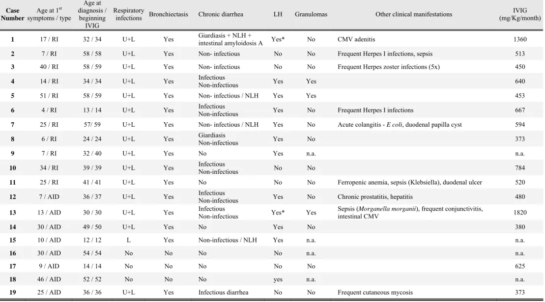
Twenty-six AID were diagnosed, including Evans´s Syndrome (1), immune thrombocytopenia (ITP) (5), autoimmune hemolytic anemia (1), pernicious anemia (4), eritroblastopenia (1), rheumatoid arthritis (3), vitiligo (1), alopecia areata (3), psoriasis (2), Sjogren’s Syndrome (1), autoimmune hepatitis (1), Crohn’s Disease (2) and primary hypothyroidism (1).
Table 1 details AID diagnosis, age at presentation and AID treatment of the 19 patients. Six patients had more than one AID, with autoimmune cytopenias being the most frequently diagnosed (26.3%). In eight patients (patients 12 to 19), AID was diagnosed before CVID diagnosis (mean delay 13.6 ± 10.3 years; maximum delay 29 years). After starting symptoms of AID, patients 13, 14, 15, 16, 17, 18 and 19 were treated with oral steroids during variable periods but not in the year before CVID diagnosis were made. Investigations that lead to CVID were mainly prompted either by frequent respiratory infections (patients 12, 13, 14, 15 and 19) or recurrent bouts of autoimmune cytopenias (patients 16 and 18). Patient 17 was diagnosed CVID when she was 14 years old. She presented with vitiligo and autoimmune hepatitis when she was nine years old. IgA deficiency was then diagnosed and small doses of oral steroids and azathioprine were prescribed during one year.
At the time of the present study laboratory evaluation (including CD4+CD25high), patients 6 and 13 were the only ones on immunosuppressive therapy - cyclosporine for Crohn’s disease,
3.2 Other clinical features of patients with AID
First symptoms of CVID were recurrent respiratory infections in 11 cases and AID in the
other eight, with mean age at the beginning of symptoms of 20.9 ± 15.6 and 21.2 ± 13.8 years old respectively, as described in Table 2. Mean age at CVID diagnosis was 46.5 ± 15.0 years old (minimum 10; maximum 58). There were no significant differences in age at the beginning of symptoms or at diagnosis between patients whose first symptoms were of AID and those who initially presented with upper or lower respiratory infections and developed AID during evolution (n=11).
Sixteen patients presented recurrent upper and lower respiratory infections and bronchiectasis during evolution in association with chronic productive cough in eight and sinusitis in 11 patients.
Twelve patients reported intermittent periods of diarrhea with no infectious cause identified in stool cultures and intestinal biopsies. In four of these patients lymphoid nodular hyperplasia was found in intestinal biopsy, possibly justifying chronic diarrhea. Eight patients had recurrent infectious diarrhea and Giardia lamblia was the most frequent cause. Only six patients did not present gastrointestinal symptoms.
Lymphoid hyperplasia, defined as the presence of splenomegaly and / or lymphadenopaties, was found in 12 patients. Patients 1 and 13 were splenectomized for uncontrolled autoimmune cytopenias at 39 and 13 years old, respectively, and presented lymphadenopaties in both cases. Three patients had granulomatous disease, which may have been underdiagnosed, as biopsies were not performed in all patients. There were no reports of malignancy during clinical evolution.
All patients were on IVIG replacement therapy, with highly variable doses and periodicity, individually adapted to each patient’s weight and clinical condition (668 ± 402 mg/Kg/month;