I
Avaliação in vitro do potencial de regeneração
óssea do osso de choco utilizando co-culturas de
osteoblastos e osteoclastos
Monografia de Investigação
Mestrado Integrado em Medicina Dentária da Universidade do Porto
Teresa Brandão da Silva
Porto, 2017
I
Avaliação in vitro do potencial de regeneração
óssea do osso de choco utilizando co-culturas de
osteoblastos e osteoclastos
Monografia de Investigação
Mestrado Integrado em Medicina Dentária da Universidade do Porto
Teresa Brandão da Silva
Aluno do 5º ano do Mestrado Integrado da FMDUP
teresabsilva@live.com.pt
Orientadora: Professora Doutora Ana Isabel Pereira Portela
Professora Auxiliar da FMDUP
Co-orientadora: Professora Doutora Meriem Lamghari Moubarrad
Professora Auxiliar do ICBAS
Porto 2017
I
Sometimes you just have to believe in yourself, follow your heart and do the best as you can, even if is not the easy way.
I
ACKNOWLEDGEMENTS
I would like to thank:
To my advisor Professor Ana Isabel Pereira Portela from Faculdade de Medicina Dentária, Universidade do Porto, who was always very friendly and helpful. Thank you for all the support and for believing in me.
To my co-advisor Professor Meriem Lamghari Moubarrad from Instituto de Ciências Biomédicas Abel Salazar, Universidade do Porto who was extremely kind for accepting my project, for being diligent, interested and for sharing her scientific knowledge and humanism.
To Professor José Maria da Fonte Ferreira from Materials and Ceramic Engineering Departement, Universidade de Aveiro who was generous and helpful. I really appreciated the time spent in providing and preparing the scaffolds and all the experience and knowledge shared.
To Professor Pedro Lopes Granja from Instituto de Ciências Biomédicas Abel Salazar, Universidade do Porto for the opportunity of developing all of this work in i3S, for believing in me and in the potential of this material and for being gentle and a good person.
To Francisco for being so persistent, generous, kind, understanding, calm and a very good friend. For teaching me all the protocols and procedures performed and for helping me with all aspects of this study which couldn’t be done without his help.
To the members of Nanobiomaterials for Targeted Therapies group (Daniela, Estrela, Inês Juliana, Luís, Rita) for providing all the support and friendship.
To Cláudia for being so persistent, calm, kind and positive.
To Helena for being a very good friend, for all the support, generosity, understanding and for believing in me.
To Sofia who was extremely gentle and helpful and for providing and preparing the scaffolds.
To André for providing me with unfailing support and continuous encouragement. To my brother for the friendship and support.
To my parents for believing in my capacities and effort, for supporting me all of these years and for providing the opportunities to achieve my goals.
II CONTENTS
ACKNOWLEDGEMENTS ... I CONTENTS ... II List of figures ... IV List of acronyms and abbreviations ... V List of symbols ... VI
RESUMO ... 1
ABSTRACT ... 2
INTRODUCTION ... 3
Materials and methods ... 5
1. Preparation of cuttlefish bone blocks ... 5
2. Mesenchymal stem cell culture ... 5
3. Viability ... 5
3.1. Optimization experiment without scaffolds ... 5
3.2. Optimization experiments with scaffolds ... 6
3.3. Resazurin reduction assay with scaffolds using non-treated multidishes for suspension cell culture (48 wells-plate) ... 6
4. Live/Dead assay ... 7
4.1. Optimization experiment without scaffolds ... 7
4.2. Optimization experiment with scaffolds ... 7
4.3. Live/Dead assay with scaffolds using non-treated multidishes for suspension cell culture (48 wells-plate) ... 7
5. Biocompatibility ... 8
5.1. Direct contact ... 8
5.2. Elution method ... 8
6. Cell distribution into the scaffold ... 8
6.1. Actin Staining in scaffolds ... 8
6.2. Actin Staining in scaffold’s sections ... 9
7. Osteoblast differentiation ... 9
7.1. ALP activity assay ... 10
7.2. Alizarin Red Staining ... 10
7.3. Van Kossa Staining ... 11
8. Monocyte culture... 11
9. Osteoclast induction (Osteoclastogenesis) ... 11
III
10. Statistical analysis ... 12
RESULTS ... 13
3. Viability ... 13
3.1. Optimization experiment without scaffolds ... 13
3.2. Optimization experiments with scaffolds ... 13
3.3. Resazurin reduction assay using non-treated multidishes for suspension cell culture (48 wells-plate) ... 13
4. Live/Dead assay ... 14
4.1. Optimization experiment without scaffolds ... 14
4.2. Optimization experiment with scaffolds ... 14
4.3. Live/Dead assay with scaffolds using non-treated multidishes for suspension cell culture (48 wells-plate) ... 15
5. Biocompatibility ... 16
5.1. Direct contact ... 16
5.2. Elution method ... 16
6. Cell distribution into the scaffold ... 17
6.1. Actin Staining in scaffolds ... 17
6.2. Actin Staining in scaffold’s sections ... 17
7. Osteoblast differentiation ... 18
7.1. ALP activity assay ... 18
7.2. Alizarin Red Staining ... 19
7.3. Van Kossa Staining ... 21
8. Osteoclast induction ... 21 8.1. TRAP Staining ... 21 DISCUSSION ... 23 CONCLUSIONS ... 26 REFERENCES ... 27 APPENDICES ... 30
Appendix I – Declaração de autorização da Direcção Geral de Alimentação e Veterinária ... 30
Appendix II – Declaração de autoria ... 32
IV
List of figures
Fig. 1. Viability of hMSCs at days 1, 3 and 7. Data is expressed as mean ± SEM. **p<0.01,
****p<0.0001... 13 Fig. 2. Viability of hMSCs cultured on CBS and CBSHA after 1, 3 and 7 days in culture. Data is
expressed as mean ± SEM. ... 14 Fig. 3. Live/Dead, day 1. Live cells are staining in green and dead cells presented in red. ... 14 Fig. 4. Representative images of Live/Dead with scaffolds, day 1. A: CBS. B: CBSHA. C: Cells in the bottom of the well of CBS. D: Cells in the bottom of the well of CBSHA. ... 15 Fig. 5. Representative images of Live/Dead with scaffolds using non-treated multidishes for suspension cell culture, day 1. A: CBS. B: CBSHA. ... 15 Fig. 6. Elution method - Viability of cells cultured on CBSM and CBSHAM after 3 and 7 days in culture. Data is expressed as mean ± SEM. *p<0.05, **p<0.01, ****p<0.0001 ... 16 Fig. 7. Representative images of Actin Staining showing Human mesenchymal stem cells attached to the scaffolds, day 7 (x40 A, x40 B and x10 C) ... 17 Fig. 8. Representative images of Actin Staining in scaffold's sections of day 1 (x10 A and x10 B). A: CBSHA. B: CBS. Dashed line: define the approximate limit of the scaffolds. ... 17 Fig. 9. Representative images of Actin Staining in scaffold's section of day 7 (x10 A1, cells in detail in A2 and x10 B). A1: CBSHA. A2: CBSHA. B: CBS. Dashed line: define the approximate limit of the scaffolds. ... 18 Fig. 10. Representative wells of ALP activity assay at day 7 ... 18 Fig. 11. Representative wells of ALP activity assay at day 14 ... 19 Fig. 12. Alizarin Red Staining performed in 2D at day 14. Data is expressed as mean ± SEM. *p<0.0007 ... 20 Fig. 13. Representative wells of Alizarin Red Staining at days 14 and 21 ... 20 Fig. 14. Representative wells of Van Kossa Staining at days 14 and 21 ... 21 Fig. 15. TRAP Staining performed in samples. Arrows are pointing to osteoclasts which are on the surface of the scaffold (A) and in the bottom of the well (B) ... 21 Fig. 16. TRAP Staining performed on samples. Arrows showing osteoclasts in the surface of the scaffold (image A in detail) ... 22
V
List of acronyms and abbreviations
ALP Alkaline phosphatase
ARP Alveolar ridge preservation
BSA Bovine serum albumin
Ca Calcein
CB Cuttlefish bone
CBS Cuttlefish bone scaffold
CBSM Cuttlefish bone scaffold medium
CBHAS Cuttlefish bone - hydroxyapatite scaffold
CBHASM Cuttlefish bone - hydroxyapatite scaffold medium
CPC Cetylpyridinium chloride
DMEM Dulbecco's Modified Eagle Medium
DMEM complete DMEM Low Glucose medium (Gibco, Thermo Fisher Scientific, USA) containing 10% FBS and 1% P/S
FBS Fetal bovine serum
hMSC Human Mesenchymal stem cell
HT Hydrothermal transformation
M-CSF Macrophage colony-stimulating factor
OS Osteogenic stimulant
PBS Phosphate buffer saline (1x)
PFA Paraformaldehyde
PI Propidium Iodide
P/S Penicillin/streptomycin
RANKL Receptor activator of nuclear factor kappa-B ligand TBST Tris Buffered Saline with Tween
VI List of symbols mL Millilitre mg Milligram ng Nanogram 𝑁𝑎2𝑃𝑂4 Sodium phosphate 𝜇𝐿 Microlitre 𝜇𝑚 Micrometer 𝜇𝑀 Micromolar mM Milimolar M Molar ºC Degrees Celsius 𝐶𝑎𝐶𝑂3 Calcium carbonate 𝐶𝑎10(𝑃𝑂4)6(𝐻𝑂)2 Hidroxyapatite 𝐶𝑂2 Carbon dioxide min. Minutes mm3 Cubic millimetre Pb Lead Zn Zinc Cu Cooper Cd Cadmium Co Cobalt Sb Antimony Hg Mercury
1
RESUMO
Introdução: A extração dentária está associada a modificações nos tecidos duros e moles.
Estas alterações levam a uma diminuição da altura e do volume da crista alveolar com a sua atrofia. A reabilitação protética destas áreas pode constituir um desafio. No sentido de minimizar as modificações ósseas, diversos materiais têm sido apresentados. A utilização do osso de choco como material de preservação e regeneração óssea tem sido estudada.
Objetivos: Pretendeu-se avaliar o potencial de regeneração óssea do osso de choco através
da monitorização da atividade e diferenciação celulares de osteoblastos e osteoclastos.
Materiais e métodos: A viabilidade das células estaminais mesenquimais humanas foi
avaliada através dos testes da resazurina e Live/Dead. A biocompatibilidade foi testada usando resazurina, quando as células estavam em contacto direto com o material e quando o meio, que esteve em contacto com o material, foi adicionado às células. A distribuição das células nas amostras foi analisada através do staining da actina. A diferenciação das células estaminais em osteoblastos, nos blocos, foi avaliada em três experiências, fosfatase alcalina, Alizarin Red e Van Kossa. O tartrate-resistant acid phosphatase staining foi realizado para confirmar o desenvolvimento de osteoclastos a partir de monócitos/macrófagos, células percussoras adicionadas às amostras.
Resultados: Os resultados do estudo mostraram a viabilidade das células estaminais
mesenquimais humanas nas amostras. A adesão e migração celular ao longo das amostras parece ter ocorrido. A diferenciação dos osteoblastos nos blocos foi observada. A osteoclastogénese nas amostras parece ter ocorrido.
Conclusão: As amostras apresentaram biocompatibilidade e permitiram a viabilidade,
adesão, proliferação e diferenciação celular. Apesar da sua fragilidade, este biomaterial revelou propriedades interessantes que podem levar a considera-lo um excelente candidato para a preservação da crista alveolar e regeneração óssea.
Em trabalhos futuros sugere-se a aplicação do material in vivo, utilizando um modelo animal similar à cavidade oral humana.
Palavras-chave: osso de choco, células estaminais mesenquimais, regeneração óssea,
2
ABSTRACT
Introduction: Tooth extraction is associated to several changes in hard and soft tissues.
The changes lead to a decrease of height and volume of alveolar ridge with atrophy. Prosthetic rehabilitation of this areas could be a challenge. In order to minimize bone modifications, several materials have been presented. The possibility of using cuttlefish bone as a bone preservation and regeneration material has been studied.
Objectives: The aim of this study was to evaluate the potential of cuttlefish bone as a
biomaterial with applications in bone regeneration by monitoring cell activity and differentiation in osteoblast and osteoclast.
Materials and methods: The viability of mesenchymal stem cells in scaffolds was
evaluated through resazurin reduction and Live/Dead assays. Biocompatibility was tested using resazurin, when the cells were in direct contact with the scaffolds and when the medium, which were in contact with the blocks, was added to the cells. Cells distribution in the scaffolds was analysed with the actin staining. Stem cells differentiation in osteoblasts, in the blocks, was evaluated with three different assays, Alkaline phosphatase, Alizarin Red and Van Kossa. Tartrate-resistant acid phosphatase staining was performed to confirm osteoclasts development from monocyte/macrophage precursors cells added to the scaffolds.
Results: The study results showed viability of the human mesenchymal stem cells in the
scaffolds. Adhension and cellular migration into the samples seems to occur. Osteoblast differentiation in the blocks was observed. Osteoclastogenesis, in the samples, seems to occur.
Conclusion: The scaffolds showed biocompatibility and allowed cells viability, adhension,
proliferation and differentiation. Despite its brittleness, biomaterial revealed interesting properties which may lead to considering it an excellent candidate for alveolar ridge preservation and bone regeneration.
In future works, is suggested in vivo studies, with an animal model similar to human oral cavity.
Key Words: cuttlefish bone, mesenchymal stem cells, bone regeneration, alveolar ridge
3
INTRODUCTION
After tooth extraction, a natural bone remodelling process begins (1-11), with most changes occurring in the first 3 months (8, 10, 12-16). In first 6 months post-extraction, is observed a mean reduction of 1.24 mm in height and 3.8 mm in width (3, 12, 17). Mandibular bone resorption is faster than on the maxillary bone (2, 8, 12), resorption in width is more significant than in height (1-4, 7, 8, 11-14, 16) and the buccal surface is the most affected (2-4, 7, 8, 12, 14). Prosthodontic rehabilitation with implants (1, 3-7, 14, 16) and tooth-supported prostheses may be compromised (2, 12). Achieving an optimal position of rehabilitation, especially with dental implants (3, 5, 10, 12, 13) and the establishment of aesthetic and functional components could represent a challenge (1, 2, 5, 10, 18). To preserve, the bone level and the surrounding tissues, and prevent the necessity of tissue grafting, alveolar ridge preservation (ARP) should be considered a key component (1, 4, 6-9, 12, 16, 17, 19).
ARP consists in arresting or minimising the alveolar ridge resorption, after tooth extraction, maintaining bone architecture for future prosthodontic treatment, with/without dental implants (7, 12-14). ARP techniques include: grafting materials (3, 4, 6, 8, 10, 13, 17-19), with/without the use of membranes (2-4, 12, 17, 19). Recent studies have shown a significantly less reduction in alveolar ridge in vertical and horizontal dimensions, after ARP, comparing with natural socket healing (2, 4, 6, 10, 12, 13, 15). Nonetheless, some alveolar bone resorption still occurs (2-4, 6, 8, 10, 13, 15, 17).
Autogenous bone graft is obtained and applied in the same individual (2, 3, 8). The “gold standard” material for bone graft (3, 5, 8-10, 20-23), it demonstrates osteogenic (3, 5, 8, 10, 15), osteoinductive and osteoconductive potential (3, 5, 8). However, autogenous bone graft applied in the socket shows fast resorption rate (3, 5) and a reduction of osteoconduction (3). Several studies have shown that, when applied to extraction sockets, autologous bone chips behave the same as in sites without graft (3). Other limitations are the restricted graft availability, the morbidity (5, 8, 9, 20-24), complications associated to the donor site (5, 8, 9, 23), increased cost and operating time (8, 9). To overcome these limitations, other types of bone substitutes have been proposed (5, 9, 20, 21, 23).
Xenograft is obtained from a donor of non-human species (2, 3, 5, 8, 9). These osteoconductive materials suffer an organic components removal (2, 5), preventing immunogenic reactions (5). The inorganic mineral composition and the original architecture of the bone are
4
preserved (5). The slow resorption presented by xenografts (1, 5, 8, 10) allows their long stability (5) and the graft replacement by new formed bone (1).
Allografts are from members of same species (3, 8, 9) but genetically dissimilar (2, 8, 9). Presenting osteoconductive properties (2, 5, 8, 9) only a few shows osteoinductivity (8, 9, 15). Second surgical donor site is not needed and is readily available (1, 8). The major limitation is the immunologic response to graft protein content or risk of cross-infection which could allow disease transmission (5, 8, 15, 24).
Alloplast are synthetical graft materials (3, 5, 8, 9) or derived from a foreign inert source (2, 8). In these group of osteoconductive materials (10, 15) are included hydroxyapatite, tricalcium phosphate, some polymers and bioactive glass (2, 5, 8, 9). Disadvantages are a nonoptimal physiologic bone turnover (8), some present fragility and poor fatigue (9).
Despite all of the mentioned disadvantages, ARP procedures also add greater cost to the patient (11, 15). Therefore, is essential to find an alternative ARP material which low cost dues allows a systematic utilization in most of the dental extractions with a view to a later rehabilitation of the edentulous space.
Cuttlebone (CB) consists in the internal skeleton of cuttlefish (16, 21, 22, 24-26). This inexpensive and worldwide available (16, 22, 27-30) biomaterial presents a unique interconnective (16, 22, 25, 28, 29, 31-33) and highly porous structure, essentially composed of calcium carbonate (16, 20-23, 25, 26, 29, 32, 34-37). In order to find potential bone substitutes, several studies have analysed hydrothermal transformation (HT) of calcium carbonate from natural aragonite (𝑝𝑜𝑙𝑦𝑚𝑜𝑟𝑝ℎ 𝑜𝑓 𝐶𝑎𝐶𝑂3) to hydroxyapatite (16, 20-25, 27-30, 32-38). After HT, CB presents an
analogous crystallography and chemical composition to corals, which is similar to mineralized structure of natural bones (16, 21-24, 27-30, 33, 35). CB may have potential application as a scaffold in bone regeneration and ARP (16, 36, 38).
The aim of this study was to evaluate the potential of CB as a bone regeneration biomaterial by analysing the cell activity and differentiation, the response of CB in osteoblast and osteoclast.
5
MATERIALS AND METHODS
1. Preparation of cuttlefish bone blocks
Preparation of scaffolds was done by Materials and Ceramic Engineering Department, University of Aveiro. CB was removed from cuttlefish (Sepia officinalis, from Atlantic Sea), cleaned with running water and air-dried. The samples were cut into blocks of 4 x 4 x 3 mm3
(approximately). Some of these blocks, cuttlefish bone - hydroxyapatite scaffold (CBHAS), were submitted at hydrothermal treatment transforming calcium carbonate in HA. Both cuttlefish bone scaffold (CBS) and CBHAS were sterilized, by autoclave, prior to be used in vitro.
To optimize experimental protocols and achieve technical capacities in cell cultures, before using a co-culture model, human mesenchymal stem cells (hMSCs), osteoblasts and osteoclasts cultures were initially performed. Osteoblasts and osteoclasts co-cultures are technically more difficult to perform.
2. Mesenchymal stem cell culture
All cell culture procedures were performed under aseptic conditions. Cells were expanded into 75 mL culture flasks and maintained in DMEM complete at 37 ºC in an incubator with 5% 𝐶𝑂2. In this study, the culture medium was refreshed every 2-3 days and were used cells of passages 9-11. When confluence was reached, hMSC were trypsinized from the flasks.
3. Viability
To evaluate the viability of the cells incubated in scaffolds, resazurin reduction assay was performed at days 1, 3 and 7, using 10% Resazurin. In all experiments, 2D wells and control wells were in 96 well-plates. The plates were protected from the light. Fluorescence was measured using Synergy™ Mx Monochromator-Based Multi-Mode Microplate Reader (𝜆𝑒𝑥 = 530 𝑛𝑚, 𝜆𝑒𝑚 = 590 𝑛𝑚). Three different experiments were performed:
3.1. Optimization experiment without scaffolds
To each well were added hMSCs (1x 10⁴ cells) and 100 𝜇𝐿 of DMEM complete and incubated at 37 ºC with 5% 𝐶𝑂2. In control wells, only 100 𝜇𝐿 of
6
Resazurin (11 𝜇𝐿) was added to each well and incubate for 3h at 37 ºC with 5% 𝐶𝑂2.
Then, 100 𝜇𝐿 of each well content was transferred to the correspondent well in 96 well microplates for fluorescence and the signal was recorded.
3.2. Optimization experiments with scaffolds
CBS and CBHAS were transferred into respective well of 96 well-plate. Scaffolds were
maintained in 200
𝜇𝐿/well of DMEM complete for 30 min. at 37 ºC in an incubator with 5% 𝐶𝑂2, to promote cellular adhesion. Medium was removed and hMSCs (1x 10⁴ cells) were loaded on CBS, CBHAS and on
2D wells for 30 min. at 37 ºC in an incubator with 5% 𝐶𝑂2. After the referred period of time, was added 200 𝜇𝐿/well of DMEM complete and incubated. In control wells, only 200 𝜇𝐿/well of medium was added (no cells).
Resazurin (22 𝜇𝐿) was added to each well and incubate for 3h at 37 ºC with 5% 𝐶𝑂2. Then, only 100 𝜇𝐿 of each well content was transferred to the correspondent well in 96 well microplates for fluorescence and the signal was recorded at day 1 and 7.
The experiment was repeated twice. However, cells were allowed to adhere to the blocks and 2D wells for a longer period of time (2h) and the assay was performed at days 1, 3 and 7.
3.3. Resazurin reduction assay with scaffolds using non-treated multidishes for suspension cell culture (48 wells-plate)
CBS and CBHAS were transferred into respective well of 48 well-plate to avoid cell adhesion at the bottom of the well. To promote cellular adhesion at the samples, scaffolds were
maintained in 400
𝜇𝐿/well of DMEM complete for 30min. at 37 ºC in an incubator with 5% 𝐶𝑂2. Medium was removed and hMSCs (1x 10⁴ cells) were loaded on CBS, CBHAS and 2D wells for 2h at 37 ºC in an incubator with 5% 𝐶𝑂2. Then, 400 𝜇𝐿 and 200 𝜇𝐿 of DMEM complete were added to each
CBS, CBHAS and 2D wells, respectively, and incubated at 37 ºC with 5% 𝐶𝑂2. In control wells,
no cells were added, only 200 𝜇𝐿/well.
To each well was added 44 𝜇𝐿 (48 well-plate) or 22 (96 well-plate) of resazurin and incubate for 3h at 37 ºC with 5% 𝐶𝑂2. Then, just 100 𝜇𝐿/well was transferred to the correspondent well in 96 well microplates for fluorescence. The fluorescent signal was analysed.
7
4. Live/Dead assay
Cell viability was determined, not only with resazurin reduction assay but also using Live/Dead assay with Calcein and Propidium Iodide. Nuclei staining dye PI only achieve the nucleus of dead cells, emitting red fluorescence (𝜆𝑒𝑥= 535 𝑛𝑚, 𝜆𝑒𝑚= 617 𝑛𝑚).
Simultaneously, staining with green-fluorescent, calcein-AM indicates intracellular esterase activity presented in live cells (𝜆𝑒𝑥= 490 𝑛𝑚, 𝜆𝑒𝑚 = 515 𝑛𝑚).
4.1. Optimization experiment without scaffolds
Medium was removed and each well was gently washed with 100 𝜇𝐿 of PBS. Ca solution, containing 1 Ca:5000 PBS, was added to each well and incubate at room temperature, protected from the light, for 20min. After removing Ca solution, each well was washed with 100 𝜇𝐿 of PBS. PI solution, containing 1 PI:500 PBS, was added to the wells and incubate at room temperature, protected from the light, for 5min. Once, PI solution was removed, the wells were washed with 100 𝜇𝐿 of PBS and analysed with inverted fluorescent microscope (Carl Zeiss, Germany) coupled to a camera AxioCam HRc.
4.2. Optimization experiment with scaffolds
Both typed of scaffolds were transferred into the respective well of 96 well-plate. The
samples were maintained in 200
𝜇𝐿/well of DMEM complete for 30 min. at 37 ºC in an incubator with 5% 𝐶𝑂2, to promote cellular adhesion. Medium was removed and hMSCs (1x 10⁴ cells) were loaded on CBS, CBHAS and on 2D wells for 30 min. at 37 ºC in an incubator with 5% 𝐶𝑂2. Then, 200 𝜇𝐿/well of DMEM complete
was added and incubated.
Live/Dead protocol, described in 4.1., was repeated using CBS and CBSHA and 200 𝜇𝐿/well of PBS instead 100 𝜇𝐿/well.
4.3. Live/Dead assay with scaffolds using non-treated multidishes for suspension cell culture (48 wells-plate)
CBS and CBHAS were transferred into respective well of 48 well-plate to avoid cell adhesion at the bottom of the well. In order to allow cellular adhesion at the samples, scaffolds
were maintained in 400
𝜇𝐿/well of DMEM complete for 30min. at 37 ºC in an incubator with 5% 𝐶𝑂2. Medium was removed and hMSCs (1x 10⁴ cells) were loaded on CBS, CBHAS wells in 48 wells-plate and 2D
8
DMEM complete were added to each CBS, CBHAS and 2D wells, respectively, and incubated at 37 ºC with 5% 𝐶𝑂2. At day 1, scaffolds were moved to the 96 wells-plate and Live/Dead protocol
used in 4.2. were performed.
5. Biocompatibility
In order to evaluate biocompatibility two cell culture assays were used:
5.1. Direct contact
Tested in resazurin reduction assay described previously.
5.2. Elution method
In elution method the cellular response to medium, which was previously in contact with the material, is analysed.
CBS and CBHAS were transferred to 96 well-plate. Scaffolds were maintained in 200 𝜇𝐿/well of DMEM complete for 7 days at 37 ºC in an incubator with 5% 𝐶𝑂2. Then, medium from samples were collected in different eppendorf’s and frozen at -80 ºC.
hMSCs (1x 10⁴ cells) were loaded to each well and incubate for 2h at 37 ºC in an incubator
with 5% 𝐶𝑂2, to allow cells adhesion to the bottom of the wells. Thawed medium was applied (200 𝜇𝐿/well). At days 3 and 7 resazurin reduction assay was performed adding 22 𝜇𝐿/well of resazurin and incubate for 3h at 37 ºC with 5% 𝐶𝑂2. Then, the fluorescent signal was recorded. At day 3, after resazurin reduction assay, the culture medium was replaced by new thawed medium.
6. Cell distribution into the scaffold
In order to observe cell adherence, distribution and proliferation into the blocks, actin staining was performed. Prior to staining, scaffolds were washed twice with 200 𝜇𝐿/well of PBS, cells were fixed with 200 𝜇𝐿/well of 4% PFA for 10 min and washed again with 200 𝜇𝐿/well of PBS. Two different analyses were done using CBS and CBHAS from days 1, 3 and 7 – using the scaffolds and using thick sections of 5 𝜇𝑚 from the scaffolds. The standard protocols were as follows.
6.1. Actin Staining in scaffolds
To promote membrane permeabilization 200 𝜇𝐿/well of 0.1% Triton was loaded, for 5min. at room temperature. After removing Triton, the samples were washed with 200 𝜇𝐿/well of PBS.
9
To block cells was added 200 𝜇𝐿/𝑤𝑒𝑙𝑙 of 1% BSA and incubated for 30min. at 37 ºC with 5% 𝐶𝑂2. Subsequently, samples were washed with 200 𝜇𝐿/well of PBS and 200 𝜇𝐿/well of Alexa Fluor 488 phalloidin (Invitrogen, USA) containing 1 Alexa Fluor 488 phalloidin:100 PBS was added to each well for 20min. at room temperature (protected from the light). Samples were washed with 200 𝜇𝐿/well of PBS and 200 𝜇𝐿/well of Hoescht (Invitrogen, USA) containing 1 Hoescht:1000 PBS was added, for 5min. at room temperature (protected from light). Samples were washed with 200 𝜇𝐿/well of PBS and images were obtained with inverted fluorescent microscope (Carl Zeiss, Germany) coupled to a camera AxioCam HRc.
6.2. Actin Staining in scaffold’s sections
Each microscope slide was washed with 1mL of PBS. To achieve membrane permeabilization, 50 𝜇𝐿 𝑝𝑒𝑟 𝑚𝑖𝑐𝑟𝑜𝑠𝑐𝑜𝑝𝑒 𝑠𝑙𝑖𝑑𝑒 of 5% BSA + TBST were loaded for 30min. at the room temperature. Slides were washed with 1mL of PBS. Then, 50 𝜇𝐿/slide of Alexa Fluor 488 phalloidin (Invitrogen, USA) containing 1 Alexa Fluor 488 phalloidin:100 PBS was added to each slide for 40min. at room temperature (protected from the light). Microscope slides were washed with 1mL of PBS and 50 𝜇𝐿/slide of Hoescht (Invitrogen, USA) containing 1 Hoescht:1000 PBS was added for 5min. at room temperature (protected from the light). Samples were washed with 1mL of PBS and analysed at inverted fluorescent microscope (Carl Zeiss, Germany) associated to a camera AxioCam HRc.
This procedure was repeated with Alexa Fluor 594 Phalloidin (Invitrogen, USA) instead Alexa Fluor 488 phalloidin. To each microscope slide was added 50 𝜇𝐿/slide of Alexa Fluor 594 Phalloidin (Invitrogen, USA) containing 1 Alexa Fluor 594 Phalloidin:40 PBS for 1h at room temperature (protected from the light).
7. Osteoblast differentiation
In order to induce osteoblast differentiation of hMSC, cells were cultured in medium supplemented with an OS (100 𝜇𝑀 dexamethasone, 1 M 𝛽-glycerophosphate and 1.6 mg/mL ascorbic acid). During the experimental period, the OS-containing media was changed every 2-3 days.
CBS and CBHAS were transferred into respective well of 96 well-plate. Scaffolds were
maintained in 200
𝜇𝐿/well of DMEM complete for 30 min. at 37 ºC in an incubator with 5% 𝐶𝑂2, to promote cellular adhesion. Medium was removed and hMSCs (1x 10⁴ cells) were loaded on CBS, CBHAS and on
10
2D wells for 2h at 37 ºC in an incubator with 5% 𝐶𝑂2. Then, was added 200 𝜇𝐿/well of DMEM
complete to control wells and 200 𝜇𝐿/well of DMEM complete supplemented with an OS to differentiation wells and incubated.
7.1. ALP activity assay
In order to prove hMSC differentiation in osteoblasts, ALP activity assay was performed. During differentiation, osteoblasts express ALP. However, ALP is not limited to osteoblasts and so, it is essential to do Alizarin Red and Van Kossa Satining. The ALP assay was done at days 7 and 14 of hMSC osteogenic differentiation.
The culture plates were removed from the incubator and the medium was replaced by 200 𝜇𝐿/well of PBS. After removing PBS, 200 𝜇𝐿/well of 4% PFA solution was added and the plates incubate for 15min. at 4 ºC. Then, all wells were washed with 200 𝜇𝐿/well of distilled and deionized water, which left for 1min. The process was repeated but leaving the water for 15min. AP solution, containing Fast Violet and Naphtol (in proportion 1:0.04), was prepared and 200 𝜇𝐿 was added to each well. The plate was protected from the light and incubated at room temperature for 45min. After this time, all wells were gently washed twice with 200 𝜇𝐿/well of distilled and deionized water. The water was removed and the plates were checked under the stereomicroscope (SZX10, Olympus, Center Valley, PA, USA) connected to a digital camera (DP21, Olympus).
7.2. Alizarin Red Staining
During mineralization, osteoblasts can be induced to produce vast extracellular calcium deposits, which can be stained by Alizarin Red S. Calcium deposits represent a positive
indication of in vitro bone formation and will be stained orange-red. Alizarin Red Staining was performed at days 14 and 21 of hMSC osteogenic differentiation.
After culture medium was removed, wells were washed twice with 200 𝜇𝐿/well of distilled and deionized water. Then, cells were fixed in ice-cold 70% ethanol for 1h at -20 ºC. The ethanol was removed and wells were allowed to air dry. All wells were washed twice with 200 𝜇𝐿/well of distilled and deionized water and the satin was eluted with 10% (w/v) CPC, containing 10g CPC diluted in 100 mL of 10 mM of 𝑁𝑎2𝑃𝑂4 solution (dilute 0.142 g of 𝑁𝑎2𝑃𝑂4 in 100 mL of distilled
and deionized water), at the rotatory shaker for 20min. with gentle agitation. The samples were photographed under the stereomicroscope (SZX10, Olympus, Center Valley, PA, USA) coupled to a digital camera (DP21, Olympus) and the absorvence was measure at 570 nm, comparing to alizarin red standard curve.
11
7.3. Van Kossa Staining
Van Kossa Satining is not specific for the calcium ion, revelling calcium or calcium salt deposits. The cells are treated with a silver nitrate solution and the replacement of reduced calcium by silver occurs visualized as metallic silver. Van Kossa Staining was performed at days 14 and 21 of hMSC osteogenic differentiation.
In brief, culture plates were removed from the incubator and the medium was replaced by 200 𝜇𝐿/well of PBS. After removing PBS, 200 𝜇𝐿/well of 4% PFA solution was added and the plates incubate for 15min. at 4 ºC. Then, all wells were washed with 200 𝜇𝐿/well of distilled and deionized water, which was left for 1min. The referred procedure was repeated, leaving the water for 15min. The 2.5% silver nitrate solution was added (200 𝜇𝐿/well) and the plates were placed under ultra-violet light for 30min. After rinsed with 200 𝜇𝐿/well of distilled and deionized, 200 𝜇𝐿/well of 5% sodium thiosulfate solution was added for 2min. The wells were gently washed with 200 𝜇𝐿/well of distilled and deionized water and checked under the stereomicroscope (SZX10, Olympus, Center Valley, PA, USA) coupled to a digital camera (DP21, Olympus).
8. Monocyte culture
The primary human monocytes culture was obtained from human donor blood as previously reported (39). Cells were isolated and maintained in α-MEM (Gibco, Thermo Fisher Scientific, USA) containing 10% FBS and 1% P/S for 7 days, at 37 ºC in an incubator with 5% 𝐶𝑂2.
9. Osteoclast induction (Osteoclastogenesis)
To induce expression of genes that characterize the osteoclast lineage and promote the development of mature osteoclasts from monocyte/macrophage precursors cells, CSF-1 and RANKL are required.
Cells were maintained 2 days in α-MEM (Gibco, Thermo Fisher Scientific, USA) containing 25ng/mL M-CSF (PeproTech, USA) and 5 days in α-MEM (Gibco, Thermo Fisher Scientific, USA) containing 25ng/mL M-CSF (PeproTech, USA) and 25ng/mL RANKL (PeproTech, USA) at 37 ºC in an incubator with 5% 𝐶𝑂2. In this study, the culture medium was refreshed every 2-3 days.
12
9.1. TRAP Staining
TRAP is expressed by osteoclasts. Following the manufacturer’s instructions, an Acid phosphatase, Leukocyte (TRAP) kit (Sigma-Aldrich) was used. Images were obtained using a stereomicroscope (SZX10, Olympus, Center Valley, PA, USA) associated to a digital camera (DP21, Olympus).
10. Statistical analysis
The results are presented as mean ± standard error of the mean (SEM). Statistical analysis was performed using one-way and two-way ANOVA and Student’s t-test. Statistical significance was considered as p < 0.05.
13
RESULTS
3. Viability
3.1. Optimization experiment without scaffolds
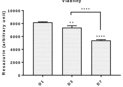
Resazurin values show a decrease corresponding to a reduction in cells metabolic activity in days 1, 4 and 7 (Fig. 1). The referred aspect could be explained by the decrease of free space for cell expansion and for some arbitrary factors. However, this experiment was performed in order to optimize the subsequence experiments and the technical issues associated to cell manipulation.
D1 D3 D7 0 2 0 0 0 4 0 0 0 6 0 0 0 8 0 0 0 1 0 0 0 0 R e s a z u r in ( a r b it r a r y u n it ) V ia b ility * * * * * * * * * *
Fig. 1. Viability of hMSCs at days 1, 3 and 7. Data is expressed as mean ± SEM. **p<0.01, ****p<0.0001
3.2. Optimization experiments with scaffolds
In order to optimize the experiments with the scaffolds and the technical issues associated to cell and scaffolds manipulation, optimization experiments with scaffolds were performed. The values of resazurin assays are not presented due the lack of normalization factors, which could allow the comparison between 2D and 3D results.
3.3. Resazurin reduction assay using non-treated multidishes for suspension cell culture (48 wells-plate)
As shown in Fig. 2, no statistical differences are observed between 2D, CBS and CBSHA in days 1, 3 and 7. This result supports the idea of viability of the cells in CBS and CBSHA.
14 D1 D3 D7 0 2 4 6 V ia b ilit y R e s a z u r in ( u n it s /c e ll ) 2 D C B S C B S H A
Fig. 2. Viability of hMSCs cultured on CBS and CBSHA after 1, 3 and 7 days in culture. Data is expressed as mean ± SEM.
4. Live/Dead assay
4.1. Optimization experiment without scaffolds
The fluorescence images show huge number of live cells with some few dead cells (Fig. 3). This experiment was performed in order to optimize subsequence experiments and technical issues associated to cell manipulation.
4.2. Optimization experiment with scaffolds
The results of live/dead assay images show live and dead cells in the surface of both scaffolds (Fig. 4). However, due the short period of time used for cells adherence to the samples (30 min.) a vast number of cells were found in the bottom of the wells. This experiment allowed the optimization of the live/dead protocol when using CBS and CBSHA.
A B C
15
4.3. Live/Dead assay with scaffolds using non-treated multidishes for suspension cell culture (48 wells-plate)
The resultant images reveal scaffolds fluorescence, which proves unfeasible for the detection of fluorescence of the cells in the surface of the samples (Fig. 5).
A B
C D
A B
Fig. 4. Representative images of Live/Dead with scaffolds, day 1. A: CBS. B: CBSHA. C: Cells in the bottom of the well of CBS. D: Cells in the bottom of the well of CBSHA.
Fig. 5. Representative images of Live/Dead with scaffolds using non-treated multidishes for suspension cell culture, day 1. A: CBS. B: CBSHA.
16
5. Biocompatibility 5.1. Direct contact
The results are presented in viability results (section 3.3.) where no differences were found between conditions.
5.2. Elution method
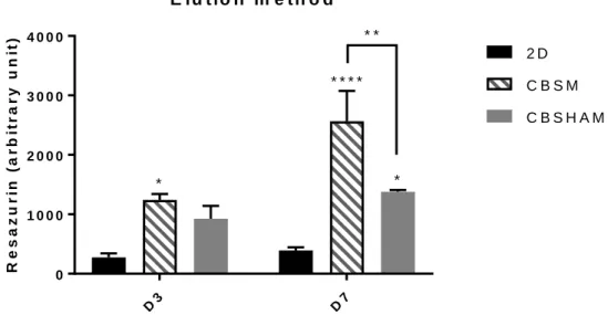
Statistical differences are observed between 2D and CBSM at day 3 with a high resazurin level in CBSM (Fig. 6). Moreover, concerning day 7, these differences are equally significant and are observed between 2D and CBSM, 2D and CBSHAM, CBSM and CBSHAM, with CBSM achieving the highest resazurin value followed by CBSHAM. The results presented seem to show that both, CBSM and CBSHAM, may increase cells metabolic activity.
D3 D7 0 1 0 0 0 2 0 0 0 3 0 0 0 4 0 0 0 E lu t io n m e t h o d R e s a z u r in ( a r b it r a r y u n it ) 2 D C B S M C B S H A M * * * * * * * *
Fig. 6. Elution method - Viability of cells cultured on CBSM and CBSHAM after 3 and 7 days in culture. Data is expressed as mean ± SEM. *p<0.05, **p<0.01, ****p<0.0001
17
6. Cell distribution into the scaffold 6.1. Actin Staining in scaffolds
The attachment of the cells to the scaffolds were observed with the Actin Staining (Fig. 7). The cells cytoskeleton is stained in green and the nuclei is observed stained in blue.
6.2. Actin Staining in scaffold’s sections
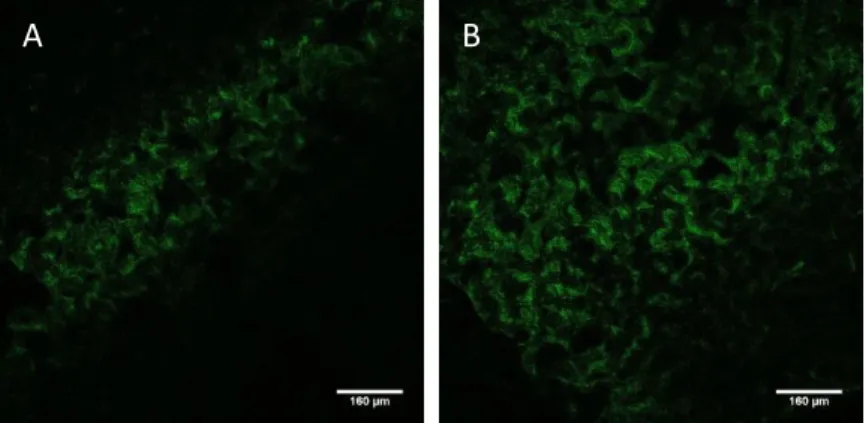
The fluorescence images of day 1 demonstrated hMSCs attached to CBS and CBSHA (Fig. 8). In Fig. 9, cells are attached to the scaffolds at day 7 and cells position in different parallel sheets seems to show cells migration into the samples.
A B C
Fig. 7. Representative images of Actin Staining showing Human mesenchymal stem cells attached to the scaffolds, day 7 (x40 A, x40 B and x10 C)
Fig. 8. Representative images of Actin Staining in scaffold's sections of day 1 (x10 A and x10 B). A: CBSHA. B: CBS. Dashed line: define the approximate limit of the scaffolds.
18
7. Osteoblast differentiation 7.1. ALP activity assay
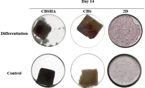
Intensive staining was seen in differentiation wells compared with the controls (Fig. 10 and Fig. 11). When comparing the results at day 7 and day 14, an increase of the staining is presented at day 14. The hMSC differentiation in osteoblast occurred and scaffolds seems to allow this process.
Fig. 10. Representative wells of ALP activity assay at day 7
Fig. 9. Representative images of Actin Staining in scaffold's section of day 7 (x10 A1, cells in detail in A2 and x10 B). A1: CBSHA. A2: CBSHA. B: CBS. Dashed line: define the approximate limit of the scaffolds.
19
Fig. 11. Representative wells of ALP activity assay at day 14
7.2.Alizarin Red Staining
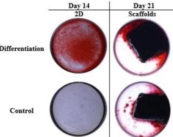
The representative graph of Alizarin Red in 2D wells shown higher values of extracellular calcium deposits in differentiation wells, when compared with control wells (Fig. 12). The referred results proved that the differentiation of hMSCs in osteogenic cells occurred with a significant expression in differentiation wells, in comparison with control wells. The representative images (Fig.13) of this staining revealed a significant difference between 2D wells (differentiation and control). Intensive red stain, showing calcium deposits, is presented in 2D differentiation wells which contrast with the stain in 2D control well. The wells with scaffolds showed a complete stain of the blocks due to its calcium composition. This result does not allow any conclusions about hMSCs differentiation in scaffolds.
20 Fig. 12. Alizarin Red Staining performed in 2D at day 14. Data is expressed as mean ± SEM. *p<0.0007
21
7.3. Van Kossa Staining
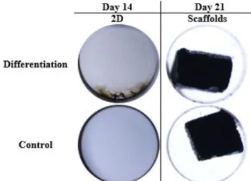
The images obtained from Van Kossa Staining (Fig. 14) were consistent with the results obtained in Alizarin Red Staining. The 2D differentiation wells showed a metallic silver deposit, correspondent to calcium deposits, which proved the osteogenic differentiation of hMSCs. In contrast, in 2D control wells no deposit is visible. The wells with scaffolds showed a complete stain of the blocks with the metallic silver deposit due to its composition of calcium. This result does not allow any conclusions about hMSCs differentiation in scaffolds.
Fig. 14. Representative wells of Van Kossa Staining at days 14 and 21
8. Osteoclast induction 8.1. TRAP Staining
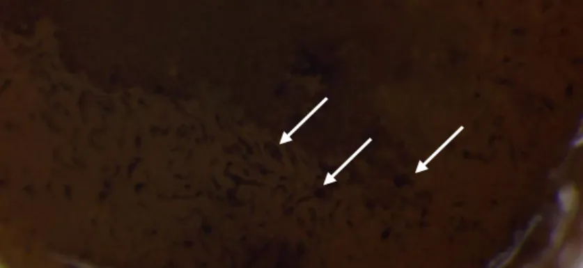
Multinucleated TRAP-positive cells (osteoclasts) are presented in the surface of the scaffold and in the bottom of the wells (Fig. 15 and Fig. 16) suggesting that osteoclastogenesis occurred. However, they are hard to visualize and so additional tests should be performed such as gene expression of specific osteoclastic markers.
Fig. 15. TRAP Staining performed in samples. Arrows are pointing to osteoclasts which are on the surface of the scaffold (A) and in the bottom of the well (B)
22
Fig. 16. TRAP Staining performed on samples. Arrows showing osteoclasts in the surface of the scaffold (image A in detail)
23
DISCUSSION
Bone regeneration and ARP could be achieved through application of several materials, isolated or combined, in bone defects. In what concerns socket filling, the grafting material should present some essential properties: allow the maintenance of the space without affecting the normal healing process, present osteoconduction with formation of dense bone (1-3, 8, 9), which allow stability of the implants, resorption of the material should occur with its substitution for bone, availability, safety and relatively inexpensive (1, 8, 9). We should take in consideration the association between the sequence of the treatment and the resorption rate of the material (1, 2, 9). Slow resorbing materials maintain its presence for a long time and allow new bone formation (3, 7).
CB is formed by two different parts (16, 21). The dorsal shield which is the thick external wall and the other part is the internal lamellar matrix, an extremely porous parallel structure in which the different sheets distance from each other 200-600 𝜇𝑚 (16, 21). Ideal pore size (80 𝜇𝑚 in width and 100 𝜇𝑚 in height) and interconnectivity presented by CBSHA seem to support and promote vascularization and the growth of hard and soft tissues (24, 27). The possibility to obtain HA from CB aragonite through hydrothermal reaction associated to the maintenance of the porous architecture with low cost and availability, has led investigations to its application in bone preserving and regenerating processes (16, 27, 31).
Over the last decades, human activities have been introducing several pollutants in marine ecosystems (16, 40). Heavy metals are an important group of environmental pollutants (40, 41), commonly found in waste water (16, 40). CB presents a strong capacity for bioaccumulation of several contaminants, such as metals, in their tissues (16, 34, 41-44). A high removal capacity for divalent heavy metal ions (such as Pb, Zn, Cu, Cd, Co and Sb) from water is also presented by HA (16, 45-50). Therefore, given the biological origin of HA produced from cuttlebone, whose marine habitat has heavy metals, doubts arise as to the amount of these metals that may be present in the product of the reaction, as well as the risk that these metals may cause when implanting the material in the human bone and its possible migration to the biological medium.
Testing the material in cell culture is essential for the establishment of viability, cytotoxicity, adhesion, proliferation and migration of the cells in scaffolds.
The viability values shown that scaffolds allowed viability of the cells, presenting levels of cell metabolic activity similar to 2D. In literature, viability was analysed using different techniques but the analysis through resazurin assay was not found.
24
Biocompatibility results shown that CBSM and CBSHAM increased cells metabolic activity which could be explained by the fact that if heavy metals are present in the medium, their concentration is considered non-toxic to the cells. However, cellular activity could be stimulated and increased by the presence of some heavy metals and/or calcium dissolved from the CB scaffolds (51). Therefore, more experiments are essential to prove scaffold’s biocompatibility and to quantify the type and the amount of these metals. In literature, no articles were found testing biocompatibility of this material through elution method.
The quantification of heavy metals in the scaffolds before and after HT were performed in recent master’s dissertation (16). Several metals were found with Pb presenting the highest values (over the recommended levels) (16). Cu, Hg and Cd were found in the range of recommended levels but regarding the parenteric administration only Cd is over the stablished values (16). The HT process seems to be favourable to the different values analysed specially in the decrease of Pb levels (16).
Actin Staining proved cells adherence to the scaffolds and the viability of the cells in the scaffolds. The migration of the cells was observed with this staining, showing cells distribution through different sheets of the scaffolds. The referred staining was not performed in other studies with CB.
ALP activity revealed that scaffolds allowed the development of osteogenic differentiation. However, the differences between CBS and CBSHA are not significantly to allow any illation since the results are not quantitative. Quantitative gene expression studies may reveal subtle differences between materials. The results also suggest that the scaffold without OS supplements may promote osteogenic differentiation since a certain degree of ALP activity was observed in control scaffolds.
Hongmin et al. (24) quantified ALP activity and observed an increase of it in CBS and CBSHA during 13 days with 35.4% higher activity on CBSHA than on CBS. Rocha et al. (27, 28) shown that CB provided scaffolds that enhanced osteoblasts viability presenting ALP values similar to the control.
TRAP Staining showed that osteoclastogenesis possibly occurred in the surface of the scaffolds. However, would be interesting to evaluate the osteoclast activity into the scaffolds. Studies with osteoclast applied to CB were not found.
Several studies tested structural and chemical modifications in CB in order to improve cell growth and proliferation, compressive strength (22, 25, 29, 35), reduce the kinetics and the yield of the reaction (33).
25
Some in vivo studies have been described using CB scaffolds.
Hongmin et al. (24) tested CBS and CBSHA in dorsal subcutaneous pockets of mice and observed that blood invasion occurred and new bone was formed in a CBSHA in contrast with CBS in which no bone was formed.
Li et al. (23) implanted CBSHA, with different times of HT, into rabbit femurs. The results showed that biocompatibility and slowly absorption of the scaffolds with new bone formation, due to the osteoblasts infiltration.
Nevertheless, more studies in vivo are necessary before testing the material in Humans. Using a model which could simulate the human oral cavity conditions is required to conclude the effect of this biomaterial in ARP and in bone regeneration.
26
CONCLUSIONS
In this study, CBS and CBSHA obtained from Sepia officinalis exhibit biocompatibility allowing cells adhesion, proliferation and differentiation. The worldwide availability, low cost production and the easy machining to obtain ideal shapes for each demand are some of the few characteristics that make this material so unique. Osteoinductive capacity associated to chemical and structural characteristics of CB appoints it to be an excellent candidate for ARP. Nevertheless, the major disadvantage of CBSHA is its brittleness.
In future works, would be interesting evaluate the material response in osteoblast and osteoclast co-cultures and its application in vivo, with a model, which could simulate the human oral cavity.
27
REFERENCES
1. Block MS. Dental Extractions and Preservation of Space for Implant Placement in Molar Sites. Oral Maxillofac Surg Clin North Am. 2015;27(3):353-62.
2. Jambhekar S, Kernen F, Bidra AS. Clinical and histologic outcomes of socket grafting after flapless tooth extraction: a systematic review of randomized controlled clinical trials. The Journal of prosthetic dentistry. 2015;113(5):371-82.
3. Kassim B, Ivanovski S, Mattheos N. Current perspectives on the role of ridge (socket) preservation procedures in dental implant treatment in the aesthetic zone. Australian dental journal. 2014;59(1):48-56.
4. Vittorini Orgeas G, Clementini M, De Risi V, de Sanctis M. Surgical techniques for alveolar socket preservation: a systematic review. Int J Oral Maxillofac Implants. 2013;28(4):1049-61.
5. Sanz M, Vignoletti F. Key aspects on the use of bone substitutes for bone regeneration of edentulous ridges. Dent Mater. 2015;31(6):640-7.
6. Avila-Ortiz G, Elangovan S, Kramer KW, Blanchette D, Dawson DV. Effect of alveolar ridge preservation after tooth extraction: a systematic review and meta-analysis. J Dent Res. 2014;93(10):950-8.
7. De Risi V, Clementini M, Vittorini G, Mannocci A, De Sanctis M. Alveolar ridge preservation techniques: a systematic review and meta-analysis of histological and histomorphometrical data. Clinical oral implants research. 2015;26(1):50-68.
8. Haggerty CJ, Vogel CT, Fisher GR. Simple bone augmentation for alveolar ridge defects. Oral Maxillofac Surg Clin North Am. 2015;27(2):203-26.
9. Pilipchuk SP, Plonka AB, Monje A, Taut AD, Lanis A, Kang B, et al. Tissue engineering for bone regeneration and osseointegration in the oral cavity. Dent Mater. 2015;31(4):317-38.
10. Chan HL, Lin GH, Fu JH, Wang HL. Alterations in bone quality after socket preservation with grafting materials: a systematic review. Int J Oral Maxillofac Implants. 2013;28(3):710-20.
11. Horowitz R, Holtzclaw D, Rosen PS. A review on alveolar ridge preservation following tooth extraction. The journal of evidence-based dental practice. 2012;12(3 Suppl):149-60.
12. Atieh MA, Alsabeeha NH, Payne AG, Duncan W, Faggion CM, Esposito M. Interventions for replacing missing teeth: alveolar ridge preservation techniques for dental implant site development. The Cochrane database of systematic reviews. 2015(5):CD010176.
13. Horvath A, Mardas N, Mezzomo LA, Needleman IG, Donos N. Alveolar ridge preservation. A systematic review. Clinical oral investigations. 2013;17(2):341-63.
14. Wang RE, Lang NP. Ridge preservation after tooth extraction. Clinical oral implants research. 2012;23 Suppl 6:147-56.
15. Morjaria KR, Wilson R, Palmer RM. Bone healing after tooth extraction with or without an intervention: a systematic review of randomized controlled trials. Clinical implant dentistry and related research. 2014;16(1):1-20.
16. Veiga C. Osso de choco como biomaterial na Medicina Dentária: Faculdade Medicina Dentária da Universidade do Porto; 2016.
17. Masaki C, Nakamoto T, Mukaibo T, Kondo Y, Hosokawa R. Strategies for alveolar ridge reconstruction and preservation for implant therapy. J Prosthodont Res. 2015;59(4):220-8.
18. Avila-Ortiz G, Bartold PM, Giannobile W, Katagiri W, Nares S, Rios H, et al. Biologics and Cell Therapy Tissue Engineering Approaches for the Management of the Edentulous Maxilla: A Systematic Review. Int J Oral Maxillofac Implants. 2016;31 Suppl:s121-64.
19. Clementini M, Tiravia L, De Risi V, Vittorini Orgeas G, Mannocci A, de Sanctis M. Dimensional changes after immediate implant placement with or without simultaneous regenerative procedures: a systematic review and meta-analysis. Journal of clinical periodontology. 2015;42(7):666-77.
28
20. Kim BS, Kang HJ, Yang SS, Lee J. Comparison of in vitro and in vivo bioactivity: cuttlefish-bone-derived hydroxyapatite and synthetic hydroxyapatite granules as a bone graft substitute. Biomedical materials. 2014;9(2):025004.
21. Kim BS, Kim JS, Sung HM, You HK, Lee J. Cellular attachment and osteoblast differentiation of mesenchymal stem cells on natural cuttlefish bone. Journal of biomedical materials research Part A. 2012;100(7):1673-9.
22. Kim BS, Kang HJ, Lee J. Improvement of the compressive strength of a cuttlefish bone-derived porous hydroxyapatite scaffold via polycaprolactone coating. Journal of biomedical materials research Part B, Applied biomaterials. 2013;101(7):1302-9.
23. Li X, Zhao Y, Bing Y, Li Y, Gan N, Guo Z, et al. Biotemplated syntheses of macroporous materials for bone tissue engineering scaffolds and experiments in vitro and vivo. ACS applied materials & interfaces. 2013;5(12):5557-62.
24. Hongmin L, Wei Z, Xingrong Y, Jing W, Wenxin G, Jihong C, et al. Osteoinductive nanohydroxyapatite bone substitute prepared via in situ hydrothermal transformation of cuttlefish bone. Journal of biomedical materials research Part B, Applied biomaterials. 2015;103(4):816-24.
25. Kim BS, Yang SS, Lee J. A polycaprolactone/cuttlefish bone-derived hydroxyapatite composite porous scaffold for bone tissue engineering. Journal of biomedical materials research Part B, Applied biomaterials. 2014;102(5):943-51.
26. Jia X, Qian W, Wu D, Wei D, Xu G, Liu X. Cuttlebone-derived organic matrix as a scaffold for assembly of silver nanoparticles and application of the composite films in surface-enhanced Raman scattering. Colloids and surfaces B, Biointerfaces. 2009;68(2):231-7.
27. Rocha JH, Lemos AF, Agathopoulos S, Kannan S, Valerio P, Ferreira JM. Hydrothermal growth of hydroxyapatite scaffolds from aragonitic cuttlefish bones. Journal of biomedical materials research Part A. 2006;77(1):160-8.
28. Rocha JH, Lemos AF, Agathopoulos S, Valerio P, Kannan S, Oktar FN, et al. Scaffolds for bone restoration from cuttlefish. Bone. 2005;37(6):850-7.
29. Milovac D, Gallego Ferrer G, Ivankovic M, Ivankovic H. PCL-coated hydroxyapatite scaffold derived from cuttlefish bone: morphology, mechanical properties and bioactivity. Materials science & engineering C, Materials for biological applications. 2014;34:437-45.
30. Battistella E, Mele S, Foltran I, Lesci IG, Roveri N, Sabatino P, et al. Cuttlefish bone scaffold for tissue engineering: a novel hydrothermal transformation, chemical-physical, and biological characterization. Journal of applied biomaterials & functional materials. 2012;10(2):99-106.
31. Cadman J, Chang CC, Chen J, Chen Y, Zhou S, Li W, et al. Bioinspired lightweight cellular materials--understanding effects of natural variation on mechanical properties. Materials science & engineering C, Materials for biological applications. 2013;33(6):3146-52.
32. Ivankovic H, Gallego Ferrer G, Tkalcec E, Orlic S, Ivankovic M. Preparation of highly porous hydroxyapatite from cuttlefish bone. Journal of materials science Materials in medicine. 2009;20(5):1039-46.
33. Kannan S, Rocha JH, Agathopoulos S, Ferreira JM. Fluorine-substituted hydroxyapatite scaffolds hydrothermally grown from aragonitic cuttlefish bones. Acta biomaterialia. 2007;3(2):243-9.
34. Kim BS, Yang SS, Yoon JH, Lee J. Enhanced bone regeneration by silicon-substituted hydroxyapatite derived from cuttlefish bone. Clinical oral implants research. 2017;28(1):49-56.
35. Milovac D, Gamboa-Martinez TC, Ivankovic M, Gallego Ferrer G, Ivankovic H. PCL-coated hydroxyapatite scaffold derived from cuttlefish bone: in vitro cell culture studies. Materials science & engineering C, Materials for biological applications. 2014;42:264-72.
36. Ivankovic H, Tkalcec E, Orlic S, Ferrer GG, Schauperl Z. Hydroxyapatite formation from cuttlefish bones: kinetics. Journal of materials science Materials in medicine. 2010;21(10):2711-22.
37. Tkalcec E, Popovic J, Orlic S, Milardovic S, Ivankovic H. Hydrothermal synthesis and thermal evolution of carbonate-fluorhydroxyapatite scaffold from cuttlefish bones. Materials science & engineering C, Materials for biological applications. 2014;42:578-86.
29
38. Mizuno M, Fukunaga K. Analysis of tissue condition based on interaction between inorganic and organic matter in cuttlefish bone. J Biol Phys. 2013;39(1):123-30.
39. Kleinhans C, Schmid FF, Schmid FV, Kluger PJ. Comparison of osteoclastogenesis and resorption activity of human osteoclasts on tissue culture polystyrene and on natural extracellular bone matrix in 2D and 3D. J Biotechnol. 2015;205:101-10.
40. Jaishankar M, Tseten T, Anbalagan N, Mathew BB, Beeregowda KN. Toxicity, mechanism and health effects of some heavy metals. Interdiscip Toxicol. 2014;7(2):60-72.
41. Pereira P, Raimundo J, Vale C, Kadar E. Metal concentrations in digestive gland and mantle of Sepia officinalis from two coastal lagoons of Portugal. Sci Total Environ. 2009;407(3):1080-8.
42. Miramand P, Bustamante P, Bentley D, Koueta N. Variation of heavy metal concentrations (Ag, Cd, Co, Cu, Fe, Pb, V, and Zn) during the life cycle of the common cuttlefish Sepia officinalis. Sci Total Environ. 2006;361(1-3):132-43.
43. Raimundo J, Pereira P, Vale C, Canario J, Gaspar M. Relations between total mercury, methylmercury and selenium in five tissues of Sepia officinalis captured in the south Portuguese coast. Chemosphere. 2014;108:190-6.
44. Le Pabic C, Caplat C, Lehodey JP, Milinkovitch T, Koueta N, Cosson RP, et al. Trace metal concentrations in post-hatching cuttlefish Sepia officinalis and consequences of dissolved zinc exposure. Aquat Toxicol. 2015;159:23-35.
45. Corami A, Mignardi S, Ferrini V. Copper and zinc decontamination from single- and binary-metal solutions using hydroxyapatite. J Hazard Mater. 2007;146(1-2):164-70.
46. Mignardi S, Corami A, Ferrini V. Evaluation of the effectiveness of phosphate treatment for the remediation of mine waste soils contaminated with Cd, Cu, Pb, and Zn. Chemosphere. 2012;86(4):354-60. 47. Zhang Z, Li M, Chen W, Zhu S, Liu N, Zhu L. Immobilization of lead and cadmium from aqueous solution and contaminated sediment using nano-hydroxyapatite. Environ Pollut. 2010;158(2):514-9. 48. Gomez del Rio JA, Morando PJ, Cicerone DS. Natural materials for treatment of industrial effluents: comparative study of the retention of Cd, Zn and Co by calcite and hydroxyapatite. Part I: batch experiments. J Environ Manage. 2004;71(2):169-77.
49. Wang YM, Chen TC, Yeh KJ, Shue MF. Stabilization of an elevated heavy metal contaminated site. J Hazard Mater. 2001;88(1):63-74.
50. del Rio JG, Sanchez P, Morando PJ, Cicerone DS. Retention of Cd, Zn and Co on hydroxyapatite filters. Chemosphere. 2006;64(6):1015-20.
51. Contreras L, Drago I, Zampese E, Pozzan T. Mitochondria: the calcium connection. Biochim Biophys Acta. 2010;1797(6-7):607-18.
30
APPENDICES
Appendix I – Declaração de autorização da Direcção Geral de Alimentação e Veterinária
Exmo.(s) Senhor(es),
Relativamente ao pedido de autorização que nos foi formulado para a recolha, transporte e utilização de subprodutos animais de categoria 3, provenientes de peixarias locais da cidade de Aveiro, nomeadamente, osso/casca de choco para fins específicos de investigação no Instituto Nacional de Engenharia Biomédica da Universidade do Porto, informa-se V.ª Ex.ª, que ao abrigo do disposto no Artigo 17.º do Regulamento (CE) n.º 1069/2009 de 21 de Outubro, pode ser autorizado o manuseamento e utilização de subprodutos animais de categoria 3, destinados a fins de investigação, desde que, para garante do controlo dos riscos para a saúde pública e animal, sejam cumpridas as seguintes condições:
• O operador dos subprodutos animais para diagnóstico e investigação, deve tomar todas as medidas necessárias para evitar a propagação de doenças transmissíveis aos seres humanos ou aos animais durante o manuseamento das matérias sob a sua responsabilidade, sobretudo através da aplicação de boas práticas de laboratório.
• É proibida qualquer utilização subsequente dos subprodutos animais, para outros fins que não o exame no âmbito das atividades autorizadas.
• O transporte até ao destino final deve ser efetuado em embalagem, veículo ou contentor adequado para o efeito e identificado com a menção «Categoria 3 – Destinados à investigação e ao diagnóstico»;
• A menos que sejam conservadas para efeitos de referência, as amostras para diagnóstico e investigação, e quaisquer produtos derivados da utilização dessas amostras, devem ser eliminados:
a) Como resíduos, por incineração ou coincineração;
b) No caso dos subprodutos animais ou produtos derivados referidos no artigo 8.º, alínea a), subalínea iv), no artigo 8.º, alínea c) e alínea d), no artigo 9.º e no artigo 10.º do Regulamento (CE) n.º 1069/2009 que fazem parte de culturas de células, kits de laboratório ou amostras de laboratório, através de um tratamento em condições que são pelo menos equivalentes ao método validado para autoclaves a vapor[1] e subsequente eliminação como resíduos ou águas residuais, em conformidade com a legislação pertinente da União.
[1] CEN TC/102 - Esterilizadores para fins médicos - EN 285:2006 + A2:2009 - Esterilização
– Esterilizadores a vapor – Grandes esterilizadores; referência publicada no JO C 293 de 2.12.2009, p. 39.
c) Por esterilização sob pressão e subsequente eliminação ou utilização, em conformidade com os artigos 12.º, 13.º e 14.º do Regulamento (CE) n.º 1069/2009. • O utilizador deve proceder a um registo datado dos subprodutos animais utilizados, que deve especificar a descrição das matérias, espécie animal, categoria, quantidade, data, local de origem,
31
nome do expedidor, nome do utilizador e método de eliminação das amostras e de quaisquer produtos derivados.
Mais se informa que, nos termos do disposto na alínea a), n.º 1 do Artigo 23.º do Regulamento (CE) n.º 1069/2009 de 21 de Outubro, foi atribuído ao INSTITUTO NACIONAL DE
ENGENHARIA BIOMÉDICA da UNIVERSIDADE DO PORTO, sito na Rua Alfredo Allen,
208, 4200-135 Porto, o número de registo N.12.010.UDER, como utilizador de subprodutos animais de categoria 3 para fins de investigação.
Com os melhores cumprimentos,
José M. Correia
Eng. Téc. Agr.
DGAV – Direção Geral de Alimentação e Veterinária DCCA – Divisão de Controlo da Cadeia Alimentar Quinta do Marquês, Av.ª República, 2780-155 Oeiras Tef. Geral:21 446 40 00 Tef. Secret. 21 446 40 61 Fax: 21 446 40 99 e-mail: jmcorreia@dgav.pt
