Printed in Brazil - ©2003 Sociedade Brasileira de Química 0103 - 5053 $6.00+0.00
A
r
ti
c
le
* e-mail: [email protected]
Cassane Diterpenes from
Plathymenia reticulata
Rosélia de S. Leal, Mary Anne S. Lima and Edilberto R. Silveira*
Departamento de Química Orgânica e Inorgânica, Centro de Ciências, Universidade Federal do Ceará, CP 12200, 60021-970 Fortaleza-CE, Brazil
Do lenho do caule de Plathymenia reticulata (Leguminosae) foram isolados dois novos diterpenos cassânicos: 16,18-diacetoxicass-13(15)-eno e 16-hidroxi-18-acetoxicass-13(15)-eno, além dos conhecidos diterpenos platiterpol, acetato de vinhaticila e vinhaticoato de metila. As estruturas foram determinadas com base em análise espectroscópica, envolvendo comparação com dados da literatura e transformações químicas.
From trunk heartwood of Plathymenia reticulata (Leguminosae) two novel cassane diterpenes 16,18-diacetoxycass-13(15)-ene and 16-hydroxy-18-acetoxycass-13(15)-ene, and the known diterpenes plathyterpol, vinhaticyl acetate and methyl vinhaticoate were isolated. Structure determinations were accomplished by spectroscopic analysis involving comparison with data from literature and chemical transformations.
Keywords: Plathymenia reticulata, Leguminosae, cassane diterpenes, 16,18-diacetoxycass-13(15)-ene, 16-hydroxy-18-acetoxycass-16,18-diacetoxycass-13(15)-ene, 13C NMR data
Introduction
According to Bentham,1 the genus Plathymenia
(Leguminosae) comprises two species: P. reticulata and P. foliolosa. In the course of our continuing studies of the secondary metabolites from the trunk heartwood of P. reticulata, we now report the isolation and a structural elucidation of two novel cassane diterpenes characterized as 16,18-diacetoxycass-13(15)-ene (1) and 16-hidroxy-18-acetoxycass-13(15)-ene (2), and isolation of three previously reported compounds plathyterpol1 (3),
vinhaticyl acetate (4) and methyl vinhaticoate (5).2 The
spectroscopic data of compound 5 and of the derivatives 6 to 10,are described for the first time.
Results and Discussion
Silica gel chromatography of the hexane extract obtained from the trunk heartwood of Plathymenia reticulata, yielded five compounds 1 to 5.
Compound 1, a colorless solid (mp 69-71oC), showed
a [M•+] at m/z 390, compatible with a molecular formula
C24H38O4. The 1H NMR spectrum showed resonances
corresponding to two methyl groups attached to a
quaternary carbon at δ 0.83 (s, 6H), one methyl group attached to a methine carbon at δ 0.93 (d, 3H, J 7.0 Hz) and two other methyl groups as singlets at δ 1.96 (s, 3H) and δ
1.93 (s, 3H), corresponding to two acetyl groups. The IR absorptions at 1720 and 1710 cm-1, characteristic of
carbonyl groups of ester in addition to the C-O stretching at 1230 cm-1, corroborated to identify the presence of two
acetoxy moieties.
methylene hydrogens at δ 4.36 (d, 2H, J 7.0 Hz, C-16), probably attached to a heteroatom, and vicinally coupled to the hydrogen of a trisubstituted double bond at δ 5.33 (t, 1H, J 7.0 Hz, C-15). The presence of an AB system at δ 3.55 (d, 1H, J 11.0 Hz, H-18b) and 3.70 (d, 1H, J 11.0 Hz, H-18a) was indicative of an acetylated carbinolic methylene group adjacent to an asymmetric center (C-4). These observations, in conjunction with the cross-peak correlation observed on the 1H-1H COSY experiment between the hydrogen at
δ 4.36 with the hydrogen at δ 5.33, and the hydrogen at
δ 3.55 with the hydrogen at δ 3.70 respectively, were all consistent with this proposition.
Chemical shifts and comparative analysis of BB and DEPT – 13C NMR (Table 1) spectra revealed 24 lines in
agreement with the suggested molecular formula. From these data one can easily deduce the presence of acetate portions at δ 171.1 and δ 171.3, the unsaturated carbons at
δ 151.8 and δ 113.7, two carbinolic methylenes at δ 60.8 and δ 73.3, and 18 saturated carbon signals (5 methyls, 7 methylenes, 4 methines and 2 quaternaries). The unambiguous assignment of all carbon (Table 1) and hydrogens was possible by the HETCOR spectrum analysis. The correlations of the carbinolic methylenes at
δ 60.8 with the hydrogen at δ 4.36 and at δ 73.3 with the hydrogens at δ 3.55 and δ 3.70, as well as the correlation between the unsaturated methine at δ 113.7 with the hydrogen at δ 5.33 were visualized.
The molecular formula C24H38O4 requires six degrees of unsaturation. Since two carbonyls and a double bond account for three of them, the remaining three unsaturation indicate the tricyclic character of 1.
Based on the above evidences, as well as analysis of analogous substances isolated from this genus,1,2 the
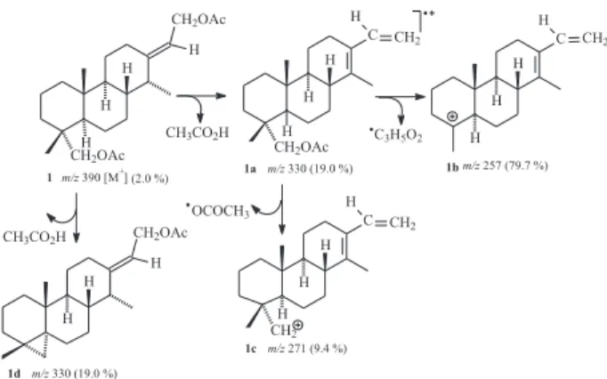
structure of a cassane diterpene does fit the data for compound 1.The observed mass spectrumfragmentation pattern is in agreement with the proposed structure for 1. The fragment at m/z 330 (1a) could be explained by elimination of acetic acid from either the allylic moiety or the carbinolic methylene, while the most stable fragment at m/z 257 (1b) confirmed the diacetylated character of 1 (Scheme 1).
Reduction of 1 withLiAlH4 yielded solid 6 whose IR spectrum showed a broad band at 3400 cm-1 corresponding
to the free hydroxyls, and the absence of carbonyl absorptions at 1720 and 1710 cm-1. The 1H NMR spectrum
of 6 indeed revealed the expected protection of the carbinol methylene of the AB system at δ 3.00 (1H, d, J 11.0 Hz) and 3.20 (1H, d, J 11.0 Hz) and of the allylic methylene at
δ 4.03 (2H, d, J 7.0 Hz). Except for the absence of methyl groups at δ 1.96 and δ 1.93, all other signals of the 1H
NMR spectrum of 6 resembles that of observed for 1. These results, in addition to the biogenetic pattern observed for the major compound, 4,2 of this plant, support the
assignment of structure of 1 as 16,18-diacetoxycass-13 (15)-ene, a new cassane diterpene.
The final structure for 1, including the relative stereochemistry, was based on comparison of the 13C NMR
data (Table 1) of 1 and 4, the later isolated previously from the same source which had its structure established by X-ray crystallography.3 Moreover the chemical shift for
C-18 (δ 73.3) is in agreement with the equatorial orientation of the acetylated oxymethylene (C-18). An axial orientation
Table 1. 13C NMR (75 MHz) spectral data for diterpenes 1, 4, 5 and 8 to 10 in CDCl3
C 1a 4b 5c 8 9d 10e
1 39.2 39.2 35.7 38.9 38.0 36.9
2 18.1 18.1 18.2 18.1 18.0 18.0
3 35.9 35.9 36.9 35.3 35.8 35.0
4 36.8 37.4 47.6 37.7 36.6 47.6
5 49.6 49.5 49.8 48.7 49.0 49.9
6 31.2 31.1 31.0 31.7 30.4 31.5
7 21.7 21.7 24.2 21.7 21.5 24.5
8 40.4 35.7 35.8 38.4 36.9 35.0
9 48.3 45.8 45.8 42.1 45.1 42.3
1 0 36.6 36.6 37.1 36.6 38.9 36.4 1 1 23.8 22.4 22.2 26.5 21.5 26.2 1 2 26.4 149.8 149.8 78.3 106.5 78.1 1 3 151.8 122.6 122.6 35.2 173.6 35.0 1 4 44.1 31.6 31.6 41.9 40.7 41.9 1 5 113.7 109.7 109.7 30.0 113.1 30.0 1 6 60.8 140.5 140.5 66.5 173.6 66.2 1 7 14.3 17.7 17.8 16.7 13.0 11.0 1 8 73.3 73.5 179.0 72.4 73.2 179.3 1 9 17.8 18.0 17.2 18.1 18.0 17.1 2 0 14.9 15.1 14.8 15.0 14.8 14.5
aδ 171.1 and δ 171.3 (H
3C-C=O), δ 21.1 and δ 21.0 (CH3-C=O); bδ 171.6 (CH
3-C=O) and δ 21.2 (CH3-C=O);
c δ 51.9 (OCH 3); d δ 171.5 (H
3C-C=O) and δ 21.0 (CH3-C=O);
eδ 51.7 (OCH 3).
for that moiety would shield C-19 (~δ 65.3) as already observed by Godoy et al. for vouacapenol.4 The chemical
shift differences just for C-8, C-9, C-14 and C-17 in 1 relative to 4 could be explained by a conjunction of effects of the ring strain release after opening of the furan ring, the correspondent disappearance of the γ-effect of the oxygen at C-12 or yet by the “endocyclic homoallyl effect”, alleged by Wenkert et al.5 The suggested 13C NMR data assignment
was done based on the HMQC and HMBC spectral analysis. For example, the methyl group at δH 0.96 (δ 15.1, C-20) did correlate to a quaternary carbon at δ 36.6 (C-10), a methylene at δ 39.2 (C-1), and two methynes at δ 45.8 (C-9) and δ 49.5 (C-5). The methyl group at δH 0.84 (δ 18.0, C-19) correlated to a quaternary carbon at δ 37.4 (C-4), a methylene at δ 35.9 (C-3) and also to the methyne at
δ 49.5 (C-5), and finally the methyl at δH 0.98 (δ 17.2, C-17) correlated to two methynes at δ 35.7 (C-8) and 31.6 (C-14). Table 1 does present all other observed correlations. Compound 2, obtained as a colorless oil, showed [M•+]
at m/z 348 (2.0 %). The IR spectrum revealed the presence of a free hydroxyl group as a broad band at 3400 cm-1, one
carbonyl group at 1715 cm-1and a double bond at 1660
cm-1. The 1H NMR data of 2 was similar to that recorded for
1, except for the presence of only one acetoxy group at δ
2.01 (3H, s). The downfield region in the 1H NMR spectrum
was crowded as in the spectrum of compound 1, except for the absorption at δ 3.93 (2H, d, J 7.0 Hz), corresponding to the allylic methylene H-16 attached to a hydroxyl group, as observed previously in 6.
These results allowed to establish 2 as the 16-hydroxy-18-acetoxycass-13(15)-ene, a new cassane diterpene which contains an allylic alcohol and an acetylated carbinolic methylene group attached to an asymmetric center. In order to confirm the structure proposal, compound 2 was acetylated with acetic anhydride and pyridine to yield a product identical to 1. Reduction of 2 with LiAlH4 yielded also the diol 6, already obtained from 1, confirming the presence of the monoacetate group.
The known diterpenes previously described for the same plant species, plathyterpol (3), vinhaticyl acetate (4), and the major constituent methyl vinhaticoate (5),2 whose
pharmacological investigations have demonstrated its spasmolitic effect,3 were also isolated. Their physical and
spectral data were in agreement with those reported in the literature,1-4 and the 13C NMR data of compound 5 are
described for the first time (Table 1).
Pursuing the preparation of new cassane derivatives, compounds 7 to 10 were also prepared for biological tests purposes and for spectral analysis (Table 1).
Hydrogenation of 4 yielded compounds 7 and 8, that were separated by SiO2 chromatography. The less polar
product obtained as an yellow oil, showed IR absorption at 1710 cm-1 characteristic of carbonyl group of ester
moiety and the disappearance of the absorptions related to the furan ring, in comparison with 4. This information was confirmed by 1H NMR analysis by absence of the two
doublets in the downfield region characteristic of the disubstituted furan ring. The presence of fragments at m/z 97 (7a) and m/z 165 (7b) in the mass spectrum does agree with compound 7 as the tetrahydrogenated derivate (Scheme 2). The more polar product obtained as a colorless solid, mp 124-125 oC, revealed the presence of a free
hydroxyl group as a broad band at 3400 cm-1 and the
absence of signals corresponding to the carbonyl group and furan ring in the IR spectrum when compared with 4. This information in conjunction with the absence of one acetoxy group at δ 2.0 and the presence of additional signals in the saturated region, in the 1H NMR spectrum,
led to the structure of compound 8 as the tetrahydrogenated alcohol derivative of vinhaticyl acetate 4.
Oxidation of compound 4 with Jones’ reagent yielded a colorless solid, mp 181-184 oC. The IR spectrum showed a
free hydroxyl at 3330 cm–1, two carbonyl groups at 1730
and 1715 cm-1, and one trisubstituted double bond at
1640 cm-1. The 13C NMR data at δ 173.6 and δ 113.1, and
δ 5.56 in the 1H NMR, and yet a representative resonance
related to the non hydrogenated hemiketal carbon at δ 106.5, confirmed a γ-hydroxy-γ-butenolide moiety instead of a furan ring as in 4. All these inferences were supported by the chemical shifts and comparative analysis with resonance data of active lactols derivatives from furan cassane diterpenes reported in the literature.6-8 Therefore, the structure
of this product was determined to be the lactol 9.
Upon hydrogenation, compound 5 yielded a colorless solid, mp 74-76 oC. The absence of signals in the lowfield
region in 1H NMR spectrum confirmed the reduction of
the furan ring when compared with 5. The formation of a tetrahydrofuran derivative was confirmed through the 13C
NMR spectrum where was observed the absence of unsaturated carbon signals and the appearance of four additional signals in the region of saturated carbons: two methines at δ 78.1 (C-12) and δ 35.0 (C-13) and two methylenes at δ 30.0 (C-15) and δ 66.2 (C-16). Thus, the product must be the tetrahydrofuran derivative of methyl vinhaticoate, 10.
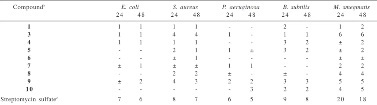
Compounds 2 to 10 were submitted to antibacterial and antifungal tests according to the general qualitative antimicrobial activity bioassay described by Hufford et al.,9 as modified by Clark et al.10 A standard antibacterial
agent, streptomycin sulfate, and a standard antifungal agent, amphotericin B, were included in each assay as positive controls. All of them showed only weak antimicrobial activity (Table 2 and 3).
Experimental
General procedures
Melting points were obtained on a Mettler PF-5. IR spectra were recorded using a Perkin Elmer 720 or 1320 spectrometers. The mass spectra were obtained on a HP 5995-A mass spectrometer by electron impact ionization (70 eV). 1H NMR spectra at 60 MHz where recorded on a
Varian EM-360 while the 1H NMR spectra at 300 MHz and 13C NMR spectra at 75 MHz were recorded on a Varian
XL-300 spectrometer; chemical shifts were given in δ
(ppm) relative to TMS as an internal standard.
Plant material
Trunk heartwood of Plathymenia reticulata was collected in Crato, Ceará and the plant material was
Table 2. Antibacterial activitya of compounds 1, 3 to 10
Compoundb E. coli S. aureus P. aeruginosa B. subtilis M. smegmatis
2 4 4 8 2 4 4 8 2 4 4 8 2 4 4 8 2 4 4 8
1 1 1 1 1 - - 2 - 1 2
3 1 1 4 4 1 - 1 1 6 6
4 1 1 1 1 - - 3 2 ± 2
5 - - 2 1 1 ± 3 2 ± 2
6 - - ± 1 - - - - ± ±
7 ± 1 ± ± 1 1 - - 2 2
8 - - 2 2 ± - ± - 4 4
9 ± 2 4 3 2 2 3 3 5 5
1 0 - - - 3 2 2 4 5
Streptomycin sulfatec 7 6 8 7 6 5 9 8 2 0 1 8
a Antibacterial activity was recorded as the width of the inhibition zone measured from the edge of the agar well to the edge of the inhibition zone after 24 h and 48 h of incubation using the following code: (-): no activity; (±) questionable; (numerals): average radius, in millimeters, of definite inhibition; b Compounds were prepared in a solution 1 mg mL-1 EtOH; c 1mg mL-1 25% EtOH.
Table 3. Antifungal activitya of compounds 1, 3 to 10
Compoundb S. cerevisiae C. neoformans A. flavus T. mentagrophytes
4 8 7 2 4 8 7 2 4 8 7 2 4 8 7 2
1 2 2 - - - - 1 1
3 2 2 5 3 ± - 4 3
4 2 2 ± - - - 1 1
5 3 3 - - - - 2 1
6 1 1 - - - - 1 1
7 3 3 ± ± - - 4 3
8 3 3 3 3 - - 3 2
9 3 3 ± - - - 3 2
1 0 2 2 1 1 - - 1 2
Amphotericin B c 1 1 8 1 3 1 1 5 4 9 7
identified by Dr. Afrânio G. Fernandes (Universidade Federal do Ceará). A voucher specimen number 9014 has been deposited in the Herbário Prisco Bezerra of the Departamento de Biologia, Universidade Federal do Ceará, Brazil.
Extraction and isolation of the constituents
The air dried heartwood (4.0 kg) was powdered and extracted with hexane (3 x 12.0 L) at room temperature. The solvent was removed under reduced pressure yielding a viscous yellow oil (130.0 g). Part of the hexane extract (100.0 g) was coarsely partitioned on silica gel (400.0 g) column chromatography and eluted with hexane, CHCl3 and EtOAc. The fractions obtained were combined according to the TLC analysis and the less polar fractions, F1-5, F-8, F-10 and F-11, showed to be of further interest. Filtration under vacuum of the F1-5 fraction yielded compound 4 (5.6 g) as a pale yellow solid. Upon re-chromatography of the filtrate on silica gel using hexane, compound 5 (0.3 g) was obtained as a pale yellow solid.
Fraction F-8 (19.1 g) was re-chromatographed on silica gel (20.0 g), using hexane-CHCl3, CHCl3 and EtOAc as eluting solvents. The fractions obtained were combined according to the TLC analysis and from F-8 (3-23) fraction the compound 3 (1.56g) was isolated as a colorless oil.
After successive chromatography on silica gel (75.0 g) of fraction F-10 (4.1 g), and elution with hexane, CHCl3 and AcOEt, the fractions obtained were combined according to the TLC analysis. Compound 1 (0.3 g) was obtained from fraction F-10 (38-61) (0.86 g) as a colorless solid.
16,18-diacetoxycass-13(15)-ene (1). Colorless solid; mp 69-71 oC; IR ν
max/cm
-1 2900, 2840, 1720, 1710, 1660,
1440, 1360, 1230, 1215, 1020, 785, 760 (KBr); 1H NMR
(300 MHz, CDCl3) δ 5.33 (t, J 7.0 Hz,1H), 4.36 (d, J 7.0 Hz, 2H), 3.70 (d, J 11 Hz, 1H), 3.55 (d, J 11.0 Hz, 1H), 1.96 (s, 3H), 1.93 (s, 3H), 0.93 (d, J 7.0Hz, 3H), 0.83 (s, 6H); 13C
NMR (CDCl3, 75 MHz): see Table 1; GC/EIMS (70 eV) m/z 390 (M•+, 2 %), 330 (19), 257 (79), 255 (19), 175 (28),
161 (35), 159 (19), 148 (23), 135 (62), 133 (62), 131 (20), 123 (27), 121 (44), 119 (35), 109 (44), 107 (78), 105 (53), 97 (21), 95 (52), 93 (77), 91 (60), 81 (100), 79 (78), 69 (43), 67 (69), 55 (81), 53 (22).
Fraction F-11 (21.63 g), was re-chromatographed on silica gel (100.0 g) using hexane-CHCl3, CHCl3 and CHCl3 -EtOAc as eluting solvents. After combination of the fractions according to the TLC analysis, compound 2 (0.8 g) was obtained from fraction F-11 (22-76) (5.7 g), as a colorless oil.
16-hydroxy-18-acetoxycass-13(15)-ene (2). Yellow oil;
IR νmax/cm-1: 3400, 2920, 2860, 1715, 1660, 1460, 1440,
1380, 1235, 1030, 795 (film); 1H NMR (60 MHz, CDCl 3):
5.23 ( t, J 7.0 Hz, 1H), 3.93 (d, J 7.0 Hz, 2H), 3.66 (ABq, J 11.0 Hz, 2H), 2.01 (s, 3H), 1.16 (d, J 7.0 Hz, 3H), 0.86 (s, 6H); GC/EIMS (70 eV) m/z 348 (M•+ 2.0 %), 346 (23), 328
(26), 253 (50), 251 (35), 221 (22), 189 (26), 187 (58), 175 (49), 173 (37), 171 (26), 161 (59), 159 (53), 148 (23), 147 (64), 135 (62), 133 (78), 132 (78), 131 (63), 121 (87), 120 (58), 119 (78), 117 (25), 109 (75), 108 (41), 107 (89), 105 (51), 85 (91), 93 (58), 81 (100), 60 (93).
Reduction of 1. LiAlH4 (0.03 g) was added to a solution of 1 (0.1 g) in dry Et2O (2.0 mL) under stirring at room temperature for 3 h. The mixture was treated following usual work-up and the reaction products were chromatographed on a silica gel column using hexane/ CHCl3 (1:1) as eluting solvents, to afford 6 as colorless needles (0.056 g): mp 122-125 oC; IR ν
max/cm
-1: 3400,
2970, 2890, 1660, 1480, 1400, 1060 (film); 1H NMR (60
MHz, CDCl3): 5.33 (t, J 7.0 Hz, 1H), 4.03 (d, J 7.0 Hz, 2H), 3.20 (d, J 11.0 Hz, 1H), 3.00 (d, J 11.0 Hz, 1H), 0.93 (d, J 7.0 Hz, 3H), 0.83 (s, 3H), 0.77 (s, 3H).
Acetylation of 2. Compound 2 (0.3 g) was dissolved with pyridine (2.0 mL) and Ac2O (2.0 mL) and stirred at room temperature overnight. Subsequent workup afforded a colorless solid (0.32 g) that was chromatographed using hexane/EtOAc (3:1) as eluent. The acetylated product was identified by direct comparison (mp, 1H NMR) with
16,18-diacetoxycass-13(15)- ene (1).
Reduction of2. Compound 2 (0.8 g) was dissolved in dry Et2O (10.0 mL), LiAlH4 (0.3 g) was added to a solution and the mixture was stirred at room temperature for 3 h. After usual workup, the reaction products were chroma-tographed on a silica gel column using hexane/EtOAc (3:1) as eluting solvents, to afford 6 (0.62 g).This compound was characterized after direct comparison (mp, TLC co-chromatography and 1H NMR) with those obtained
previously.
Hydrogenation of4. Compound 4 (0.1 g) was dissolved in MeOH (10.0 mL) and added to a suspension containing Pd/C (0.015 g), that has been saturated with H2. The mixture was stirred at room temperature during overnight. Workup in the usual way afforded a residue (0.09 g) containing two products that were separated by SiO2 chromatography using hexane/CHCl3 (3:1) as eluting solvents. The less polar product, compound 7 (36.0 mg), was obtained as a pale yellowish oil. IR νmax/cm-1: 2880, 2800, 1710, 1440,
1360, 1220, 1080, 1040 (film); 1H NMR (60 MHz, CDCl 3):
3.66 (q, J 11.0 Hz, 2H), 2.03 (s, 3H), 0.90 (d, J 7.0 Hz, 3H), 0.90 (s, 6H); GC/EIMS (70 eV) m/z (M•+, absent), 275 (42),
(64), 107 (65), 97 (100), 95 (82), 93 (65), 55 (95). The more polar product, compound 8 (45.0 mg), was obtained as a colorless solid: mp 124-125 oC; IR ν
max/cm
-1: 3390, 2900,
2890, 1480, 1470, 1370 1080 (KBr); 1H NMR (60 MHz,
CDCl3): 3.53 (m,2H), 3.03 (q, 2H, J 11.0 Hz), 0.83 (d, 3H, J 11.0 Hz), 0.83 (s, 3H), 0.72 (s, 3H); 13C NMR (75 MHz): see
Table 1; GC/EIMS (70 eV) m/z 306 (M+, 9.3%), 275 (65),
193 (35), 175 (29),161 (31), 149 (81), 147 (34), 121 (42), 97 (100), 79 (36), 69 (53), 55 (62).
Oxidation of 4. 5.0 mL of Jones’ reagent was added slowly to a solution of 4 (0.5 g) in acetone (10.0 mL). The mixture was stirred at 0 oC during 30 minutes. Usual workup
and SiO2 column chromatography using hexane/CHCl3 (2:1), yielded compound 9 (0.2 g) as a colorless solid:mp 181-183 oC; IR ν
max/cm
-1: 3330, 2940, 2900, 1730, 1715,
1640, 1430, 1220, 1120 (KBr); 1H NMR (60 MHz, CDCl 3):
5.56 (s, 1H), 3.66 (q, 2H, J 11.0 Hz), 2.50 (t, 2H, J 10.0 Hz), 2.10 (s, 3H), 1.15 (d, 3H, J 7.0 Hz), 0.86 (s, 6H); 13C NMR
(75 MHz): see Table 1; GC/EIMS (70 eV) m/z: (M•+, absent),
189 (20), 162 (22), 160 (100), 149 (29), 121 (27), 109 (43), 95 (60), 93 (41), 91 (50), 81 (78), 79 (45), 69 (3), 53 (32). Hydrogenation of 5. Compound 5 (0.47 g) was dissolved in MeOH and added to a suspension containing Pd/C (0.05 g) in MeOH (30.0 mL), that was previously saturated with H2. The mixture was stirred at room temperature during overnight. Usual work up and SiO2 column chromatography using hexane/CHCl3 1:1 yielded compound 10 (0.35 g) as a colorless solid: mp 74-76 oC;
IR νmax/cm-1: 2870, 2850, 1715, 1450, 1395, 1260, 1150
(KBr); 1H NMR (60 MHz, CDCl
3): 3.63 (s, 3H), 1.20 (s,
3H), 0.86 (d, J 7.0 Hz, 3H), 0.88 (s, 3H); 13C NMR (75
MHz): see Table 1.
Acknowledgements
The autors are grateful to Dr. Francisco J. A. Matos and Dr. Afranio G. Fernandes (Universidade Federal do Ceará) for plant collection and identification and to Dr. J. D. McChesney and Dr. C. D. Hufford (University of Mississipi-USA) for the antimicrobial tests and 300/75 MHz NMR, data respectively. This work was financially supported by grants from CNPq/CAPES/FINEP/PADCT/FUNCAP/ PRONEX.
References
1. King, T.J.; Rodrigo, S.; Chem. Commun. 1967, 575. 2. Matos, F.J.A.; Craveiro, A.A., Maurera, M.A.M.A.; J. Nat.
Prod. 1984, 47, 581.
3. Maurera, M.A.M.A.; Matos, F.J.A.; Craveiro, A.A.; Proc.Anais do VI Simpósio de Plantas Medicinais do Brasil, Fortaleza, Brazil, 1980.
4. Godoy, R. L.; Lima, P. D. D. P.; Pinto, A. C.; Aquino Neto, F. R.; Phytochemistry1989, 28, 642.
5. Wenkert, E.; Cochran, D. W.; Hagaman, E. W.; Schell, F. M.; Neuss, N.; Katner, A. S.; Potier, P.; Kan, C.; Plat, M.; Koch, M.; Mehri, H.; Poisson, J.; Kunesch, N.; Rolland, Y.; J. Am. Chem. Soc. 1973,95, 4990.
6. Fang, J.; Lang, C.; Chen, W.; Cheng, Y.; Phytochemistry1991,
30, 2793.
7. Ahmed, M.; Ahmed, A. A.; Phytochemistry1990, 29, 2715. 8. Mahajan, J. R.; Monteiro, M. B.; J. Chem. Soc., Perkin Trans I
1973, 520.
9. Hufford, C. D.; Funderburk, M. J.; Robertson, L. W.; J. Pharm. Sci.1975, 64, 789.
10. Clark, A. M.; El-Feraly, F. S.; Li, W., S.; J. Pharm. Sci 1981,
70, 951.
Received: December 3, 2001