1 Physical Therapy Department, Universidade de Pernambuco (UFP), Petrolina, PE, Brazil
2 Departament of Public Health, Escola de Enfermagem de Ribeirão Preto, Universidade de São Paulo (USP), Ribeirão Preto, SP, Brazil Received: 04/01/2013 Revised: 07/31/2013 Accepted: 09/01/2013
a r t i c l e
Low and high-frequency TENS in post-episiotomy pain
relief: a randomized, double-blind clinical trial
Ana C. R. Pitangui1, Rodrigo C. Araújo1, Michelle J. S. Bezerra1,
Camila O. Ribeiro1, Ana M. S. Nakano2
ABSTRACT | Objective: To evaluate the effectiveness of low-frequency TENS (LFT) and high-frequency TENS (HFT) in post-episiotomy pain relief. Method: A randomized, controlled, double-blind clinical trial with placebo composed of 33 puerperae with post-episiotomy pain.TENS was applied for 30 minutes to groups: HFT (100 Hz; 100 µs), LFT (5 Hz; 100 µs),and placebo (PT). Four electrodes were placed inparallel near the episiotomy and four pain evaluations were
performed with the numeric rating scale. The irst and the second evaluation took place before TENS application and
immediately after its removal and were done in the resting position and in the activities of sitting and ambulating. The
third and fourth evaluation took place 30 and 60 minutes after TENS removal, only in the resting position. Intragroup differences were veriied using the Friedman and Wilcoxon tests, and the intergroup analysis employed the Kruskal-Wallis test.Results: In the intragroup analysis, there was no signiicant difference in the PT during rest, sitting, and
ambulation (P>0.05). In the HFT and LFT, a signiicant difference was observed in all activities (P<0.001). In the intergroup analysis, there was a signiicant difference in the resting position in the HFT and LFT (P<0.001). In the sitting activity, a signiicant difference was veriied in the second evaluation in the HFT and LFT (P<0.008). No signiicant difference was veriied among the groups in ambulation (P<0.20). Conclusions: LFT and HFT are an effective resource that may be included in the routine of maternity wards.
Keywords: physical therapy;vaginal delivery;postpartum period; transcutaneous electrical nerve stimulation; perineal pain.
Registration Australian New Zealand Clinical Trials Registry: ACTRN12610000529044.
HOW TO CITE THIS ARTICLE
Pitangui ACR, Araújo RC, Bezerra MJS, Ribeiro CO, Nakano AMS. Low and high-frequency TENS in post-episiotomy pain relief: a randomized, double-blind clinical trial. Braz J Phys Ther. 2014 Jan-Feb; 18(1):72-78. http://dx.doi.org/10.1590/ S1413-35552012005000143
Introduction
Transcutaneous electrical nerve stimulation (TENS) consists of a non-invasive, easily handled, safe and low-cost resource that sends electrical
impulses through the skin1-3. It has typically biphasic
waves containing positive and negative phases, which may be either symmetric or asymmetric4,
with the main purpose of relieving pain1-5. Although
its mechanism of electroanalgesia production is still
controversial, its effectiveness would be explained by
the gate control theory of pain and by the activation of a system of endogenous opioids1-3,6,7.
Perineal pain is manifested in the postpartum period mostly due to tissue lesions that may occur spontaneously (lacerations) or due to the surgical incision (episiotomy)8. Episiotomy is a lesion resulting
from a surgical cut to the perineum with scissors or scalpel to help deliver the baby and avoid severe
tears that can be dificult to repair. Nevertheless,
there is evidence to recommend restricted rather than routine use of episiotomy because the restrictive policy appears to have more benefits9. Women
submitted to episiotomy have a greater prevalence of pain complaints10-13 and have dificulty performing
functional activities13,14. However, despite the
recommendations for restrictive use of episiotomy, there is still a high prevalence of this procedure in
Brazilian maternities, with rates of up to 60.7%15,
a statistic that supports the development of studies that analyze the techniques that reduce the morbidity caused by this practice.
In the literature, it is possible to verify the use
of several pharmacologic and non-pharmacologic resources aimed at reducing pain from perineal trauma11,14. The most common resources in obstetric
practice include non-hormonal anti-inlammatory
oral8 and rectal16 analgesics, and non-pharmacologic
resources, such as cryoanalgesia11,17.
Despite the existence of clinical investigations
regarding these therapeutic practices in perineal pain14, there is still a lack of methodological quality
and several gaps in the studies regarding their effectiveness. Therefore, new trials are necessary so that decisions can be made regarding the real effectiveness of these modalities. Evaluating the effect of high and low-frequency TENS in post-episiotomy pain relief of puerperae is a great advance
for studies in this area and it is justiied by the need
to broaden the usable resources and behaviors in puerperal care.
Method
This was a randomized, controlled, double-blind clinical trial with placebo carried out with puerperae submitted to post-episiotomy vaginal delivery at a public hospital in Petrolina, Pernambuco, Brazil. Data
were collected between August 2009 and July 2010.
The puerperae were included according to the
following criteria: (1) low-risk pregnancy; (2) age above 15 years; (3) ability to read, write, and speak in Portuguese; (4) awareness of time and space; (5) between six and 24 hours post-vaginal delivery; (6)
midline or mediolateral episiotomy with stitches;
(7) post-episiotomy pain; (8) absence of any
genitourinary pathology.
The participants who were excluded presented: (1) obesity (Body Mass Index [BMI] ≥ 30 kg/m2); (2)
puerperal complications; (3) instrumental delivery
(use of forceps); (4) perineal lacerations, (5) epidural anesthesia; (6) use of analgesic resources during data
collection.
This study was approved by the Research Ethics
Committee of Universidade de Pernambuco (CEP/
UPE), Petrolina, PE, Brazil, under protocol number
145/09. All participants and legal guardians, in case of participants under 18 years of age, voluntarily
signed an informed consent form.
A pilot test with 12 patients was carried out to
verify the comprehension and effectiveness of the data collection instruments and to calculate sample size. Reduction in the pain scores post-intervention was applied as a parameter, considering clinically
relevant a reduction of 1.39 points in the numeric
rating scale (NRS)18-22. Sample size was estimated
through simple sample equation, with power of
80% and standard deviation of 1.0, which required a
sample size of nine volunteers in each group.
The participants were randomized into groups according to a spreadsheet generated in a computer program by a researcher who was not involved in the selection of participants. Randomization occurred in the order in which each patient was enrolled in the
study. It was established that the puerperae should be within six to 24 hours post-vaginal delivery. The minimum limit of six hours post-delivery was
determined because this is the period recommended
for women to leave their bed, whereas the 24 hours is related to the acute phase of the lesion and the peak of the inlammatory process12. In the case of medication
use and based on the drug dose, the waiting time was counted from the time when the last dose of
analgesic/anesthesia was administered, considering
the possible interferences that could cause biases in the initial pain assessment.
The main researcher trained two examiners, one responsible for the pain assessments and for illing
out evaluation forms and the other responsible
for applying TENS. The device KINESIS New Microcontrolled (KW Eletrônica Ltda., Amparo, SP,
Brazil) was used with two pairs of silicone-carbon electrodes (5.5cm × 3cm), hypoallergenic conductive gel, and hypoallergenic microporous surgical tapes
(25 mm × 10 m). The device was calibrated before
data collection.
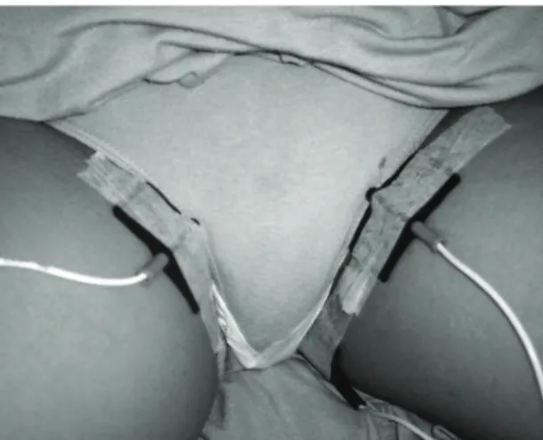
TENS was applied to the three study groups: high-frequency TENS (HFT), low-frequency TENS (LFT), and placebo TENS (PT). The electrodes were placed in parallel, near the episiotomy, in the region of the pudendal and genitofemoral nerves, both responsible for the perineal area (Figure 1).
The HFT group received frequency of 100 Hz and 100 µs pulse, and the LFT group received frequency of 5 Hz and 100 µs pulse for 30 minutes. The electrical impulse intensitywas controlled by the participants and adjusted when necessary. They were instructed that the sensation of strong and tolerable pulses should remain, although it should be sensorially comfortable.At the end of the application, the researcher recorded the intensity employed by each participant.
The participants of the PT group had their electrodes placed similarly to the HFT and LFT groups, and although the device remained on for 30
minutes with the light on to simulate it was working,
it did not send any electrical stimulation. At the end of the study, due to ethical reasons, the participants in the PT group were offered HFT to treat the pain. HFT was chosen because it has already been used in a previous study14 with effective results.
analyses, Kruskal-Wallis’s test was employed. The
level of signiicance adopted was p≤0.05.
Results
Out of the 50 patients evaluated for eligibility
in this study, 16 were excluded because they
did not meet the inclusion criteria, and one was
excluded from randomization, totaling 33 evaluated puerperae (Figure 2). The three groups were
homogenous as to sociodemographic variables:
age (P=0.10), marital status (P=0.41), ethnicity (P=0.37), education (P=0.10) and occupation (P=0.20); obstetric and labor variables: prenatal
care (P=1.00), number of appointments attended
(P=0.47), number of pregnancies (P=0.51), deliveries (P=0.62), miscarriages (P=0.89), gestational age (P=0.56), type of episiotomy (P=0.34); and neonatal variables: gender (P=0.31), weight (P=0.09), height (P=0.20) and Apgar score in the irst minute (P=0.21) and the fifth minute (P=0.08). Among
the pain relief resources used by the participants, non-pharmacological therapeutic methods were not
observed, but oral dipyrone was taken by 65.6% (n=21) of the participants (P=0.64).
The initial mean of pain intensity was similar in the
three groups. In the intragroup analysis, there was no signiicant difference in the PT group regarding pain
intensity in the resting position and in the activities of sitting and ambulating in any evaluation (P>0.05).
In the HFT and LFT groups, in the resting position, a signiicant difference was observed between the irst
and all following evaluations, and between the second
and the third and fourth evaluations (P<0.001). There was also a signiicant difference in the sitting and ambulating activities (P<0.001).
In the results of the intergroup analysis, there was a signiicant difference in the resting position
in the comparison among the groups in the second
(P<0.046), third (P<0.001), and fourth (P<0.001)
evaluations, evidencing the reduction in pain scores
in the HFT and LFT groups. In the sitting activity, a signiicant difference was veriied in the second
evaluation, indicating reduction in the pain scores of the HFT and LFT groups when compared to the
PT group (P<0.008). No signiicant difference was veriied among the three groups while ambulating (P<0.20; Table 1).The amplitude in the HFT group
was 21.77±2.11 mA with variation between 19 and 26 mA, whereas in the LFT group it was 24.08±2.55 mA with variation between 21 and 30 mA.
In the HFT and LFT groups, 100% of the
puerperae referred to TENS as comfortable and Figure 1. Schematic representation of the positioning of the
electrodes.
history, labor and newborn data. Afterwards, four pain assessments were performed. The initial
evaluation took place prior to the use of TENS. The
participants were questioned regarding the pain from episiotomy and those that mentioned the presence of pain answered the NRS (in the resting position) regarding the movements of sitting and ambulating. At the end of this stage, TENS was applied to the three groups.
The second evaluation began immediately after the removal of TENS. The NRS was applied once again (in the resting position) regarding the sitting and ambulating movements. A questionnaire was applied to the three groups regarding the TENS, with the following questions: (1) is TENS comfortable or
uncomfortable?; (2) would you use it again?; (3) were you dissatisied, slightly satisied, satisied or very satisied?
The third evaluation was performed 30 minutes after removing TENS, and the fourth evaluation was
performed 60 minutes after removing TENS. These
evaluations measured pain through the NRS only in the resting position.
Data were analyzed in the program SPSS version
16.0. The Kruskal-Wallis test and Dunn’s post-test
were used for the following continuous variables: age, number of pregnancies, deliveries, abortions, number of prenatal appointments, gestational age,
weight, height, and newborn’s Apgar score. The
chi-square test was used for the analysis of the categorical variables: marital status, ethnicity,
education, occupation, prenatal care, pudendal block,
episiotomy, gender, use of pain relief resources, type
Figure 2. Flow diagram of the included patients.
Table 1. Post-episiotomy pain intensity.
Mean (SD)
Group HFT (n = 11) Group LFT (n = 12) Group PT (n = 10) P-value
First evaluation
Rest 4.54±2.38a,b,c 4.50±2.02a,b,c 4.11±1.69 .940
Sitting 6.81±1.60a 6.08±1.88a 6.77±1.78 .504
Ambulating 6.18±2.31a 5.66±2.57a 5.44±1.87 .695
Second evaluation
Rest 1.72±2.19d,e,f 2.25±1.60d,e,f 3.88±2.08 .046
Sitting 3.18±2.04f 3.75±1.65f 6.44±2.18 .008
Ambulating 3.36±2.33 3.83±2.16 5.22±2.16 .208
Third evaluation
Rest 0.81±1.66f 1.66±1.43f 4.44±2.18 .001
Fourth evaluation
Rest 0. 27±0.64f 1.16±1.46f 4.11±2.20 .001
P<0.05. Data are reported as mean (SD). aIntragroup difference between the irst and second evaluation. bIntragroup difference between the irst
stated they would use it again, whereas in the PT
group, the answers were 89% (n=8) and 56% (n=5),
respectively. Regarding the satisfaction with the use
of TENS, 45% (n=5) of the participants in the HFT group stated they were satisied and 45% (n=5) very satisied, whereas in the LFT group 75% (n=9) were satisied and 17% (n=2) very satisied. As for the PT group, 45% (n=3) mentioned they were satisied and 11% (n=1) were very satisied.
Discussion
These indings suggest that both HFT and LFT
cause clinically relevant reduction in pain intensity immediately after its application, with a residual effect lasting for one hour after use. TENS in the HFT and LFT groups reduced the intensity of initial pain scores, measured as moderate in both groups,
and later as weak.
In the four evaluations performed in the resting
position, in the HFT group, there was pain reduction
measured by the NRS (4.54/10; to 1.72/10; to 0.81/10; to 0.27/10), and this pattern was also observed in the LFT group (4.50/10; to 2.25/10; to 1.66/10; to 1.16/10). In the sitting activity, the pain intensity scores decreased from the irst to the second evaluation in the HFT group (6.81/10 to 3.18/10) and in the LFT group (6.08/10 to 3.75/10).
Similar data were observed in the ambulating activity, evidencing the reduction in pain scores
in both groups, HFT (6.18/10 to 3.36/10) and LFT (5.66/10 to 3.83/10). No signiicant difference was veriied in the pain score of the PT group in any
evaluation.
Therefore, it is clear that both HFT and LFT reduced the levels of pain in the resting position.
In both groups that employed TENS, there was
a clinically relevant difference in the intragroup analysis in all situations evaluated, i.e. TENS reduced
pain in the puerperae by more than 1.39 units in
the NRS, which is considered a relevant value for moderate pain intensity18,19,21,22.
In all evaluations the reduction in pain scores was higher than 1.8 units in the NRS, which is considered a signiicant value for severe pain. In the HFT group, all scores were higher than 2.4, indicating great
improvement in the pain treatment20.
In the intergroup analysis, there was a signiicant
difference in the pain scores in the resting position between the HFT and LFT groups versus the PT group in the second, third, and fourth evaluation.
There was also a signiicant difference in the second
evaluation in the sitting activity, however, there
was no difference among the three groups in the ambulating activity.
Similarly to this study,other authors observed a reduction in the post-operative pain score after the application of TENS through the NRS14,23,24.
Nevertheless, the exact mechanism of action of TENS
in different conditions of pain is still uncertain1,25,26.
Studies suggest that the analgesic effect obtained by LFT and HFT may be produced by the interference
of the therapeutics in the blocking of the transmission
of the nociceptive input at the level of the spinal cord through the activation of δ-opioids and GABAA receptors,subsequently reducing input through the spinothalamic tract3,27-29. LFT would induce the
antihyperalgesia mediated by the release of serotonin and δ-opioid receptors in the spinal cord dorsal horn, whereas HFT would release δ-opioid receptors27-29.
Furthermore, it is important to make the right
adjustment to the position of the electrodes and the intensity of the electric stimulation to ensure ideal pain relief. Normally, there may be differences in the intensity of the stimulation between HFT and LFT. HFT is applied at the level of sensorial stimulation and LFT at the motor level1,3. In HFT, the stimulation
must be increased until the point that the patient feels a comfortable tingling sensation, being increased
until the maximum level tolerated by the patient
without being harmful, whereas in LFT, the patient will report the sensation of “tapping”, but with no muscle contraction1,30.
In the present study, the electrodes were placed
in parallel near the episiotomy site, thus, the current generated paresthesia in the entire perineal area,
relieving the pain. In general, HFT is applied at low
intensities and LFT at high intensities1, and some
authors suggest that the amplitude of the current must be over 15 mA31. Agreeing with the literature, in this
study the puerperae of the LFT group was submitted to higher intensities than those of the HFT group and, the amplitude of the stimulation in the groups varied
between 19-30 mA.
Except for some reports of uncomfortable stimulation, TENS has no acute side effects. In the
long term, an adverse effect that may be observed
in some patients is skin irritation32. However, no
side effect was veriied in this study, and the authors believe this fact may be explained by the short time of
application (30 minutes). Similar results were found by other studies also with no side effects resulting from the use of TENS14,23,31.
Several authors state that TENS is more effective when applied at lower intensities of pain than in severe pain23, and its application is indicated when
situation analyzed in this study. Despite the fact that the results have showed the effectiveness of TENS, it is important to highlight that the evaluation of its effect lasted one hour after its application, and the authors suggest that future studies verify the residual effect of TENS for longer periods.
Regarding the effect of TENS during movement and in functional activities, authors confirm the improvement of pain scores in ambulation6,14,
respiratory function, and movement6. In the intragroup
analysis of this study, pain decreased signiicantly in
all activities, i.e. resting, sitting, and ambulating. However, when comparing HFT and LFT groups to the PT group, there was no difference observed in the ambulating activity, which may have resulted from
the inluence of other variables of the pain complaint.
Regarding patient satisfaction, in the HFT and
LFT groups, 100% of the puerperae reported that they
found TENS comfortable and they would use it again,
and in the PT group most of the patients conirmed these indings as well. Hence, it is possible to observe that even though there was a conirmed reduction in
pain scores only in the groups that used active TENS and a higher percentage of puerperae claimed to be
satisied with the treatment in these groups, good
satisfaction levels were also observed in the PT group,
a fact that was also veriied in other studies24.
Unfortunately, it is not possible to know the inluence of the placebo effect on the satisfaction
of these patients, since the referred satisfaction could be associated with the attention offered by the professionals to these women during collection. However, it is possible to guarantee that in this study an appropriate methodology was used with placebo and the blinding of the patients.
Some limitations may be found in this study. Due to the routine of the service, ethical aspects and practical reasons of the collection, it was not possible to verify the use of drugs by the puerperae after the application of TENS. Other limitations are related to the sample size and the duration of follow-up in the effect of TENS (one hour after its application), thus the authors suggest the development of future studies
with larger samples aimed at verifying the beneits
of this treatment for longer periods.
Conclusion
HFT and LFT are safe and effective resources without side effects and presenting good acceptance, which may be included in the routine of maternity wards, thus contributing to the improvement of the care provided to puerperae.
Acknowledgements
To Coordenação de Aperfeiçoamento de Pessoal de Nível Superior (CAPES), Brasília, DF, Brazil.
References
1. Sluka KA, Walsh D. Transcutaneous electrical nerve stimulation: basic science mechanisms and clinical effectiveness. J Pain. 2003;4:109-21. PMid:14622708. http://dx.doi.org/10.1054/jpai.2003.434
2. Radhakrishnan R, Sluka KA. Deep tissue afferents, but not cutaneous afferents, mediate transcutaneous electrical nerve stimulation-Induced antihyperalgesia. J Pain. 2005;6:673-80. http://dx.doi.org/10.1016/j. jpain.2005.06.001
3. Sluka KA, Lisi TL, Westlund KN. Increased release of serotonin in the spinal cord during low, but not high, frequency transcutaneous electric nerve stimulation in rats with joint inflammation. Arch Phys Med Rehabil. 2006;87:1137-40. http://dx.doi.org/10.1016/j. apmr.2006.04.023
4. Hingne PM, Sluka KA. Differences in waveform characteristics have no effect on the anti-hyperalgesia produced by transcutaneous electrical nerve stimulation (TENS) in rats with joint inlammation. J Pain. 2007;8:251-5. http://dx.doi.org/10.1016/j.jpain.2006.08.008 5. Maeda Y, Lisi TL, Vance CG, Sluka KA. Release of GABA
and activation of GABA(A) in the spinal cord mediates the effects of TENS in rats. Brain Res. 2007;1136:43-50. http://dx.doi.org/10.1016/j.brainres.2006.11.061 6. Rakel B, Frantz R. Effectiveness of transcutaneous
electrical nerve stimulation on postoperative pain with movement. J Pain. 2003;4:455-64. http://dx.doi. org/10.1067/S1526-5900(03)00780-6
7. Breit R, Van der Wall H. Transcutaneous electrical nerve stimulation for postoperative pain relief after total knee arthroplasty. J Arthroplasty. 2004;19:45-8. http://dx.doi. org/10.1016/S0883-5403(03)00458-3
8. Chou D, Abalos E, Gyte GM, Gülmezoglu AM. Paracetamol/acetaminophen (single administration) for perineal pain in the early postpartum period. Cochrane Database Syst Rev. 2010;(3):CD008407. PMid:20238369. http://dx.doi.org/10.1002/14651858.CD008407 9. Carroli G, Mignini L. Episiotomy for vaginal birth.
Cochrane Database Syst Rev. 2009;(1):CD000081. 10. Pitangui ACR, Sousa L, Ferreira CHJ, Gomes FA, Nakano
AMS. Measurement and characteristics of perineal pain in primiparous undergoing episiotomy. Acta Paul Enferm. 2009;22(1):77-82.
11. Steen M, Cooper K, Marchant P, Griffiths-Jones M, Walker J. Randomised controlled trial to compare the effectiveness of icepacks and Epifoam with cooling maternity gel pads at alleviating postnatal perineal trauma. Midwifery. 2000;16:48-55. http://dx.doi.org/10.1054/ midw.1999.0188
2004;191:1199-204. http://dx.doi.org/10.1016/j. ajog.2004.02.064
13. Beleza ACS, Ferreira CHJ, De Sousa L, Nakano AMS. Mensuração e caracterização da dor após episiotomia e sua relação com a limitação de atividades. Rev Bras Enferm. 2012;65(2): 264-8. http://dx.doi.org/10.1590/ S0034-71672012000200010
14. Pitangui ACR, Sousa L, Gomes FA, Ferreira CHJ, Nakano AMS. High-Frequency TENS in Post-Episiotomy Pain Relief in Primiparous Puerperae: A randomized, controlled trial. J Obstet Gynaecol Res. 2012;38:980-7. http://dx.doi. org/10.1111/j.1447-0756.2011.01824.x
15. Francisco AA, Oliveira SMJV, Silva FMB, Bick D, Riesco MLG. Women’s experiences of perineal pain during the immediate postnatal period: A cross-sectional study in Brazil. Midwifery. 2011;27(6):e254-9. http://dx.doi. org/10.1016/j.midw.2010.10.012
16. Hedayati H, Parsons J, Crowther CA. Topically applied anaesthetics for treatment of perineal pain after childbirth. Cochrane Database Syst Rev. 2005;(2):CD004223. PMid:15846702. http://dx.doi.org/10.1002/14651858. CD004223.pub2
17. East CE, Begg L, Henshall NE, Marchant PR, Wallace K. Local cooling for relieving pain from perineal trauma sustained during childbirth. Cochrane Database Syst Rev. 2012;(5):CD006304. PMid:22592710. http://dx.doi. org/10.1002/14651858.CD004223.pub2
18. Todd KH, Funk KG, Funk JP, Bonacci R. Clinical signiicance of reported changes in pain severity. Ann Emerg Med. 1996;27:485-9. http://dx.doi.org/10.1016/ S0196-0644(96)70238-X
19. Gallagher EJ, Liebman M, Bijur PE. Prospective validation of clinically important changes in pain severity measured on a visual analog scale. Ann Emerg Med. 2001;38:633-8. http://dx.doi.org/10.1067/mem.2001.118863
20. Cepeda MS, Africano JM, Polo R, Alcala R, Carr DB. What decline in pain intensity is meaningful to patients with acute pain? Pain. 2003;105:151-7. http://dx.doi. org/10.1016/S0304-3959(03)00176-3
21. Kendrick DB, Strout TD. The minimum clinically signiicant difference in patient-assigned numeric scores for pain. Am J Emerg Med. 2005;23:828-32. http://dx.doi. org/10.1016/j.ajem.2005.07.009
22. Bernstein SL, Bijur PE, Gallagher EJ. Relationship between intensity and relief in patients with acute severe pain. Am J Emerg Med. 2006;24:162-6. http://dx.doi. org/10.1016/j.ajem.2005.08.007
23. DeSantana JM, Santana-Filho VJ, Guerra DR, Sluka KA, Gurgel RQ, Da Silva WM Jr. Hypoalgesic effect of the transcutaneous electrical nerve stimulation following inguinal herniorrhaphy: a randomized, controlled trial. J Pain. 2008;9:623-9. http://dx.doi.org/10.1016/j. jpain.2008.01.337
24. DeSantana JM, Sluka KA, Lauretti GR. High and low frequency TENS reduce postoperative pain intensity after laparoscopic tubal ligation: a randomized controlled trial. Clin J Pain. 2009;25:12-9. http://dx.doi.org/10.1097/ AJP.0b013e31817d1070
25. Han JS. Acupuncture and endorphins. Neurosci Lett. 2004;361:258-361. http://dx.doi.org/10.1016/j. neulet.2003.12.019
26. DeSantana JM, Walsh DM, Vance C, Rakel BA, Sluka KA. Effectiveness of transcutaneous electrical nerve stimulation for treatment of hyperalgesia and pain. Curr Rheumatol Rep. 2008;10:492-9. http://dx.doi.org/10.1007/ s11926-008-0080-z
27. Sluka KA, Deacon M, Stibal A, Strissel S, Terpstra A. Spinal blockade of opioid receptors prevents the analgesia produced by TENS in arthritic rats. J Pharmacol Exp Ther. 1999;289:840-6. PMid:10215661.
28. Kalra A, Urban MO, Sluka KA. Blockade of opioid receptors in rostral ventral medulla prevents antihyperalgesia produced by transcutaneous electrical nerve stimulation (TENS). J Pharmacol Exp Ther. 2001;298:257-63. PMid:11408550.
29. Radhakrishnan R, King EW, Dickman JK, Herold CA, Johnston NF, Spurgin ML, et al. Spinal 5-HT(2) and 5-HT(3) receptors mediate low, but not high, frequency TENS-induced antihyperalgesia in rats. Pain. 2003;105:205-13. http://dx.doi.org/10.1016/ S0304-3959(03)00207-0
30. Chesterton LS, Foster NE, Wright CC, Baxter GD, Barlas P. Effects of TENS frequency, intensity and stimulation site parameter manipulation on pressure pain thresholds in healthy human subjects. Pain. 2003;106:73-80. http:// dx.doi.org/10.1016/S0304-3959(03)00292-6
31. Bjordal JM, Johnson MI, Ljunggreen AE. Transcutaneous electrical nerve stimulation (TENS) can reduce postoperative analgesic consumption. A meta-analysis with assessment of optimal treatment parameters for postoperative pain. Eur J Pain. 2003;181-8. http://dx.doi. org/10.1016/S1090-3801(02)00098-8
32. Platon B, Andréll P, Raner C, Rudolph M, Dvoretsky A, Mannheimer C. High-frequency, high-intensity transcutaneous electrical nerve stimulation as treatment of pain after surgical abortion. Pain. 2010;148:114-19. http://dx.doi.org/10.1016/j.pain.2009.10.023
33. Benedetti F, Amanzio M, Casadio C, Cavallo A, Cianci R, Giobbe R, et al. Control of postoperative pain by transcutaneous electrical nerve stimulation after thoracic operations. Ann Thorac Surg. 1997;63:773-6. http:// dx.doi.org/10.1016/S0003-4975(96)01249-0
Correspondence
Ana Carolina Rodarti Pitangui Universidade de Pernambuco Departamento de Fisioterapia
Br 203 Km 2, s/n, Campus Universitário, Vila Eduardo CEP 56328-903, Petrolina, PE, Brazil