Effect of iron overload on the severity
of liver histologic alterations and on
the response to interferon and ribavirin
therapy of patients with hepatitis C
infection
1Serviço de Gastro-Hepatologia, Hospital Prof. Edgard Santos,
Universidade Federal da Bahia, Salvador, BA, Brasil
2Serviço de Gastro-Hepatologia, Hospital São Rafael, Salvador, BA, Brasil 3CPqGM, Fundação Oswaldo Cruz, Salvador, BA, Brasil
R.M. Souza1,2, L.A.R. Freitas3,
A.C. Lyra1,2, C.F. Moraes2,
E.L. Braga1,2
and L.G.C. Lyra1,2
Abstract
The objective of the present study was to determine the presence of hepatic iron overload in patients with chronic HCV infection and to correlate it with histologic alterations, HCV genotype and response to therapy. Liver tissue samples from 95 patients with chronic hepatitis C were divided into two groups: group I, presence of iron overload in hepatic tissue (Perls’ staining) and group II, no iron overload. Hepatic iron overload was detected in 30 (31.6%) of 95 patients. Of the 69 patients tested by genotyping, 49 (71.01%) were genotype 1 and 20 (28.99%) genotype non-1. Iron overload was detected in 14 (28.6%) patients with genotype 1 and in 6 (30%) with genotype non-1 (P = 0.906). There was a significant difference in fibrosis stage between groups (P = 0.005). In group I (N = 30), one patient had stage F0/F1 of fibrosis, while in group II (N = 65), 22 (33.8%) patients had minimal or no fibrosis. Fibrosis stage F2/F3 was observed in 70% of group I patients compared to 46.2% of group II. Eighty-five patients were treated with a combination of interferon and ribavirin; 29 of them (34.1%) had a sustained virologic response and 8 (27.6%) of them had hepatic iron overload. Iron overload was detected in 18 (32.1%) of the 56 non-responders (P = 0.73). Hepatic iron overload was frequent among patients with chronic hepatitis C and was associated with a more severe stage of liver fibrosis. There was no association between iron overload and HCV genotype and response to interferon and ribavirin therapy.
Correspondence
L.G.C. Lyra
Av. Juracy Magalhães Jr., 1855/501 41940-060 Salvador, BA Brasil
Fax: +55-71-276-2106 or 399-6266 E-mail: [email protected] [email protected]
Received September 28, 2004 Accepted September 9, 2005
Key words
•Hepatitis C infection •Iron overload •Genotype
•Inflammatory activity •Fibrosis
•Antiviral therapy
Elevated iron parameters and mild iron overload are common in the liver of patients with chronic hepatitis C. It has been sug-gested that ferritin and serum iron might be correlated with liver inflammation and se-rum markers of fibrogenesis (1,2). Bassett et
production of profibrogenic cytokines such as tumor growth factor ß (TGF-ß) (2). Of note, there appears to be a discrepancy be-tween the frequency of altered iron param-eters in serum and in liver tissue. Riggio et al. (4) found that 40% of patients had in-creased iron in serum compared to 10% in tissue.
The presence of iron overload has been reported to possibly be involved in fibrosis progression and in the development of hepa-tocellular carcinoma, although the studies are controversial (5,6). Over the last few years there has also been much interest in the role of iron in the outcome of antiviral thera-py in patients with chronic HCV infection (7,8).
Several studies have demonstrated that iron overload is associated with lower re-sponse rates to interferon-α (IFN-α) mono-therapy. Little is known about whether iron overload also has an impact on the response rate to combination therapy with interferon and ribavirin.
The aims of the present study were to determine the presence of hepatic iron over-load in patients with chronic HCV infection and to correlate it with histologic liver alter-ations, HCV genotype and the response to combination therapy with interferon and ribavirin.
We evaluated patients who were anti-HCV positive by commercial second- or third-generation ELISA and HCV-RNA posi-tive by PCR (Amplicor, Roche, Indianapo-lis, IN, USA). Thirty-two patients had been referred to Hospital São Rafael and 63 to Hospital Universitário Prof. Edgard Santos, both in Salvador, BA, Brazil, during the period from 1997 to 2000.
These 95 patients had participated in a previous protocol for the treatment of hepa-titis C and fulfilled the following criteria:
18-65 years old, ALT level ≥1.5 times the
upper normal limit, compensated liver dis-ease, and negative serum markers for HIV and HBV. All subjects had an appropriate
amount of liver tissue for analysis and none had been submitted to blood transfusion dur-ing the previous 30 days or had other clinical condition that might lead to iron overload.
Eighty-five of 95 patients had completed therapy with interferon using one of the following two schedules: induction therapy
with 3 MU IFN-α daily during the first 3
months, followed by treatment three times per week until 1 year was completed, or 3
MU IFN-α three times per week. In both
schedules ribavirin was given orally in com-bination at the dose of 1 g/day.
Patients were divided into two groups according to the presence (group I) or ab-sence (group II) of iron overload in liver biopsies.
Hepatic tissue fragments were fixed in 10% formaldehyde, embedded in paraffin and stained with a) hematoxylin and eosin, b) picrosirius red, c) Gomori’s silver im-pregnation, d) PAS with or without diasta-sis, and e) Perls’ staining (Prussian blue reaction). Iron overload was defined as the presence of any amount of iron detected by Perls’ staining. Necro-inflammatory activ-ity and stage of liver fibrosis were deter-mined using the METAVIR system (The French METAVIR Cooperative Study Group, 1994).
The Hospital São Rafael Ethics Commit-tee approved the study and the protocol con-formed to the ethical guidelines of the 1975 Declaration of Helsinki.
were divided into two groups: with or with-out iron detected in liver tissue.
Group I contained 30 patients (31.6%) with iron overload in hepatic tissue and group II consisted of 65 (68.4%) patients with no iron detected in liver tissue. There was no significant difference between groups in terms of age (45.4 ± 8.3 years in group I vs
45.9 ± 8.9 years in group II) or gender. Histological analysis showed stage F0/ F1 of liver fibrosis in 23 of 95 (24.2%) patients, stage F2/F3 in 51 (53.7%) and stage F4 in 21 (22.1%). Necro-inflammatory ac-tivity was A0/A1 in 41 of 95 (43.2%) sub-jects, A2 in 41 (43.2%) and A3 in 13 (13.6%). Hepatic steatosis was detected in 53 of 95 (55.8%) subjects.
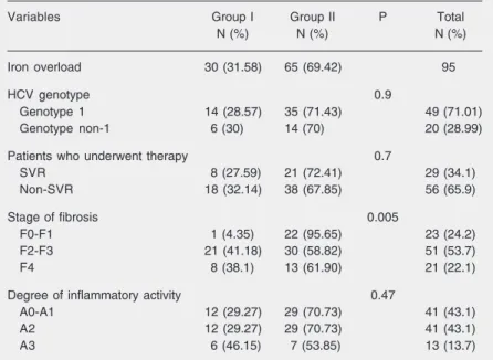
The data in Table 1 show a statistically significant difference in stage of fibrosis between subjects with hepatic iron overload (group I) and patients with no iron detected in the liver (group II) (P= 0.005). In group I, only 1 patient (3.4%) had stage F0/F1 of liver fibrosis, while in group II, 33.8% of patients had minimal or no liver fibrosis (F0/ F1). F2/F3 fibrosis was observed in 70% of group I patients compared to 46.2% of group II patients. However, there was no differ-ence between groups in terms of inflamma-tory activity, ALT levels or presence of he-patic tissue steatosis. A liver section with hepatocellular iron overload in a patient with chronic hepatitis C without treatment is shown in Figure 1.
HCV genotype had been determined in 69 individuals; 49 (70%) were infected with HCV genotype 1, and 20 (30%) with non-1 HCV genotype. There was no statistically significant difference between groups re-garding HCV genotype.
Among the 85 patients who underwent combination therapy, 29 (34.1%) were sus-tained responders while 56 (65.9%) were non-responders or relapsers. Regarding iron overload and sustained virological response, there was also no statistically significant difference between groups.
Table 1. Characteristics of patients with hepatitis C infection.
Variables Group I Group II P Total
N (%) N (%) N (%)
Iron overload 30 (31.58) 65 (69.42) 95
HCV genotype 0.9
Genotype 1 14 (28.57) 35 (71.43) 49 (71.01)
Genotype non-1 6 (30) 14 (70) 20 (28.99)
Patients who underwent therapy 0.7
SVR 8 (27.59) 21 (72.41) 29 (34.1)
Non-SVR 18 (32.14) 38 (67.85) 56 (65.9)
Stage of fibrosis 0.005
F0-F1 1 (4.35) 22 (95.65) 23 (24.2)
F2-F3 21 (41.18) 30 (58.82) 51 (53.7)
F4 8 (38.1) 13 (61.90) 21 (22.1)
Degree of inflammatory activity 0.47
A0-A1 12 (29.27) 29 (70.73) 41 (43.1)
A2 12 (29.27) 29 (70.73) 41 (43.1)
A3 6 (46.15) 7 (53.85) 13 (13.7)
SVR = sustained virological response.
Figure 1. Hepatocellular iron overload in patients with untreated chronic hepatitis C: liver section stained by Perls’ method for iron and with sirius red for collagen shows iron granules within the hepatocyte cytoplasm and thin perisinusoidal fibrosis. 400X. Bar = 25 µm.
1 2 3
chronic HCV infection. The relationship be-tween HCV infection and serum levels of iron and ferritin was described soon after HCV was cloned. Later other investigators evaluated the association between iron lev-els in hepatic tissue and the severity of viral infection or the response to treatment with antiviral drugs (9,10). In our study, in which 95 samples of hepatic biopsy were analyzed, the presence of iron overload in hepatic tis-sue was observed in 31.6% of the cases. Other investigators have reported different frequencies of iron overload in patients with chronic hepatitis C. Ikura et al. (11) found that 73% of 63 patients treated with interfer-on had irinterfer-on overload. On the other hand, Riggio et al. (4) found that 10% of 81 sub-jects infected with HCV had iron overload. Our results are similar to those reported by Kazemi-Shirazi et al. (12) who evaluated the relationship between the presence of iron and mutation of the hemochromatosis gene in patients with chronic hepatitis C. They detected iron overload in 33.3% of 149 pa-tients subjected to liver biopsy. In 1999, Hezode et al. (13) reported a frequency of iron overload in 42.1% of 209 individuals with chronic hepatitis C.
We used Perls’ staining to detect iron overload in tissue. A good correlation has been reported between atomic absorption spectroscopy and semi-quantitative deter-minations in hepatic tissue using Perls’ stain-ing (11,12). The biopsy samples from 95 patients were divided into two groups: group I, individuals with iron overload and group II, subjects without iron overload. There was no statistically significant difference by mul-tivariate analysis in terms of clinical charac-teristics such as age and gender between groups, although there was a predominance of iron overload in male patients. Ikura et al. (11) also observed no statistically signifi-cant difference between genders. ALT lev-els were similar in both groups studied here (mean of 156 vs 138 U/L in patients with and
without overload, respectively).
Kazemi-Shirazi et al. (12), in 1999, obtained similar results when they studied iron status and the hemochromatosis mutation in 114 patients with chronic hepatitis C. Di Bisceglie et al. (9) found a difference between serum AST and ferritin levels but did not identify any difference between ALT levels and hepatic iron concentration.
HCV genotype was determined in 69 of our 95 patients. Most of them (70%) were infected with HCV genotype 1 and 30% with genotypes 2 and 3. The higher frequency of HCV genotype 1 has been reported for most Brazilian regions (14). There was no statisti-cally significant difference by multivariate analysis between the patients with iron over-load in hepatic tissue and HCV genotype, in spite of the fact that some studies have re-ported a higher frequency of iron overload in patients infected with HCV genotype 1 (15). It is not clear if the presence of iron overload interferes with the response of the liver to the therapeutic schedules with inter-feron and ribavirin. Some researchers con-sidered iron overload to be a factor contrib-uting to a decreased response to treatment (16,17). Later investigations reported tradictory results. Currently, there is no con-sensus as to the role of iron overload in the response to treatment of chronic hepatitis C or if there may be a subgroup of patients in which iron overload interferes with therapy. Recently, some investigators concluded that hepatic iron concentration does not predict the response to standard and pegylated inter-feron/ribavirin therapy in patients with chronic hepatitis C (18). Our data did not detect a difference between patients with iron overload that achieved a virological sustained response (28.57%) and non-re-sponders (32.14%).
could contribute to the development of hepatic cirrhosis and hepatocellular carcinoma. Our results are consistent with reports which found that iron overload might be associated with increased liver fibrosis (19,20).
Hepatic iron overload was observed in 31.6% of patients with hepatitis C,
espe-cially males. A greater iron accumulation was associated with a more severe stage of liver fibrosis, but there was no relationship between iron overload and response to treat-ment with interferon and ribavirin. How-ever, sample size might have been a limiting factor for definitive conclusions.
References
1. Pietrangelo A (1998). Iron, oxidative stress and liver fibrogenesis.
Journal of Hepatology, 28 (Suppl 1): 8-13.
2. Casaril M, Stanzial AM, Tognella P et al. (2000). Role of iron load on fibrogenesis in chronic hepatitis C. Hepato-Gastroenterology, 47: 220-225.
3. Bassett SE, Di Bisceglie AM, Bacon BR et al. (1999). Effects of iron loading on pathogenicity in hepatitis C virus-infected chimpanzees.
Hepatology, 29: 1884-1892.
4. Riggio O, Montagnese F, Fiore P et al. (1997). Iron overload in patients with chronic viral hepatitis: how common is it? American
Journal of Gastroenterology, 92: 1298-1301.
5. Chapoutot C, Esslimani M, Joomaye Z et al. (2000). Liver iron excess in patients with hepatocellular carcinoma developed on viral C cirrhosis. Gut, 46: 711-714.
6. Ganne-Carrie N, Christidis C, Chastang C et al. (2000). Liver iron is predictive of death in alcoholic cirrhosis: a multivariate study of 229 consecutive patients with alcoholic and/or hepatitis C virus cirrhosis: a prospective follow up study. Gut, 46: 277-282.
7. Olynyk J, Reddy K, Di Bisceglie A et al. (1995). Hepatic iron concen-tration as a predictor of response to interferon alpha therapy in chronic HCV. Gastroenterology, 108: 1104-1109.
8. Hayashi H, Takikawa T, Nishimura N et al. (1994). Improvement of serum aminotransferase levels after phlebotomy in patients with chronic active hepatitis C and excess hepatic iron. American
Jour-nal of Gastroenterology, 89: 986-988.
9. Di Bisceglie AM, Axiotis C, Hoofnagle J et al. (1992). Measurements of iron status in patients with chronic hepatitis. Gastroenterology, 102: 2108-2113.
10. Arber N, Konikoff F & Moshkowitz M (1994). Increased serum iron and iron saturation without liver iron accumulation distinguish chronic hepatitis C from other chronic liver diseases. Digestive Diseases
and Sciences, 39: 2656-2659.
11. Ikura Y, Morimoto H, Johmura H et al. (1996). Relationship between hepatic iron deposits and response to interferon in chronic hepatitis
C. American Journal of Gastroenterology, 91: 1367-1373.
12. Kazemi-Shirazi L, Datz C, Maier-Dobersberger T et al. (1999). The reaction of iron status and hemochromatosis gene mutations in patients with chronic hepatitis C. Gastroenterology, 116: 127-134. 13. Hezode C, Cazeneuve C, Coué O et al. (1999). Liver accumulation
in patients with chronic active hepatitis C: prevalence and role of hemochromatosis gene mutations and relationship with hepatic his-tology lesions. Journal of Hepatology, 31: 979-984.
14. Busek S & Oliveira G (2003). Molecular epidemiology of the hepati-tis C virus in Brazil. Genetics and Molecular Research, 2: 117-123. 15. Gehrke SG, Stremmel W, Mathes I et al. (2003). Hemochromatosis and transferrin receptor gene polymorphisms in chronic hepatitis C: impact on iron status, liver injury and HCV genotype. Journal of
Molecular Medicine, 81: 780-787.
16. Kageyama F, Kobayashi Y, Murohisa G et al. (1998). Failure to respond to interferon-alpha 2a is associated with increased hepatic iron levels in patients with chronic hepatitis C. Biological Trace
Element Research, 64: 185-196.
17. Distante S, Bjoro K, Hellum KB et al. (2002). Raised serum ferritin predicts non-response to interferon and ribavirin treatment in pa-tients with chronic hepatitis C infection. Liver, 22: 269-275. 18. Hofer H, Osterreicher C, Jessner W et al. (2004). Hepatic iron
concentration does not predict response to standard and pegylated INF/ribavirin therapy in patients with chronic hepatitis C. Journal of
Hepatology, 40: 1018-1022.
19. Angelucci E, Muretto P, Nicolucci A et al. (2002). Effects of iron overload and hepatitis C virus positivity in determining progression of liver fibrosis in thalassemia following bone marrow transplanta-tion. Blood, 100: 17-21.