A Genetic Predictive Model for Canine Hip
Dysplasia: Integration of Genome Wide
Association Study (GWAS) and Candidate
Gene Approaches
Nerea Bartolomé1
*, Sergi Segarra2, Marta Artieda1, Olga Francino3, Elisenda Sánchez3,
Magdalena Szczypiorska1, Joaquim Casellas3, Diego Tejedor1, Joaquín Cerdeira4, Antonio Martínez1, Alfonso Velasco2, Armand Sánchez3
1Progenika Biopharma SA, a Grifols Company, Derio, Bizkaia, Spain,2Bioibérica SA, Barcelona, Spain, 3Servei Veterinari de Genètica Molecular, Departament de Ciència Animal i dels Aliments, Facultat de Veterinària, Universitat Autònoma de Barcelona, Barcelona, Spain,4Centro Veterinario Aluche Las Águilas, Madrid, Spain
Abstract
Canine hip dysplasia is one of the most prevalent developmental orthopedic diseases in dogs worldwide. Unfortunately, the success of eradication programs against this disease based on radiographic diagnosis is low. Adding the use of diagnostic genetic tools to the current phenotype-based approach might be beneficial. The aim of this study was to devel-op a genetic prognostic test for early diagnosis of hip dysplasia in Labrador Retrievers. To develop our DNA test, 775 Labrador Retrievers were recruited. For each dog, a blood sam-ple and a ventrodorsal hip radiograph were taken. Dogs were divided into two groups ac-cording to their FCI hip score: control (A/B) and case (D/E). C dogs were not included in the sample. Genetic characterization combining a GWAS and a candidate gene strategy using SNPs allowed a case-control population association study. A mathematical model which in-cluded 7 SNPs was developed using logistic regression. The model showed a good accura-cy (Area under the ROC curve = 0.85) and was validated in an independent population of 114 dogs. This prognostic genetic test represents a useful tool for choosing the most appro-priate therapeutic approach once genetic predisposition to hip dysplasia is known. There-fore, it allows a more individualized management of the disease. It is also applicable during genetic selection processes, since breeders can benefit from the information given by this test as soon as a blood sample can be collected, and act accordingly. In the authors’
opin-ion, a shift towards genomic screening might importantly contribute to reducing canine hip dysplasia in the future. In conclusion, based on genetic and radiographic information from Labrador Retrievers with hip dysplasia, we developed an accurate predictive genetic test for early diagnosis of hip dysplasia in Labrador Retrievers. However, further research is warranted in order to evaluate the validity of this genetic test in other dog breeds.
OPEN ACCESS
Citation:Bartolomé N, Segarra S, Artieda M, Francino O, Sánchez E, Szczypiorska M, et al. (2015) A Genetic Predictive Model for Canine Hip Dysplasia: Integration of Genome Wide Association Study (GWAS) and Candidate Gene Approaches. PLoS ONE 10(4): e0122558. doi:10.1371/journal. pone.0122558
Academic Editor:Giuseppe Novelli, Tor Vergata University of Rome, ITALY
Received:November 4, 2014
Accepted:February 22, 2015
Published:April 13, 2015
Copyright:© 2015 Bartolomé et al. This is an open access article distributed under the terms of the
Creative Commons Attribution License, which permits unrestricted use, distribution, and reproduction in any medium, provided the original author and source are credited.
Data Availability Statement:All relevant data are within the paper and its Supporting Information files.
Introduction
Canine hip dysplasia (CHD) is one of the most prevalent developmental orthopedic diseases in dogs worldwide. It is characterized by an abnormal formation of the hip joint with different de-grees of laxity and subluxation, which ultimately leads to secondary osteoarthritis (OA) and impaired animal welfare. The prevalence of CHD is particularly high among larger breeds of dogs, estimates of around 20% have been found in Labrador retrievers, one of the most popular
breeds in the world, and up to 70% in Saint Bernards [1–3].
The diagnosis of CHD is established through radiographic examination of the hip joint using different accepted scores. The FCI [Fédération Cynologique Internationale] scale is one of the most popular in Europe, while the OFA [Orthopedic Foundation for Animals] hip score
is the most commonly used in USA [4]. During the last decades a high number of selective
breeding programs based on radiographies have been implemented for different breeds with the aim of reducing CHD incidence and improving animal’s welfare. However, the phenotype-based screening programs have not been effective enough, since the prevalence of CHD
re-mains high. An improvement was described in some cases [5,6], but a slow progress or no
im-provement was achieved in others [7–9].
Current knowledge in the mode of inheritance of CHD indicates it to be a genetic complex trait with a polygenic inheritance pattern influenced by environmental factors. Both dominant
and recessive modes of inheritance have been proposed [10–13]. The results of several studies
performed in the last five years suggest that selective breeding programs based on genetic infor-mation, rather than on phenotypic selection alone, are the best alternative to achieve more
rapid improvements in CHD [14–16].
Molecular genetic studies with microsatellites led to the identification of quantitative trait
loci [QTL] for CHD and secondary OA in different breeds, such as Labrador retrievers [17,18]
or Portuguese water dogs [19,20] some years ago. The sequencing of the whole dog genome
and the characterization of more than 2.5 million single nucleotide polymorphisms [SNPs] in
2005 [21] opened the door to the development of new genotyping tools, as high-throughput
SNP genotyping microarrays and, thus, to new studies aimed at clarifying the genetic basis of CHD. The usefulness of predictive models based on combinations of SNPs, as genetic markers,
for assessing susceptibility to specific diseases has been long demonstrated in humans [22–24].
The first published studies using SNPs as genetic markers for CHD have generated interesting and promising results. A group of SNPs which confers increased risk for CHD has been
identi-fied in German shepherd dogs [25]. A Genome Wide Association Study [GWAS] with more
than 22,000 SNPs in dogs of several breeds found 4 SNPs associated to CHD and 2 SNPs to hip
OA [26]. Besides, association studies with SNPs have allowed redefining QTL intervals and
identification of the first mutation associated with canine hip dysplasia, which is located in the
fibrillin-2 gene [FBN2] [27–29]. Finally, a very recent GWAS performed with nearly 18,000
SNPs and with Labrador retrievers has identified several SNPs in genes or near genes involved
in extracellular matrix development associated to CHD [30]. A second GWAS with around
47,000 SNPs, published during the writing of this manuscript, has identified several SNPs
asso-ciated to CHD in German Shepherd Dogs [31].
Recently, a new commercial high density SNP genotyping microarray [Canine HD Bead-Chip, Illumina Inc, San Diego, CA, USA] with a genome wide coverage [170,000 SNPs evenly spaced in the dog genome] has become available at the market. The goal of the present study is to find a predictive model based on genetic markers for CHD susceptibility in Labrador retriev-ers. For that purpose, we have performed a GWAS using the Canine HD BeadChip and a can-didate gene study in which hundreds of SNPs in several cancan-didate genes related to
inflammation, bone formation and cartilage remodeling pathways, among others, were data collection and analysis, decision to publish, or
preparation of the manuscript. The specific roles of these authors are articulated in the author contributions section.
analyzed. We have developed a genetic predictive model, based on 7 SNPs, able to predict CHD development in Labrador retrievers.
Materials and Methods
Ethics Statement
The study was approved by the Ethics Committee on Animal and Human Experimentation (CEEAH) of the Universitat Autònoma de Barcelona (UAB, Spain) (Authorization reference number: DMAH 4463). All animal work has been conducted according to the national and in-ternational guidelines for animal welfare. All blood-sampling was done in veterinary clinics for small animals and with the owners' informed consent.
Study population and X-ray evaluation
A total of 775 pure breed Labrador retriever dogs (633 for the development study and 142 for the validation) were recruited from 64 Spanish veterinary clinics. Both the Labrador retriever show and field lines were equally represented in this study, including dogs from European (British and continental) and American bloodlines. This was verified by using a specific pedi-gree software (Breeders Assistant for dogs 4, Tenset Technologies Ltd., Cambridge, UK). Re-garding inbreeding rates, the animals were unrelated at least at the grandparent level.
A standard ventrodorsal hip X-ray of each dog was taken. All the X-rays were independently evaluated by three experts from the Hip Dysplasia Radiological Reading Committee at the Spanish Small Animal Veterinary Association (AVEPA) who were unaware of the dogs' geno-types. X-rays were evaluated according to the FCI (Fédération Cynologique Internationale) of-ficial scale for hip dysplasia (A = no signs of CHD, B = near normal hips, C = mild signs of CHD, D = moderate signs of CHD, E = severe CHD). The FCI grade should be coincident in the view of at least two of the three evaluators and should not differ from the evaluation of the third evaluator in more than 1 grade in the FCI scale. In case these criteria were not met, radio-graphs were re-evaluated until the criteria were fulfilled.
We followed an extreme phenotype design for the association analysis, so C dogs were not included in the sample. Dogs scored as A or B were classified into the control group (free of hip dysplasia) and dogs scored as D or E were classified into the case group (affected). To be sure that the dogs classified as A or B had developed a final phenotype, and will not evolve to other grades, a minimum age of 12 and 48 months was required for A and B dogs, respectively. No minimum age was required for dogs scored as D and E, although a maximum of 8 years old was set in order to avoid the interference of osteoarthritic changes due to aging. Among the 775 dogs 354 fulfilled the inclusion criteria and were finally genotyped (240 for the develop-ment study and 114 for the validation).
DNA extraction and SNP genotyping
Blood samples were collected in EDTA-tubes. The genomic DNA was extracted using the QIAamp DNA blood mini kit (Qiagen, Hilden, Germany) following the manufacturer instruc-tions. Purified DNA was quantified using Qubit fluorometer (Life Technologies-Molecular Probes, Oregon, USA) following manufacturer instructions or Nanodrop spectrophotometer (Thermo Fisher Scientific, Wilmington, DE, USA) and adjusted to the desirable concentration. The A260/280 ratio was above 1.6 for all samples.
which analyzes more than 170,000 evenly spaced SNPs in the dog genome. On the other hand, we genotyped 768 custom SNPs located in candidate genes and quantitative trait loci (QTL) for CHD with the Illumina Golden Gate genotyping platform (Illumina Inc, San Diego, CA, USA). We selected as candidate genes, genes implicated in molecular processes involved in CHD and/or OA (cartilage degradation, inflammation, extracellular matrix metabolism and bone remodeling, among others), genes previously described as being associated with OA in humans, genes involved in cartilage and bone diseases in humans and genes located in QTLs
previously reported as associated with CHD or OA [17,19,20,28,32]. We used dbSNP (http://
www.ncbi.nih.gov/projects/SNP) and CanFam (http://www.broadinstitute.org/mammals/dog) databases for SNPs selection. We chose 2 or 3 SNPs per gene, selecting intragenic SNPs, when possible.
The dogs of the validation population were genotyped for the SNPs of interest using the KASPar chemistry (LGC Genomics, Hertfordshire, UK), which is a competitive allele-specific PCR SNP genotyping system that uses FRET (Förster Resonance Energy Transfer) quencher cassette oligonucleotides.
CHST3 sequencing and analysis
A chromosomal fragment of 5819 bp including theCHST3gene and its 5’upstream and 3’
downstream flanking regions was sequenced by Sanger method in 39 Labrador retrievers (20 unaffected individuals and 19 with hip dysplasia) using as reference the Boxer sequence of the
CHST3gene (NCBI GeneID: 489036; build 2.1). Six overlapping PCRs were performed using
the Qiagen Multiplex PCR kit (Qiagen, Hilden, Germany), with an annealing temperature of 60°C and 100 ng of DNA template. PCR products were purified using Millipore HTS filter plates (Merck Millipore, Darmstadt, Germany). Sequencing reactions were performed with BigDye Terminator v3.1 Cycle Sequencing kit (Applied Biosystems, Carlsbad, CA, USA). Sam-ples were cleaned with CleanSEQ reaction clean-up (Agencourt Bioscience Corp., Beverly, MA, USA) and analyzed on an ABI 3100 DNA Analyzer.
Statistical analysis
Monomorphic single nucleotide polymorphisms, SNPs with MAF less than 0.01, SNPs with a
call rate<99% and SNPs with severe deviation from Hardy-Weinberg equilibrium in controls
(p<0.0001) were excluded from the analyses, both in the GWAS and in the candidate gene
analyses. This filtering resulted in the selection of 449 SNPs from the candidate gene study and 130,621 SNPs from the GWAS. Principal component analysis (PCA) was carried out to assess for population stratification of the development study cohort (240 individuals). 80,116 SNPs remained after quality control, LD pruning and inactivation of the X chromosome, and were used for the PCA. Analyses were performed using the SNP & Variation Suite (SVS v7.0) soft-ware of Golden Helix.
An association test for SNP allele frequencies with CHD was performed by the chi-square test in the GWAS and the candidate genes studies. To adjust for multiple testing, we used the
Benjamini-Hochberg procedure to control False Discovery Rate (FDR) [33]. Based on the
mul-tiple test correction made in our data set, the minimum uncorrected p-value threshold that would reach statistical significance (at the 5% level) after correction for FDR would be
p<1.96x10⁻⁵in the GWAS analysis and p<5x10⁻³in the candidate gene study. We developed a
CIs. To measure the impact of the SNPs included in the model the sensitivity (Se) and specifici-ty (Sp) values were computed by means of the ROC curve.
The accuracy of the predictive model was tested on the independent validation cohort. A ROC curve was constructed with the predicted probability score for the model generated by the equation derived in the development cohort for each subject of the validation cohort. The statistical difference between the AUCs of the ROC curves of the two cohorts was calculated by means of a two-sample Z-test.
To determine the contributing chi-square value of each predictor, the predictive model was calculated by eliminating one predictor at a time. The chi-square value for the reduced model was recorded for each factor. The square value of each factor was subtracted from the
chi-square value of the full model to determine its percentage contribution [34].
The univariate statistical analysis was performed with the softwares SVS v7.3.1 (Golden Helix Inc.) for the candidate gene study and PLINK v1.07 for the GWAS. The software SPSS v15.0 was used for the multivariate analysis. Measures of pairwise linkage disequilibrium (LD)
(r2>0.8) were determined using SVS v7.3.1 software (Golden Helix Inc.).
Results
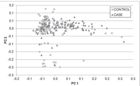
PCA plot of the 240 Labrador included at the development study and 80,116 SNPs revealed no
stratification among the affected dogs and free of hip dysplasia dogs (Fig 1). The distribution
according to the FCI scale for hip dysplasia and the average age of the Labrador retrievers
in-cluded in the development (n = 240) and validation (n = 114) cohorts are depicted inTable 1.
In the development cohort, we found 250 SNPs significantly associated to CHD after FDR
correction for multiple testing in the GWAS analysis (p1.96x10-5) and 33 SNPs in the
candi-date genes study (p5x10-3) (Fig 2,S1 TableandS2 Table).
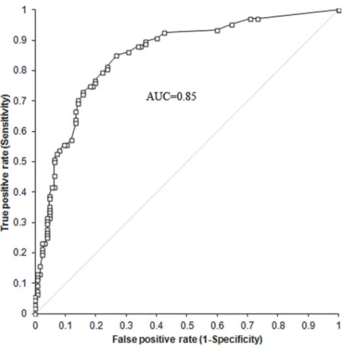
To search for a predictive model of CHD in the development population, the SNPs statisti-cally significant after controlling for FDR both in the GWAS and in the candidate gene studies were entered together into multivariate forward logistic regression analysis. We obtained a pre-dictive model with a good accuracy as indicated by a ROC AUC of 0.85 (95% CI 0.80–0.90)
Fig 1. Principal Component Analysis (PCA) plot of the study development cohort.80,116 SNPs
dispersed genome-wide were used. X-axis is principle component 1 and y-axis is principle component 2. Cases and controls were clustering together.
(Fig 3andTable 2). The predictive model was developed in a population of 235 dogs, since 5 of the 240 individuals of the development cohort had missing genotypes. The logistic model com-bines 7 SNPs, namely BICF2P772455, BICF2G630227898, BICF2G630339806,
BICF2G630558239, BICF2P548082, BICF2S230609 and BICF2S2452559 (Table 2). All the
SNPs in the model have aβcoefficient significantly different from zero, which indicates that all
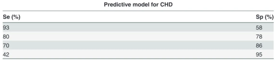
SNPs contribute to the predictive ability of the model (Table 2). The Se and Sp values of the
model are shown inTable 3. As a general data, in the cut-off point showing the best
equilibri-um between Se and Sp, the model has a Se of 80% and a Sp of 78%.
The dogs of the validation population were genotyped for the 7 SNPs included in the predic-tive model for CHD. A ROC curve was constructed with the predicted probability score for each subject of the validation cohort. A ROC curve with an AUC of 0.80 (95% CI 0.72–0.89) was found for the validation cohort. The predictive ability of the model was confirmed on a val-idation cohort. There were no statistical differences between the AUCs of the ROC curves of the two cohorts, 0.85 and 0.80, when compared using a two-sample Z test (Two-tailed p = 0.3161).
The relative contribution of each genetic variable to the predictive power of the model was calculated, BICF2G630227898 and BICF2G630339806 were the SNPs with a highest
contribu-tion, 21% and 20% respectively (S1 Fig).
Table 1. Distribution according to the FCI scale for hip dysplasia and average age of the Labrador retrievers included in the development and vali-dation cohorts.
Development cohort Validation cohort
FCI grade n Age (mean±SD) n Age (mean±SD)
Controls A 99 34±18 44 21±12
B 30 64±11 14 64±24
Cases D 64 37±21 26 48±37
E 47 45±28 30 48±48
doi:10.1371/journal.pone.0122558.t001
Fig 2. Manhattan plot for the CHD association test results obtained in the GWAS.The horizontal line indicates the FDR correction significance level (p1.96x10-5).
Fig 3. ROC curve of the predictive model for CHD.We developed a multivariate predictive model for CHD by means of forward logistic regression using as predictors the SNPs significant after controlling for FDR. We tested our model discrimination via the Hosmer_Lemeshow statistic and the receiver operating characteristic (ROC) curve with 95% CIs.
doi:10.1371/journal.pone.0122558.g003
Table 2. Predictive model for CHD.
SNP Chr: nt
position
Notable nearby gene nt change
Risk allele
Risk genotypes
β
coefficient
OR (IC 95%)
p value
BICF2P772455 chr4: 22691322 near to CHST3 A/G G GG 1.13 3.1 (1.3–
7.5)
0.012
BICF2G630227898 chr20: 2704477 near to RAB7A A/G A AA + AG 1.41 4.1 (1.8–
9.2)
<0.001
BICF2G630339806 chr3: 40302288 near to CHSY1 and ADAMTS17
A/G A AA +AG 1.69 5.4 (1.9–
15.0)
0.001
BICF2G630558239 chr7: 36171712 near to SMYD3 A/G A AA +AG 1.05 2.9 (1.4–
5.8) 0.004
BICF2P548082 chr12:
16410934 A/G G GG + AG 0.81 2.2 (1.24.4) – 0.017
BICF2S230609 chr18:
48695616 near to FGF4 A/G A AA 1.07 2.9 (1.55.7) – 0.002
BICF2S2452559 chr10:
47923623 near to PKCE A/G G GG +AG 0.79 2.2 (1.14.5) – 0.033
The chromosomic position corresponds to CanFam 3.1.
One of the SNPs included in the model is BICF2P772455, which is located very close to the
CHST3(carbohydrate chondroitin 6 sulfotransferase) gene, specifically, 14 bp upstream the
initial ATG start codon. Among the 33 SNPs associated with CHD in the candidate gene analy-sis, BICF2P772455 was the SNP more strongly associated to the disease together a second SNP,
BICF2P419109, located near the same gene, 1051 bp downstreamCHST3(S2 Table). These
two SNPs represented independent associations (LD r2<0.8). TheCHST3gene encodes a
sulfo-transferase involved in chondroitin sulfate (CS) biosynthesis. CS is a structural component of
the joint cartilage essential for its biomechanical properties, and soCHST3an interesting and
promising candidate gene for CHD. Since in the public databases there are no SNPs described
inside theCHST3canine gene, in order to know if there were other SNPs inside theCHST3
gene more strongly associated to CHD, maybe a functional variant, we decided to sequence a
chromosomic region of 5819 bp including theCHST3gene and its 5’upstream and 3’
down-stream flanking regions in 39 Labrador retrievers (20 unaffected and 19 affected individuals)
(Labrador retrieverCHST3Sequence NCBI accession number: JX402028.1). Apart from the 2
mentioned SNPs, we found other 29 SNPs located inside theCHST3gene (14 SNPs) and in its
flanking regions (15 SNPs). However, none of the 29 SNPs was more strongly associated to CHD than BICF2P419109 and BICF2P772455 (data not shown).
Discussion
CHD is an inherited disorder with a high prevalence in breeds of large size, such as Labrador retrievers, that ultimately leads to impaired mobility and function, and considerably reduces the quality of life of the dogs. Phenotype-based selective breeding of dogs has not been proven to be effective in significantly reducing the prevalence of CHD. The studies performed in the last recent years suggest that prediction of CHD is feasible using genetic markers. In this study we have developed a logistic regression model, based on genetic polymorphisms, with a good predictive accuracy for CHD susceptibility in Labrador retrievers, as indicated by a ROC-AUC
of 85% [35]. The area under the ROC curve (AUC) is widely recognized as the measure of a
di-agnostic test's discriminatory power. The AUC can vary between 0.5 and 1. An area of 1 repre-sents a perfect test while an area of 0.5 reprerepre-sents no discrimination. Predictive models with an AUC above 0.75 are considered as clinically useful and models with an AUC above 0.89 are considered as excellent. Logistic models with similar values of ROC-AUC as our model for
CHD have been considered as highly predictive for some human diseases [23]. The predictive
ability of the model was validated in an independent population of Labrador retrievers. This predictive model represents a significant advance in CHD early detection. It constitutes a useful tool for veterinarians to help them choose the most appropriate therapeutic approach once ge-netic predisposition is known. Therefore, a more individualized management of the disease might be achieved. It is also applicable in selection processes, since breeders could benefit from
Table 3. Predictive accuracy of the logistic model for Canine hip dysplasia. Predictive model for CHD
Se (%) Sp (%)
93 58
80 78
70 86
42 95
Se: sensitivity; Sp: specificity
the information given by this predictive model as soon as a blood sample can be collected, and act accordingly.
The model for prediction of CHD includes 7 SNPs. The chromosomal region of 1 Mb
sur-rounding the associated SNPs was analyzed using Ensembl database (www.ensembl.org) in
search of potential candidate genes involved in CHD. Three of the seven SNPs lie within or near genes which codify proteins involved in extracellular matrix metabolism. The SNP
BICF2P772455 is located in the 5’UTR of theCHST3gene, just 14 bp upstream the initial
ATG start codon. Thus, it could be part of a regulatory element affecting the transcription of the gene. The CHST3 enzyme is involved in chondroitin sulfate biosynthesis, specifically in the sulfation process. CS is an extracellular matrix component that plays an important role in
carti-lage function, providing this tissue with resistance and elasticity. Mutations in theCHST3gene
have been previously found as associated with spondyloepiphyseal dysplasia in humans, which is a congenital skeletal development disorder characterized by joint dislocations, included hip
dislocation [36,37]. A recent GWAS study in humans has reported that a SNP located in the
CHST11gene, another gene of the same family as theCHST3gene, is associated with
preva-lence of hip OA [38]. Thus, we suggest that genes implicated in the CS synthesis could have a
role in congenital canine and human disorders related to hip joint formation and thatCHST3
could be a potential credible gene implicated in CHD and secondary OA. The second SNP is
BICF2G630339806 which is located nearCHSY1(chondroitin sulfate synthase 1) and
ADAMTS17(ADAM metallopeptidase with thrombospondin type 1 motif, 17) genes. As the
CHST3gene, the CHSY1 enzyme also plays a critical role in the biosynthesis of CS. Studies in
mice have suggested it to be an essential regulator of joint patterning [39], making a role for
CHSY1 in CHD plausible.ADAMTS17is a member of the ADAMTS family of genes that
en-code secreted metalloproteases that modify extracellular structural proteins. Polymorphisms in ADAMTS genes have been associated with human diseases related with bone metabolism,
in-cluding osteoporosis and OA [40,41], so ADAMTS17 could also be a potential gene implicated
in CHD and secondary OA. The third SNP is BICF2S230609 located near the FGF4 (fibroblast growth factor 4) gene. Fibroblasts secrete the precursors of all the components of the extracel-lular matrix, maintaining the structural integrity of connective tissues. FGF family members possess broad mitogenic and cell survival activities and are implicated in embryonic develop-ment, morphogenesis and tissue repair, among other biological processes. Members of the FGF family have been proposed to control the limb bud outgrowth. Specifically, studies in mice sug-gested that FGF4 is implicated in embryonic distal limb morphogenesis, being responsible for
the partial compensation of distal limb development in the absence of FGF8 [42]. Altogether,
these results suggest that genes implicated in extracellular matrix synthesis/degradation could have a role in CHD. These results are in line with previous findings which indicate genes of
ex-tracellular matrix components as implicated in the pathogenesis of CHD [30], and point out
the extracellular matrix metabolism as a key factor in CHD development.
Other two SNPs of the model lie very close to genes related to bone metabolism. The SNP BICF2G630227898 is located approximately 50 kb upstream the gene RAB7A which codifies for a small GTPase that is highly expressed and is predominantly localized at the ruffled border in bone-resorbing osteoclasts. During skeletal growth and remodeling the mineralized bone matrix is resorbed by osteoclasts, it has been described that downregulation of Rab7 impairs os-teoclast polarization and bone resorption, underscoring the importance of Rab7 in osos-teoclast
function and skeletal growth [43]. The gene PKCE, which codifies for the Protein Kinase C
Ep-silon, is situated approximately30 kb upstream the SNP BICF2S2452559. Protein Kinase Cs (PKCs) are a family of serine/threonine kinases involved in several cellular processes including cell proliferation, differentiation, apoptosis, and survival. Several studies have demonstrated
However, the exact role of individual PKC isoforms in the regulation of bone function is not fully clear. Our data agree with a recently published study performed with German Shepherd
dogs [31], which has found SNPs associated to CHD located in close proximity to genes which
are involved in bone formation and osteoclast activity, underlying the role of bone formation in CHD disease.
The gene SMYD3 (SET and MYND domain containing 3) is located about 0.5 Mb down-stream of the SNP BICF2G630558239. SMYD3 is a histone methyltransferase that has been
found to be implicated in muscle mass determination and skeletal muscle atrophy [46]. Thus,
SMYD3 could be a potential candidate gene involved in the atrophy of hind leg muscles associ-ated with CHD. Further research in this area is needed to confirm this hypothesis.
Finally, we have not found any potential candidate gene for CHD development located near the SNP BICF2P548082, this could be due to the fact that some of the genes near this SNP are still of unknown function or maybe because this SNP is in LD with a potential functional one in a distant genomic region.
Clearly, it is needed further research in this area to establish the link between the SNPs of the predictive model and the functional consequences for CHD and to clarify if the SNPs found are functional SNPs or SNPs in linkage disequilibrium with a nearby functional SNP.
In conclusion, we have developed a logistic model with a good accuracy for CHD prediction in Labrador retrievers based on the combination of 7 SNPs, several of them located near genes involved in extracellular matrix processes or bone metabolism. Our results in Labrador retriev-ers add evidence to the thought that genomics is the basis towards early detection of CHD. Whether our predictive model is valid or not for other dog breeds needs to be explored.
Supporting Information
S1 Fig. Relative contribution of each SNP to the predictive power of the model for CHD. (DOCX)
S1 Table. SNPs significantly associated to CHD after correction for FDR in the GWAS analysis (p1.96x10-5).
(XLSX)
S2 Table. SNPs significantly associated to CHD after correction for FDR in the candidate gene study (p5x10-3).
(XLSX)
Acknowledgments
We are grateful to Jon Fernández for his excellent technical work and Verena Blume for her help during the dog inclusion phase. We also thank the Spanish Small Animal Veterinary Asso-ciation (AVEPA) and all the clinicians and dog owners who have participated in the study.
Author Contributions
References
1. Kaneene JB, Mostosky UV, Miller R. Update of a retrospective cohort study of changes in hip joint phe-notype of dogs evaluated by the OFA in the United States, 1989–2003. Vet Surg. 2009; 38(3): 398– 405.
2. Coopman F, Verhoeven G, Saunders J, Duchateau L, Van BH. Prevalence of hip dysplasia, elbow dys-plasia and humeral head osteochondrosis in dog breeds in Belgium. Vet Rec. 2008; 163(22): 654–658. PMID:19043090
3. Leppanen M, Saloniemi H. Controlling canine hip dysplasia in Finland. Prev Vet Med. 1999; 42(2): 121–131. PMID:10551430
4. Fluckiger M. Scoring radiographs for canine hip dysplasia- the big three organizations in the world. Eu-ropean Journal of Companion Animal Practice 2007; 17: 135–140.
5. Hou Y, Wang Y, Lust G, Zhu L, Zhang Z, Todhunter RJ. Retrospective analysis for genetic
improve-ment of hip joints of cohort labrador retrievers in the United States: 1970–2007. PLoS One 2010; 5(2): e9410. doi:10.1371/journal.pone.0009410PMID:20195372
6. Janutta V, Hamann H, Distl O. Genetic and phenotypic trends in canine hip dysplasia in the German population of German shepherd dogs. Berl Munch Tierarztl Wochenschr. 2008; 121(3–4): 102–109. PMID:19086694
7. Leppanen M, Maki K, Juga J, Saloniemi H. Factors affecting hip dysplasia in German shepherd dogs in
Finland: efficacy of the current improvement programme. J Small Anim Pract. 2000; 41(1): 19–23. PMID:10713978
8. Willis MB. A review of the progress in canine hip dysplasia control in Britain. J Am Vet Med Assoc. 1997 May 15; 210(10):1480–2. PMID:9154201
9. Fluckiger M, Lang J, Binder H, Busato A, Boos J. The control of hip dysplasia in Switzerland. A retro-spect of the past 24 years. Schweiz Arch Tierheilkd. 1995; 137(6): 243–250. PMID:7481714
10. Ginja MM, Silvestre AM, Gonzalo-Orden JM, Ferreira AJ. Diagnosis, genetic control and preventive management of canine hip dysplasia: a review. Vet J. 2010; 184(3): 269–276. doi:10.1016/j.tvjl.2009. 04.009PMID:19428274
11. Janutta V, Hamann H, Distl O. Complex segregation analysis of canine hip dysplasia in German shep-herd dogs. J Hered. 2006; 97(1): 13–20. PMID:16267165
12. Maki K, Janss LL, Groen AF, Liinamo AE, Ojala M. An indication of major genes affecting hip and elbow dysplasia in four Finnish dog populations. Heredity 2004; 92(5): 402–8. PMID:14997179
13. Silvestre AM, Ginja MM, Ferreira AJ, Colaco J. Comparison of estimates of hip dysplasia genetic pa-rameters in Estrela Mountain Dog using linear and threshold models. J Anim Sci. 2007; 85(8): 1880– 1884. PMID:17468417
14. Guo G, Zhou Z, Wang Y, Zhao K, Zhu L, Lust G, et al. Canine hip dysplasia is predictable by
genotyp-ing. Osteoarthritis Cartilage 2011; 19(4): 420–429. doi:10.1016/j.joca.2010.12.011PMID:21215318 15. Sanchez-Molano E, Woolliams JA, Blott SC, Wiener P. Assessing the impact of genomic selection
against hip dysplasia in the Labrador Retriever dog. J Anim Breed Genet. 2013; 131(2): 134–145. doi: 10.1111/jbg.12056PMID:24134497
16. Stock KF, Distl O. Simulation study on the effects of excluding offspring information for genetic evalua-tion versus using genomic markers for selecevalua-tion in dog breeding. J Anim Breed Genet. 2010; 127(1): 42–52. doi:10.1111/j.1439-0388.2009.00809.xPMID:20074186
17. Todhunter RJ, Mateescu R, Lust G, Burton-Wurster NI, Dykes NL, et al. Quantitative trait loci for hip dysplasia in a cross-breed canine pedigree. Mamm Genome. 2005; 16(9): 720–730. PMID:16245029
18. Mateescu RG, Burton-Wurster NI, Tsai K, Phavaphutanon J, Zhang Z, Murphy KE, et al. Identification of quantitative trait loci for osteoarthritis of hip joints in dogs. Am J Vet Res. 2008; 69(10): 1294–1300. doi:10.2460/ajvr.69.10.1294PMID:18828685
19. Chase K, Lawler DF, Adler FR, Ostrander EA, Lark KG. Bilaterally asymmetric effects of quantitative trait loci (QTLs): QTLs that affect laxity in the right versus left coxofemoral (hip) joints of the dog (Canis familiaris). Am J Med Genet A. 2004; 124A(3): 239–247. PMID:14708095
20. Chase K, Lawler DF, Carrier DR, Lark KG. Genetic regulation of osteoarthritis: A QTL regulating cranial and caudal acetabular osteophyte formation in the hip joint of the dog (Canis familiaris). Am J Med Genet A. 2005; 135(3): 334–335. PMID:15887284
22. Ehret GB, Munroe PB, Rice KM, Bochud M, Johnson AD, Chasman DI, et al. Genetic variants in novel pathways influence blood pressure and cardiovascular disease risk. Nature 2011; 478(7367): 103– 109. doi:10.1038/nature10405PMID:21909115
23. Eleftherohorinou H, Wright V, Hoggart C, Hartikainen AL, Jarvelin MR, Balding D, et al. Pathway analy-sis of GWAS provides new insights into genetic susceptibility to 3 inflammatory diseases. PLoS one 2009; 4(11): e8068. doi:10.1371/journal.pone.0008068PMID:19956648
24. Gudmundsson J, Sulem P, Manolescu A, Amundadottir LT, Gudbjartsson D, Helgason A, et al. Ge-nome-wide association study identifies a second prostate cancer susceptibility variant at 8q24. Nat Genet. 2007; 39(5): 631–637. PMID:17401366
25. Distl O. Genetics of hip dysplasia. Proceedings of the 14 th European Society of Veterinary Orthopae-dics and Traumatology (ESVOT) Congress. 2008; S61.
26. Zhou Z, Sheng X, Zhang Z, Zhao K, Zhu L, Guo G, et al. Differential genetic regulation of canine hip dysplasia and osteoarthritis. PLoS one 2010; 5(10): e13219. doi:10.1371/journal.pone.0013219 PMID:20949002
27. Friedenberg SG, Zhu L, Zhang Z, Foels WB, Schweitzer PA, Wang W, et al. Evaluation of a fibrillin 2 gene haplotype associated with hip dysplasia and incipient osteoarthritis in dogs. Am J Vet Res. 2011; 72(4): 530–540. doi:10.2460/ajvr.72.4.530PMID:21453155
28. Zhu L, Zhang Z, Feng F, Schweitzer P, Phavaphutanon J, Vernier-Singer M, et al. Single nucleotide polymorphisms refine QTL intervals for hip joint laxity in dogs. Anim Genet. 2008; 39(2): 141–146. doi: 10.1111/j.1365-2052.2007.01691.xPMID:18261189
29. Zhu L, Zhang Z, Friedenberg S, Jung SW, Phavaphutanon J, Vernier-Singer M, et al. The long (and winding) road to gene discovery for canine hip dysplasia. Vet J. 2009; 181(2): 97–110. doi:10.1016/j. tvjl.2009.02.008PMID:19297220
30. Lavrijsen IC, Leegwater PA, Martin AJ, Harris SJ, Tryfonidou MA, Heuven HC, et al. Genome wide analysis indicates genes for basement membrane and cartilage matrix proteins as candidates for hip dysplasia in labrador retrievers. PLoS one 2014; 9(1): e87735. doi:10.1371/journal.pone.0087735 PMID:24498183
31. Fels L, Distl O. Identification and Validation of Quantitative Trait Loci (QTL) for Canine Hip Dysplasia (CHD) in German Shepherd Dogs. PLoS one 2014; 9(5): e96618. doi:10.1371/journal.pone.0096618 PMID:24802516
32. Marschall Y, Distl O. Mapping quantitative trait loci for canine hip dysplasia in German Shepherd dogs. Mamm Genome. 2007; 18(12): 861–870. PMID:18027024
33. Benjamini Y, Hochberg Y. Controlling the False Discovery Rate: a Practical and Powerful Approach to
Multiple Testing. J R Statist Soc. 1995; B 57 (1): 289–300.
34. Balsa A, Del Almo J, Blanco F, Caliz R, Silva L, Sanmarti R, et al. Prediction of functional impairment and remission in rheumatoid arthritis patients by biochemical variables and genetic polymorphisms. Rheumatology (Oxford) 2010; 49(3): 458–66. doi:10.1093/rheumatology/kep380PMID:20032229 35. Nguyen MT, Devarajan P. Biomarkers for the early detection of acute kidney injury. Pediatr Nephrol.
2008; 23(12): 2151–2157. PMID:17394022
36. Hermanns P, Unger S, Rossi A, Perez-Aytes A, Cortina H, Bonafé L, et al. Congenital joint dislocations caused by carbohydrate sulfotransferase 3 deficiency in recessive Larsen syndrome and humero-spi-nal dysostosis. Am J Hum Genet. 2008; 82(6): 1368–1374. doi:10.1016/j.ajhg.2008.05.006PMID: 18513679
37. Thiele H, Sakano M, Kitagawa H, Sugahara K, Rajab A, Höhne W, et al. Loss of chondroitin 6-O-sulfo-transferase-1 function results in severe human chondrodysplasia with progressive spinal involvement. Proc Natl Acad Sci USA. 2004; 101(27): 10155–10160. PMID:15215498
38. Zeggini E, Panoutsopoulou K, Southam L, Rayner NW, Day-Williams AG, Lopes MC, et al. Identifica-tion of new susceptibility loci for osteoarthritis (arcOGEN): a genome-wide associaIdentifica-tion study. Lancet 2012; 380(9844): 815–823. doi:10.1016/S0140-6736(12)60681-3PMID:22763110
39. Wilson DG, Phamluong K, Lin WY, Barck K, Carano RA, Diehl L, et al. Chondroitin sulfate synthase 1 (Chsy1) is required for bone development and digit patterning. Dev Biol. 2012; 363(2): 413–425. doi: 10.1016/j.ydbio.2012.01.005PMID:22280990
40. Rodriguez-Lopez J, Pombo-Suarez M, Loughlin J, Tsezou A, Blanco FJ, Meulenbelt I, et al. Association of a nsSNP in ADAMTS14 to some osteoarthritis phenotypes. Osteoarthritis Cartilage 2009; 17(3): 321–327. doi:10.1016/j.joca.2008.07.012PMID:18790654
42. Boulet AM, Moon AM, Arenkiel BR, Capecchi MR. The roles of Fgf4 and Fgf8 in limb bud initiation and outgrowth. Dev Biol. 2004; 273(2): 361–372. PMID:15328019
43. Zhao H, Laitala-Leinonen T, Parikka V, Vaananen HK. Downregulation of small GTPase Rab7 impairs osteoclast polarization and bone resorption. J Biol Chem. 2001; 276(42): 39295–39302. PMID: 11514537
44. Moonga BS, Stein LS, Kilb JM, Dempster DW. Effect of diacylglycerols on osteoclastic bone resorption.
Calcif Tissue Int. 1996; 59(2): 105–108. PMID:8687978
45. Wang C, Steer JH, Joyce DA, Yip KH, Zheng MH, Xu J. 12-O-tetradecanoylphorbol-13-acetate (TPA) inhibits osteoclastogenesis by suppressing RANKL-induced NF-kappaB activation. J Bone Miner Res. 2003; 18(12): 2159–2168. PMID:14672351