The Expression of TALEN before Fertilization
Provides a Rapid Knock-Out Phenotype in
Xenopus laevis
Founder Embryos
Kei Miyamoto1☯¤*, Ken-ichi T. Suzuki2☯
*, Miyuki Suzuki2, Yuto Sakane2,
Tetsushi Sakuma2, Sarah Herberg1, Angela Simeone1, David Simpson1, Jerome Jullien1, Takashi Yamamoto2, J. B. Gurdon1
1Wellcome Trust/Cancer Research UK Gurdon Institute, University of Cambridge, Cambridge, United Kingdom,2Department of Mathematical and Life Sciences, Graduate School of Science, Hiroshima University, Higashi-Hiroshima, Japan
☯These authors contributed equally to this work.
¤ Current address: Laboratory of Molecular Developmental Biology, Faculty of Biology-Oriented Science and Technology, Kinki University, Wakayama, Japan
*[email protected](KM);[email protected](KTS)
Abstract
Recent advances in genome editing using programmable nucleases have revolutionized gene targeting in various organisms. Successful gene knock-out has been shown in Xeno-pus, a widely used model organism, although a system enabling less mosaic knock-out in founder embryos (F0) needs to be explored in order to judge phenotypes in the F0 genera-tion. Here, we injected modified highly active transcription activator-like effector nuclease (TALEN) mRNA to oocytes at the germinal vesicle (GV) stage, followed byin vitro matura-tion and intracytoplasmic sperm injecmatura-tion, to achieve a full knock-out in F0 embryos. Unlike conventional injection methods to fertilized embryos, the injection of TALEN mRNA into GV oocytes allows expression of nucleases before fertilization, enabling them to work from an earlier stage. Using this procedure, most of developed embryos showed full knock-out phe-notypes of the pigmentation genetyrosinaseand/or embryonic lethal genepax6in the founder generation. In addition, our method permitted a large 1 kb deletion. Thus, we describe nearly complete gene knock-out phenotypes inXenopus laevisF0 embryos. The presented method will help to accelerate the production of knock-out frogs since we can bypass an extra generation of about 1 year inXenopus laevis. Meantime, our method pro-vides a unique opportunity to rapidly test the developmental effects of disrupting those genes that do not permit growth to an adult able to reproduce. In addition, the protocol shown here is considerably less invasive than the previously used host transfer since our protocol does not require surgery. The experimental scheme presented is potentially appli-cable to other organisms such as mammals and fish to resolve common issues of mosai-cism in founders.
OPEN ACCESS
Citation:Miyamoto K, Suzuki K-iT, Suzuki M, Sakane Y, Sakuma T, Herberg S, et al. (2015) The Expression of TALEN before Fertilization Provides a Rapid Knock-Out Phenotype inXenopus laevis
Founder Embryos. PLoS ONE 10(11): e0142946. doi:10.1371/journal.pone.0142946
Editor:Christoph Winkler, National University of Singapore, SINGAPORE
Received:August 3, 2015
Accepted:October 28, 2015
Published:November 18, 2015
Copyright:© 2015 Miyamoto et al. This is an open access article distributed under the terms of the
Creative Commons Attribution License, which permits unrestricted use, distribution, and reproduction in any medium, provided the original author and source are credited.
Data Availability Statement:All relevant data are within the paper and its Supporting Information files.
Introduction
Programmable nucleases such as TALENs and CRISPR/Cas9 have been used to efficiently pro-duce gene knock-out animals in various species including those previously regarded as non-per-missive by conventional gene disruption methods [1–9].Xenopusis one of the model organisms that is widely used in studying developmental biology, cell biology and nuclear reprogramming [10], and hence a sophistication of genome editing technologies inXenopusis very desirable. Zinc finger nucleases were originally used to disrupt genes inXenopus tropicalisembryos [11]. Notably TALENs-mediated genome editing has been shown to be a convincing route to achieve gene knock-out inXenopus tropicalisandXenopus laevisalthough the resulting knock-out embryos often exhibit a mosaic of mutant and wild-type phenotypes in founders (F0) [12–16]. Gene knock-out inXenopus laevisis supposed to be more challenging than that inXenopus tropicalis
due to the allotetraploid genome althoughXenopus laevishas unique characteristics such as larger oocytes, extensively characterized egg extracts and established reprogramming assays [17,18].
TALENs consist of a customized DNA-binding domain and a nuclease domain derived from Fok I endonuclease. A pair of customized TALENs (right and left side TALENs) binds to their target sites and then Fok I is dimerized to introduce DNA double strand breaks. This is followed by DNA repair mainly mediated by non-homologus end joining, thus resulting in deletion and/or insertion mutations in targeted genes. Such TALEN activity has recently been enhanced by modifying repeat sequences in the TALE DNA-binding modules, referred to as Platinum TALEN [15]. Moreover, it has recently been shown that Platinum TALEN fits well with the efficient gene knock-in system based on microhomology-mediated end-joining at the TALEN-targeted site [19].
Several genes have been knocked out using TALENs inXenopus[12–14].tyrosinase(tyr), a gene involved in melanin synthesis, is one of the most targeted genes to discern the effect of TALENs since the resulting knock-out frogs show the albino phenotype. Embryonic lethal genes have also been disrupted by TALENs.pax6knock-out by TALEN mRNA injection to one-cell stage embryos results in eye deformation in F0 embryos [12,20], and null mutants for this gene die at the tadpole stage due to axial defects [20]. In addition to the disruption of TALEN-targeted short sequences, two sets of TALENs have been used to cut out a large genetic locus inXenopus
tropicalis pax6, resulting in 10 kb deletion [20]. In theory this approach is useful for disrupting
long non-coding sequences such as promoters/enhancers and non-coding RNA [21].
In this study, we used Platinum TALENs to establish a less-mosaic gene knock-out system
inXenopus laevisto observe full knock-out phenotypes in F0 embryos. We first hypothesized
that mosaicism of F0 embryos is attributable to the injection of TALEN mRNAs into fertilized embryos since injected mRNAs are not readily distributed evenly throughout embryos by the time when the embryo has already divided into several cells. Therefore, we have performed the injection of TALEN mRNAs intoXenopusoocytes at the germinal vesicle (GV) stage by taking advantage of the oocyte injection and culture system [18,22,23]. By this means, TALE nucleases are expressed before fertilization, and maternal and paternal genomes are exposed to TALENs for a much longer time than when they are injected into fertilized embryos. This experimental setup enabled multiple gene disruption up to 100% efficiency and allowed us to obtain founder embryos with full knock-out phenotypes.
Materials and Methods
Animals
All experimentation with frogs was carried out following requirements of the UK Home Office and following the guidelines of Hiroshima University for the care and use of experimental
no role in study design, data collection and analysis, decision to publish, or preparation of the manuscript.
animals. This research project was approved by the UK Home Office (licence reference num-ber: PPL80/2548) and by the committee of Hiroshima University for the use and care of experi-mental animals (approval number: G13-2). For collecting oocytes, frogs were anesthetized by subcutaneous injection of 120 mg (in 400μl) of Tricaine methanesulfonate (MS222).
Subse-quently, the frogs were slaughtered by exsanguination under anesthesia, followed by freezing for appropriate disposal. Importantly, our refined protocol does not require frog surgery unlike the previously published host transfer method [24], thereby addressing the Home Office Three R’s guidelines (Replacement, Reduction and Refinement).
Oocyte preparation, TALEN mRNA injection,
in vitro
maturation and ICSI
Injection to immature oocytes, oocyte maturation and intracytoplasmic sperm injection (ICSI) were performed by following our previously published protocol [18]. Moreover, a very detailed protocol with video instruction for this method has recently been published [23]. A change to this published protocol is that TALEN mRNAs were injected instead of antisense oligonucleo-tides. A brief protocol with the minor change incorporated is shown below.
Oocytes were collected from frogs injected with pregnant mare's serum gonadotropin (PMSG) and defolliculated using liberase (Roche). Oocytes were injected with 250 pg each of right and left TALEN mRNAs (tyrandpax6) and cultured in Modified Barth’s Solution (MBS) with 0.1% BSA at 18°C. Formars2-lknock-out experiments, two sets of right and left TALEN mRNA pairs were injected. Control oocytes were injected with 500 pg of right TALEN mRNA only. After a few hours incubation, TALEN mRNA-injected oocytes were transferred into MBS with 3μM progesterone. After overnight treatment with progesterone (16 h at 16°C),
proges-terone-containing solution was washed away andin vitromatured oocytes were moved onto agarose-coated plates filled with the injection solution [23].In vitromatured eggs were then subjected to ICSI using a frozen sperm stock diluted with the sperm dilution buffer [23]. Embryonic cleavage was examined 5–6 h after ICSI and cleaved embryos were transferred to the incubation solution [23]. Next morning, surviving embryos were transferred to 0.1x Marc’s Modified Ringer (MMR) and further incubated.
Injection to fertilized one-cell stage embryos was performed by following our previous reports [12,18].
Construction of TALEN plasmids
A two-step Golden Gate assembly method using the Platinum Gate TALEN Kit (Addgene; cat#1000000043) [15] was used to construct Platinum TALEN plasmids containing the homo-dimer-type FokI nuclease domain. The assembled repeat arrays were subsequently inserted into the final destination vector, ptCMV-153/47-VR. For double knock-out experiments, pre-viously reported TALENs forpax6were used [12]. Target sequences for TALENs are summa-rized inS1 Table.
Identification of
mars2-like
gene
The transcript sequence ofmars2-like(mars2-l) was originally found in transcriptome assem-bly (accession ID: SRA279316), which was mapped to theXenopus laevisgenome 6.1 [18,25].
Themars2-likesequence is also found inXenopus laevis6.0 genome using BLAST (http://www.
xenbase.org/genomes/blast.do). The scaffold position of the exon that was knocked out in this study is at Scaffold189860:120472.121801 (+ strand) and the parent_id is MARS2|
NM_001086369.1).mars2-ldoes not seem to encode a meaningful protein in any frames, although it carries 87% sequence homology tomars2.
Genotyping of embryos
TALENs-expressed or control embryos at the indicated stages were collected and at least 3 sets of a single embryo in each treatment were transferred into Eppendorf tubes. DNA was extracted using DNeasy Blood and Tissue kit (QIAGEN). For knock-out analyses ofmars2, one exon ofmars2-lor the corresponding exon ofmars2was amplified using specific PCR or qPCR primers. Importantly, primers formars2-ldo not recognizemars2, and vice versa. qPCR data were normalized by using an untargetedhoxgene (hoxb1). For genotyping oftyr knock-out embryos, extracted DNA from a single embryo was subjected to genomic PCR using home-olog-specific primer sets. PCR products were subcloned into pCR2.1/TOPO (Life Technolo-gies) by TA cloning. Colony PCR was performed to select positive clones, which were then subjected to DNA sequencing. Restriction Fragment Length Polymorphism (RFLP) analyses of
tyrandpax6were performed using HinfI (TaKaRa) or Hpy188I (NEB), respectively [12,16]. Primers used are summarized inS1 Table.
Statistical tests
Numbers of experimental replicates are shown as N. Formars2-lknock-out assays, statistical differences were calculated by two-tailed F- and T-test. The levels of significance were set as P<0.05 andP<0.01. Error bars were represented as the standard error of the mean.
Results
The full knock-out phenotype after disrupting the
tyrosinase
gene in
Xenopus laevis
founder embryos
TALEN mRNAs againstXenopus laevis tyrosinase(tyr) were injected into GV oocytes (Fig 1A). The injected oocytes werein vitromatured and fertilized by intracytoplasmic sperm injection (ICSI) (Fig 1A). Sperm was directly delivered into an egg by ICSI becausein vitromatured eggs do not carry the jelly layer, whose components are needed for thein vitrofertilization by incom-ing sperm. SinceXenopus laeviscarries allotetraploid genome, disruption of all four genomic alleles oftyraandtyrbon both maternal and paternal genome is needed to observe the full albino phenotype [26]. Sixty-three swimming tadpoles were obtained out of 326 cleaved embryos in both right and left side TALEN-expressed embryos (TALEN-RL) in 4 independent injection experiments (Fig 1B, blue bar). Tadpoles were classified into 4 groups depending on the loss of pigmentation in the retinal pigment epithelium, severe, moderate, weak and normal (unchanged) according to the previously reported criteria [12,16]. Remarkably, 93% of tadpoles exhibited the severe knock-out phenotype (Fig 1C and 1D), suggesting TALENs were able to simultaneously induce mutations even in the allotetraploid genome. Embryos expressed with right side TALENs (Control R) and non-injected embryos showed normal pigmentation (Fig 1C). Finally, 8 mutant tadpoles went through metamorphosis and all showed nearly complete albino phenotype (Fig 1E). DNA sequencing analyses of all four alleles revealed that approximately 90% oftyraand
Fig 1. Injection of TALEN mRNAs into GV oocytes allows efficienttyrosinasegene disruption.(A) Schematic diagram of the experiment to express TALENs inXenopus laeviseggs before fertilization. (B) Development oftyrosinase(tyr) TALENs-expressed oocytes. TALEN mRNA-injected GV oocytes, followed byin vitromaturation and ICSI, were able to develop to the swimming tadpole stage although mRNA injection itself seems to decrease the developmental capacity ofin vitromatured eggs. Actual numbers of embryos that were injected with sperm and that developed to each developmental stages are indicated next to the corresponding bars. (C-E) Expression oftyrTALENs in GV oocytes allowed almost complete albino phenotypes in F0 embryos and frogs. Enlarged pictures of two TALEN-RL-expressed tadpoles are shown in Fig 1D. Control R represents control embryos and frogs in which only right side TALEN was expressed.
Knock-out of an embryonic lethal gene in the F0 generation
The full knock-out phenotype aftertyrtargeting prompted us to test whether an embryonic lethal phenotype can be recapitulated in the F0 generation by disrupting such a gene using our strategy. We selectedpax6as a target because the null mutantXenopusembryos ofpax6die at the swimming tadpole stage [20] and because the phenotype ofpax6out or knock-down has been characterized [12,20,27]. We designed TALENs that recognize the paired domain in exon 6 of bothpax6homeologs,pax6aandpax6b. Thepax6TALEN mRNAs were injected into oocytes, and the injected oocytes werein vitromatured and fertilized as described (Fig 1A). An eighty percent of swimming tadpoles showed thepax6mutant phenotype; deformed eyes or small eyes (Fig 2A). Furthermore, severe knock-out embryos (60% of all tad-poles) also showed delayed development of digestive tract [27] and axial defects, which are observed inpax6null mutant embryos [20]. Interestingly, these severe mutant phenotypes were not seen whenpax6TALEN mRNAs were injected into one-cell stage embryos (Fig 2B), suggesting that oocyte injection allows the stronger knock-out phenotype. We then tried dou-ble knock-outs ofpax6andtyr. The tadpoles obtained showed almost complete loss of pigmen-tation in eyes and on the trunk region (Fig 3A). The loss of pigmentation in eyes made it difficult to judgepax6mutant phenotypes, but Restriction Fragment Length Polymorphism (RFLP) analysis confirmed successful mutagenesis in all 9 tadpoles examined (Fig 3B). Further-more, DNA sequencing analysis of double knock-out embryos revealed that 3 out of 3 embryos examined showed mutations in 100% of sequenced alleles (Fig 3C). In conclusion the oocyte injection method enables us to evaluate an embryonic lethal phenotype in founder embryos. Our protocol also allows multiple knock-outs simultaneously.
Fig 2. Phenotypes of embryos that were injected withpax6TALEN mRNAs.(A) Injection ofpax6TALEN mRNAs to GV oocytes, followed byin vitro maturation and ICSI, recapitulated the null mutant phenotype ofpax6in F0 embryos. Examples of knock-out tadpoles with different magnifications (a-f;pax6 TALEN-expressed tadpoles, g-l; control tadpoles) are shown. The percentages of embryos that showed the knock-out phenotype ofpax6are summarized in the graph, as judged by the degree of eye deformation. Embryos without TALEN mRNA injection are used as a control (Non injected). (B) Different
phenotypes are observed at the tadpole stage between conventional embryo injection (Fig 2B) and the oocyte injection method (Fig 2A). TALEN mRNAs were injected into fertilized one-cell stage embryos. Examples ofpax6TALEN-expressed tadpoles with different magnifications (a-d;pax6TALEN-expressed tadpoles, e-h; control tadpoles) are shown.
Two sets of TALENs allow a deletion of long non-coding sequences
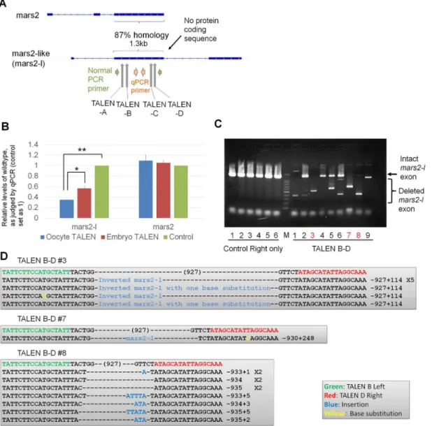
We next asked if long non-coding sequences can be deleted using our strategy (Fig 1A). In order to judge off target effects, we chose a non-coding sequence that has high homology to a protein coding gene. During bioinformatic searches for transcripts upregulated after the mid-blastula transition, we found a transcript that carries 87% sequence similarity with an exon of
mars2(NCBI: NM_001086369.1), but does not seem to encode any proteins (Fig 4A) [28].
Hereafter this transcript is referred to asmars2-like (mars2-l, seeMaterials and Methods). We designed four sets of TALEN pairs spanning an exon of themars2-lgene (TALENs-A and–B at the beginning of exon 2, TALENs-C and -D at the end of exon 2;Fig 4AandS2 Fig). Differ-ent combinations of two TALEN pairs (A-C, A-D, B-C and B-D) were expressed by mRNA injection to one-cell stage embryos (S3A Fig). While all combinations of TALEN pairs allowed deletion of an almost entire exon ofmars2-lin a part of embryos, the combination of TALENs
Fig 3. Double knock-out ofpax6andtyrinXenopusF0 embryos.(A) Double knock-out ofpax6andtyrwas achieved by TALEN mRNA injection to GV oocytes. A double knock-out tadpole is shown. (B) Detection ofpax6andtyrmutants by RFLP analysis. Wild type embryos are cut by restriction enzymes (Ui: uninjected embryo), while mutant embryos are not digested. Digested PCR products appear at lower bands (marked by open arrowheads) and the
undigested is marked by closed arrowheads. M represents 100 bp ladder marker. (C) Sequencing of three different embryos (#3, #6, #9; the numbers correspond to those in Fig 3B) revealed the 100% mutation rate inpax6andtyrdouble knock-out embryos.
B and D showed partial deletion in all samples examined. The off target effect onmars2was not observed (S3A Fig). However, we did not get embryos that have a tetraallelic knock-out of the exon by zygote injection (S3A Fig). In contrast, when TALEN mRNAs (TALEN-B and -D) were injected into oocytes, followed byin vitromaturation and ICSI (S3B Fig), rates of deleting the exon were significantly improved as measured by qPCR analyses (P<0.05,Fig 4B).
Impor-tantly, embryos that carry only mutated exon were obtained by this method (Fig 4C, Red let-ters). The presence of single major mutated bands in lanes 3, 7 and 8 suggests that the same type of deletion was introduced on all alleles at least in these samples (Fig 4C). Sequencing
Fig 4. The deletion of a large genetic locus is achieved by the expression of two sets of TALENs by oocyte injection.(A) TALENs were designed to disrupt one of the exons ofmars2-like (mars2-l) gene. PCR primers were used to confirm gene knock-out. Exons are marked by boxes. (B) The injection of TALEN mRNAs into GV oocytes (blue bar), but not into fertilized embryos (red bar), showed better knock-out of themars2-lexon, as revealed by qPCR analyses. Control represents embryos injected only with right TALEN mRNA. N = 3–4 independent experiments. Error bars are standard errors.*P<0.05. **P<0.01. (C) Expression of TALENs before fertilization allowed the production of an embryo carrying only the deletedmars2-lexon in the F0 generation (lane 3, 7 and 8 in TALEN B-D, red color). Genomic DNA was extracted from single embryos at St. 10.5–11 and subjected to PCR analysis. M represents 1.5
kp and 100 bp ladder marker. (D) Large deletions of exon 2 inmars-lwere confirmed by sequencing. Sample numbers correspond to those in Fig 4C. A part of invertedmars-lsequence was inserted in sample #3.
analysis of target sites confirmed exon deletion or the insertion ofmars2-lsequences (Fig 4D). Off target effects onmars2were not observed (S3C Fig). Together, the expression of two sets of TALEN pairs by oocyte injection enables the deletion of long non-coding sequences in F0 embryos.
Discussion
We here report a method to achieve full knock-out phenotypes inXenopus laevisF0 embryos. The presence of untargeted wild type cells in F0 genome-edited embryos has been an issue after injection of programmable nucleases to one-cell stage embryos. The injection of TALEN mRNAs from the GV oocyte stage reduced unedited cells in F0 embryos as judged bytyr
knock-out, and the embryonic lethal phenotype ofpax6knock-out was recapitulated in most of F0 embryos. These strong knock-out phenotypes seem to be caused by the presence of TALEN proteins by the time of fertilization, which then allows instant access to zygotic chro-matin and to chrochro-matin of embryos at early cleavage stages.
Although the protocol shown here is technically demanding compared to the conventional zygote injection, it instead offers a unique opportunity to obtain embryos showing full knock-out phenotypes in the F0 generation. It is also noteworthy that our strategy allows the disrup-tion of a 1 kb genomic locus, as shown bymars2-ldeletion in F0 embryos. Although the iden-tity ofmars2-lis not defined in this study,mars2-lknock-out experiments in theory suggest that our method can be used to disrupt long non-coding RNA without off target effects. The sequencing analyses of those knocked out embryos revealed several different mutation pat-terns in F0 embryos, implying that TALEN-mediated gene disruption in our system may start to work in early embryos, but not in oocytes. This result is in good agreement with the lack of non-homologus end joining activity in oocyte nuclei [29]. Alternatively, TALENs may keep cutting the target site even after the initial action [30]. In any cases, the observed stronger knock-out phenotypes by the oocyte injection suggest that more abundant and/or evenly dis-tributed TALEN proteins by oocyte injection results in a better knock-out. Considering that frogs normally take more than a year to sexual maturity, gene-manipulated frogs derived from the oocyte injection can be regarded as a rapid route to obtain frogs with full knock-out phe-notypes without waiting for the next generation. Meantime, it is still necessary to perform multi-generation experiments to obtain embryos that carry the same mutant alleles in all cells since embryos produced in this study exhibited several different mutation patterns in F0 embryos, reminiscent of some degree of mosaicism. In addition to the rapid judgement of knock-out phenotypes, our experimental system may be applied for examining the effect of gene disruption on embryonic development at very early stages such as at the midblastula transition since TALENs work from earlier embryonic stages by the oocyte injection route than by the conventional zygote injection [24]. Therefore, our method can serve as a powerful tool for studying development, enabling phenotypic judgement of gene knock-out inXenopus
laevisF0 embryos.
InXenopusresearch the oocyte host transfer technique, in whichin vitromatured oocytes
of the treated embryos [24], in good agreement with our results. It is difficult to compare the efficiency of an entire process between the host transfer and the ICSI system since the develop-ment was not scored in the host transfer report [24]. However, our papers together support the contention that expression of programmable nucleases before fertilization results in full knock-out phenotypes. In the IVM-ICSI system, we can test several combinations of TALENs in one experiment. Thus, our described system may be applicable to a large scale analysis of gene tar-geting using programmable nucleases. Importantly, the IVM-ICSI method does not require frog surgery for transferringin vitromatured eggs, therefore tackling the 3R guidelines for ani-mal care (Replacement, Reduction and Refinement).
Finally, other organisms that are used for the injection of nucleases into early embryos show mosaic knock-out phenotypes in the founder generations [33–35]. As far as the oocyte culture and maturation system is available, our strategy can be easily applied to other species. Since mammals often have established oocyte maturation systems [36], it is worth trying the injec-tion of nucleases into immature oocytes. This is especially true of animals whose life cycle is long such as livestock and non-human primates. Moreover, the CRISPR/Cas9 system might also benefit from the oocyte injection because Cas9 protein is expressed before fertilization and because guide RNA can have a greater chance of gaining access to target sites. Our experimen-tal scheme thus can potentially help the efficient production of genome-edited animals for agri-culture and medical purposes.
Supporting Information
S1 Fig. Genotyping oftyrosinaseTALENs-expressed embryos.Bothtyrosinase-a(tyra) and
tyrosinase-b(tyrb) showed approximately 90% mutation rates. Many deletion mutations show
microhomologies at junctions (red underlines). The patterns of different mutations were less in
tyrathan intyrb. This difference might be caused by recutting of target sites even after the ini-tial non-homologus end joining although it is not clear why this happened preferenini-tially in
tyrb. DNA was extracted from a single embryo (tadpole) and genomic sequences containing target sites of TALENs were amplified by PCR. Primers specific fortyraor those fortyrbwere used. Seven to 10 bacterial clones were picked up and sequenced from each tadpole.
(TIF)
S2 Fig. TALEN target sequences formars2-lknock-out.Sequences ofXenopus laevis mars2-l
andmars2are aligned and target sequences of TALENs formars2-lare marked by different
col-ours. To delete an almost entire exon ofmars2-l, each two sets of TALEN pairs were designed at the beginning (Mars2-l TALEN-A and–B) and at the end (Mars2-l TALEN-C and–D) of
mars2-l.
(TIF)
S3 Fig. Genotyping ofmars2-lTALENs-expressed embryos.(A) Expression of two sets of TALEN pairs designed at the beginning and at the end of an exon ofmars2-l, as shown inFig 4A, resulted in deletion of the exon in a part of embryos while off-target effects onmars2were not observed. The combination of TALENs-B and -D showed reproducible knock-out. TALENs were injected into fertilized embryos. Each lane represents a result from a single embryo. (B) Development of TALEN mRNAs-injected oocytes, followed byin vitromaturation and sperm injection. Actual numbers of embryos that reached each developmental stages are indicated next to the corresponding bars. (C) Off-target effects of expressing TALENs B-D from the immature oocyte stage onmars2were not observed in embryos at Stage 11. As a con-trol, only right TALENs were injected.
S1 Table. TALEN target sequences and primer sequences.Red letters represent TALEN tar-get sequences.
(DOC)
Acknowledgments
K.M. was a Research Fellow at Wolfson College and was supported by the Herchel Smith Post-doctoral Fellowship.
Author Contributions
Conceived and designed the experiments: KM KTS. Performed the experiments: KM MS YS TS SH DS JJ KTS. Analyzed the data: KM MS TS YS AS KTS. Contributed reagents/materials/ analysis tools: TS TY. Wrote the paper: KM KTS JBG. Supervised research: JBG TY.
References
1. 1. Huang P, Xiao A, Zhou M, Zhu Z, Lin S, Zhang B. Heritable gene targeting in zebrafish using custom-ized TALENs. Nat Biotechnol. 2011; 29: 699–700. doi:10.1038/nbt.1939PMID:21822242
2. Sander JD, Cade L, Khayter C, Reyon D, Peterson RT, Joung JK, et al. Targeted gene disruption in somatic zebrafish cells using engineered TALENs. Nat Biotechnol. 2011; 29: 697–698. doi:10.1038/ nbt.1934PMID:21822241
3. Tesson L, Usal C, Menoret S, Leung E, Niles BJ, Remy S, et al. Knockout rats generated by embryo microinjection of TALENs. Nat Biotechnol. 2011; 29: 695–696. doi:10.1038/nbt.1940PMID:21822240
4. Watanabe T, Ochiai H, Sakuma T, Horch HW, Hamaguchi N, Nakamura T, et al. Non-transgenic genome modifications in a hemimetabolous insect using zinc-finger and TAL effector nucleases. Nat Commun. 2012; 3: 1017. doi:10.1038/ncomms2020PMID:22910363
5. Guo X, Zhang T, Hu Z, Zhang Y, Shi Z, Wang Q, et al. Efficient RNA/Cas9-mediated genome editing in Xenopus tropicalis. Development. 2014; 141: 707–714. doi:10.1242/dev.099853PMID:24401372
6. Nakayama T, Fish MB, Fisher M, Oomen-Hajagos J, Thomsen GH, Grainger RM. Simple and efficient CRISPR/Cas9-mediated targeted mutagenesis in Xenopus tropicalis. Genesis. 2013; 51: 835–843.
PMID:24123613
7. Blitz IL, Biesinger J, Xie X, Cho KW. Biallelic genome modification in F(0) Xenopus tropicalis embryos using the CRISPR/Cas system. Genesis. 2013; 51: 827–834. doi:10.1002/dvg.22719PMID:24123579
8. Hwang WY, Fu Y, Reyon D, Maeder ML, Tsai SQ, Sander JD, et al. Efficient genome editing in zebra-fish using a CRISPR-Cas system. Nat Biotechnol. 2013; 31: 227–229. doi:10.1038/nbt.2501PMID: 23360964
9. Wang H, Yang H, Shivalila CS, Dawlaty MM, Cheng AW, Zhang F, et al. One-step generation of mice carrying mutations in multiple genes by CRISPR/Cas-mediated genome engineering. Cell. 2013; 153: 910–918. doi:10.1016/j.cell.2013.04.025PMID:23643243
10. Harland RM, Grainger RM. Xenopus research: metamorphosed by genetics and genomics. Trends Genet. 2011; 27: 507–515. doi:10.1016/j.tig.2011.08.003PMID:21963197
11. Young JJ, Cherone JM, Doyon Y, Ankoudinova I, Faraji FM, Lee AH, et al. Efficient targeted gene dis-ruption in the soma and germ line of the frog Xenopus tropicalis using engineered zinc-finger nucle-ases. Proc Natl Acad Sci U S A. 2011; 108: 7052–7057. doi:10.1073/pnas.1102030108PMID: 21471457
12. Suzuki KT, Isoyama Y, Kashiwagi K, Sakuma T, Ochiai H, Sakamoto N, et al. High efficiency TALENs enable F0 functional analysis by targeted gene disruption in Xenopus laevis embryos. Biol Open. 2013; 2: 448–452. doi:10.1242/bio.20133855PMID:23789092
13. Ishibashi S, Cliffe R, Amaya E. Highly efficient bi-allelic mutation rates using TALENs in Xenopus tropi-calis. Biol Open. 2012; 1: 1273–1276. doi:10.1242/bio.20123228PMID:23408158
14. Lei Y, Guo X, Liu Y, Cao Y, Deng Y, Chen X, et al. Efficient targeted gene disruption in Xenopus embryos using engineered transcription activator-like effector nucleases (TALENs). Proc Natl Acad Sci U S A. 2012; 109: 17484–17489. doi:10.1073/pnas.1215421109PMID:23045671
16. Sakane Y, Sakuma T, Kashiwagi K, Kashiwagi A, Yamamoto T, Suzuki KT. Targeted mutagenesis of multiple and paralogous genes in Xenopus laevis using two pairs of transcription activator-like effector nucleases. Dev Growth Differ. 2014; 56: 108–114. doi:10.1111/dgd.12105PMID:24329851
17. Jullien J, Miyamoto K, Pasque V, Allen GE, Bradshaw CR, Garrett NJ, et al. Hierarchical molecular events driven by oocyte-specific factors lead to rapid and extensive reprogramming. Mol Cell. 2014; 55: 524–536. doi:10.1016/j.molcel.2014.06.024PMID:25066233
18. Miyamoto K, Teperek M, Yusa K, Allen GE, Bradshaw CR, Gurdon JB. Nuclear Wave1 is required for reprogramming transcription in oocytes and for normal development. Science. 2013; 341: 1002–1005.
doi:10.1126/science.1240376PMID:23990560
19. Nakade S, Tsubota T, Sakane Y, Kume S, Sakamoto N, Obara M, et al. Microhomology-mediated end-joining-dependent integration of donor DNA in cells and animals using TALENs and CRISPR/Cas9. Nat Commun. 2014; 5: 5560. doi:10.1038/ncomms6560PMID:25410609
20. Nakayama T, Fisher M, Nakajima K, Odeleye AO, Zimmerman KB, Fish MB, et al. Xenopus pax6 mutants affect eye development and other organ systems, and have phenotypic similarities to human aniridia patients. Dev Biol. 2015.
21. Liu Y, Luo D, Zhao H, Zhu Z, Hu W, Cheng CH. Inheritable and precise large genomic deletions of non-coding RNA genes in zebrafish using TALENs. PLoS One. 2013; 8: e76387. doi:10.1371/journal.pone. 0076387PMID:24130773
22. Amaya E, Kroll KL. A method for generating transgenic frog embryos. Methods Mol Biol. 1999; 97: 393–414. PMID:10443381
23. Miyamoto K, Simpson D, Gurdon JB. Manipulation and in vitro maturation of Xenopus laevis oocytes, followed by intracytoplasmic sperm injection, to study embryonic development. J Vis Exp. 2015: e52496. doi:10.3791/52496PMID:25742326
24. Nakajima K, Yaoita Y. Highly efficient gene knockout by injection of TALEN mRNAs into oocytes and host transfer in Xenopus laevis. Biol Open. 2015; 4: 180–185. doi:10.1242/bio.201410009PMID: 25596277
25. James-Zorn C, Ponferrada VG, Jarabek CJ, Burns KA, Segerdell EJ, Lee J, et al. Xenbase: expansion and updates of the Xenopus model organism database. Nucleic Acids Res. 2013; 41: D865–870. doi: 10.1093/nar/gks1025PMID:23125366
26. Wang F, Shi Z, Cui Y, Guo X, Shi YB, Chen Y. Targeted gene disruption in Xenopus laevis using CRISPR/Cas9. Cell Biosci. 2015; 5: 15. doi:10.1186/s13578-015-0006-1PMID:25897376
27. Rungger-Brandle E, Ripperger JA, Steiner K, Conti A, Stieger A, Soltanieh S, et al. Retinal patterning by Pax6-dependent cell adhesion molecules. Dev Neurobiol. 2010; 70: 764–780. doi:10.1002/dneu. 20816PMID:20556827
28. Herberg S, Simeone A, Oikawa M, Jullien J, Bradshaw CR, Teperek M, et al. Histone H3 lysine 9 tri-methylation is required for suppressing the expression of an embryonically activated retrotransposon in Xenopus laevis. Sci Rep. 2015; 5: 14236. doi:10.1038/srep14236PMID:26387861
29. Hagmann M, Adlkofer K, Pfeiffer P, Bruggmann R, Georgiev O, Rungger D, et al. Dramatic changes in the ratio of homologous recombination to nonhomologous DNA-end joining in oocytes and early embryos of Xenopus laevis. Biol Chem Hoppe Seyler. 1996; 377: 239–250. PMID:8737989
30. Carroll D, Beumer KJ. Genome engineering with TALENs and ZFNs: repair pathways and donor design. Methods. 2014; 69: 137–141. doi:10.1016/j.ymeth.2014.03.026PMID:24704173
31. Heasman J, Crawford A, Goldstone K, Garner-Hamrick P, Gumbiner B, McCrea P, et al. Overexpres-sion of cadherins and underexpresOverexpres-sion of beta-catenin inhibit dorsal mesoderm induction in early Xeno-pus embryos. Cell. 1994; 79: 791–803. PMID:7528101
32. Hulstrand AM, Schneider PN, Houston DW. The use of antisense oligonucleotides in Xenopus oocytes. Methods. 2010; 51: 75–81. doi:10.1016/j.ymeth.2009.12.015PMID:20045732
33. Yen ST, Zhang M, Deng JM, Usman SJ, Smith CN, Parker-Thornburg J, et al. Somatic mosaicism and allele complexity induced by CRISPR/Cas9 RNA injections in mouse zygotes. Dev Biol. 2014; 393: 3–
9. doi:10.1016/j.ydbio.2014.06.017PMID:24984260
34. Dahlem TJ, Hoshijima K, Jurynec MJ, Gunther D, Starker CG, Locke AS, et al. Simple methods for gen-erating and detecting locus-specific mutations induced with TALENs in the zebrafish genome. PLoS Genet. 2012; 8: e1002861. doi:10.1371/journal.pgen.1002861PMID:22916025
35. Irion U, Krauss J, Nusslein-Volhard C. Precise and efficient genome editing in zebrafish using the CRISPR/Cas9 system. Development. 2014; 141: 4827–4830. doi:10.1242/dev.115584PMID: 25411213