RBCCV 44205-1497 DOI: 10.5935/1678-9741.20130075
Pressure support-ventilation versus spontaneous
breathing with “T-Tube” for interrupting the
ventilation after cardiac operations
Pressão de suporte ventilatório versus respiração espontânea em “Tubo-T” para a interrupção da
ventilação após as operações cardíacas
Isabela Scali Lourenço
1, MSc; Aline Marques Franco
1, MSc; Solange Bassetto
1; Alfredo José
Rodrigues
1, MD, PhD
1 Universidade de São Paulo, Faculdade de Medicina de Ribeirão Preto (FMRP-USP), Ribeirão Preto, SP, Brazil.
Work carried out at Hospital das Clínicas da Faculdade de Medicina de Ribeirão Preto da Universidade de São Paulo (HCFMRP-USP), Ribeirão Preto, SP, Brazil.
No inancial support.
Correspondence address: Alfredo José Rodrigues
Hospital das Clínicas da Faculdade de Medicina de Ribeirão Preto Departamento de Cirurgia e Anatomia.
Av, Bandeirantes, 3.900, Campus Universitário-Monte Alegre Ribeirão Preto, SP, Brazil - Zip code: 14048-900
E-mail: [email protected]
Article received on August, 31th, 2012
Article accepted on September 2nd, 2013 Abstract
Objective: To compare pressure-support ventilation with spontaneous breathing through a T-tube for interrupting inva-sive mechanical ventilation in patients undergoing cardiac sur-gery with cardiopulmonary bypass.
Methods: Adults of both genders were randomly allocated to 30 minutes of either pressure-support ventilation or sponta-neous ventilation with “T-tube” before extubation. Manovac-uometry, ventilometry and clinical evaluation were performed before the operation, immediately before and after extubation, 1h and 12h after extubation.
Results: Twenty-eight patients were studied. There were no deaths or pulmonary complications. The mean aortic clamping time in the pressure support ventilation group was 62 ± 35 min-utes and 68 ± 36 minmin-utes in the T-tube group (P=0.651). The mean cardiopulmonary bypass duration in the pressure-sup-port ventilation group was 89 ± 44 minutes and 82 ± 42 min-utes in the T-tube group (P=0.75). The mean Tobin index in the pressure support ventilation group was 51 ± 25 and 64.5 ± 23 in the T-tube group (P=0.153). The duration of intensive care unit stay for the pressure support ventilation group was 2.1 ± 0.36 days and 2.3 ± 0.61 days in the T-tube group (P=0.581). The atelectasis score in the T-tube group was 0.6 ± 0.8 and 0.5 ± 0.6 (P=0.979) in the pressure support ventilation group. The study
groups did not differ signiicantly in manovacuometric and ven -tilometric parameters and hospital evolution.
Conclusion: The two trial methods evaluated for interrup-tion of mechanical ventilainterrup-tion did not affect the postoperative course of patients who underwent cardiac operations with car-diopulmonary bypass.
Descriptors: Pulmonary ventilation. Extracorporeal circula-tion. Ventilator weaning.
Resumo
Objetivo: Comparar a pressão de suporte ventilatório com a respiração espontânea em “Tubo-T” para interrupção da ven-tilação invasiva em pacientes submetidos à operação cardíaca.
Métodos: Adultos de ambos os sexos foram alocados para pressão de suporte ventilatório por 30 minutos ou o mesmo pe-ríodo de ventilação espontânea com “Tubo-T” antes da extu-bação. Realizou-se manovacuometria, ventilometria e avaliação clínica antes da operação, imediatamente antes e após a extuba-ção, 1h e 12h após extubação.
Ribeirão Preto Medical School, University of São Paulo (HCFMRP-USP) and was approved by the Ethics Committee (process number 5672/2006).
Patients
We recruited 30 patients with coronary artery disease and/ or valve disease, of both genders and older than 18 years-old.
The basic protocols of perioperative care were not modiied,
with the exception of procedures for extubation. The volunteers were randomized according to a random number table generated by StatMate GraphPad 1.01 (GraphPad Software, Inc, San Diego, CA, USA). The StatMate software generated a random sequence of 15 numbers “1”
(T-tube group) and 15 numbers “2” (PSV group). The irst
patient to participate in the study was allocated to the group
corresponding to the irst number generated, the second
patient to the group corresponding to the second randomly generated number and so on.
The exclusion criteria were postoperative bleeding requiring reoperation in the immediate postoperative
period, ejection fraction ≤0.40, postoperative hemodynamic
instability precluding extubation, not understanding the procedures proposed and refusal to participate in the study at any stage.
Study groups
The PSV group was comprised of patients who underwent a period of PSV for 30 minutes before interruption of IMV. These patients were extubated immediately after the IMV period with PSV.
The T-tube group was comprised of patients who were disconnected from the ventilator when they met the criteria for interrupting the IMV then kept under spontaneous ventilation with their tracheal tube connected to a T-tube while receiving supplemental oxygen for 30 minutes before extubation.
Clinical history, baseline measurements of blood INTRODUCTION
Invasive mechanical ventilation (IMV) is often essential
in the irst hours after cardiovascular operations as patients
recover from anesthesia and reestablish homeostatic balance. When IMV is no longer required, the respiratory therapist and the physician must decide the most appropriate method to interrupt IMV.
A simple and widespread method to determine whether a patient tolerates the discontinuation of ventilatory support is a trial of spontaneous breathing [1,2]. According to the III Brazilian Consensus on Mechanical Ventilation this trial is straightforward and effective way to wean off IMV [3]. However, the spontaneous breathing trial has been replaced by other techniques, mainly by pressure support ventilation (PSV) and synchronized intermittent mandatory ventilation (SIMV) [4]. These techniques are optional modes of ventilatory support provided by modern ventilators, which are especially useful for weaning patients recovering from pulmonary dysfunctions who require prolonged IMV. Yet, there is limited evidence that such methods of transition from IMV are superior to spontaneous breathing through a T-tube followed by extubation [1], especially in patients with good cardiopulmonary reserve.
Therefore, this study aims to compare PSV with a spontaneous breathing trial using a T-tube for weaning from IMV in patients who underwent cardiovascular operations to correct valve dysfunction and/or coronary artery bypass grafting surgery. We considered weaning as the transition from controlled ventilation to spontaneous breathing before extubation.
METHODS
This prospective randomized trial was conducted in the Division of Thoracic and Cardiovascular Surgery of the
Abbreviations, acronyms & symbols
CPB Cardiopulmonary bypass EP Expiratory maximal pressure IMV Invasive mechanical ventilation IP Inspiratory maximal pressure PSV Pressure support ventilation
SIMV Synchronized intermittent mandatory ventilation
minutos e de 68 ± 36 minutos para o “Tubo-T” (P=0,651). O tempo de CEC no grupo suporte ventilatório foi 89 ± 44 minutos e para o “Tubo-T” de 82 ± 42 minutos (P=0,75). O índice de To-bin para o grupo suporte ventilatório foi 51 ± 25 e para o grupo
“Tubo-T”, 64,5 ± 23 (P=0,153). O tempo na unidade de terapia intensiva para o grupo suporte ventilatório foi 2,1 ± 0,36 dias e para o grupo “Tubo-T”, 2,3±0,61 dias (P=0,581). O escore de atelectasia para o grupo “Tubo-T” foi 0,6 ± 0,8 e para o suporte ventilatório foi 0,5 ± 0,6 (P=0,979). Não houve diferença signii -cativa na evolução clínica e nos valores de gasometria, manova-cuometria e ventilometria entre ambos os grupos.
Conclusão: O método utilizado para testar a adequação da interrupção da ventilação mecânica invasiva não afetou a evo-lução pós-operatória dos pacientes submetidos a operações car-díacas com circulação extracorpórea.
pressure, heart rate, respiratory rate, minute volume, tidal
volume, vital capacity, peak low, maximal inspiratory
pressure, and expiratory pressure were obtained from all patients before surgery. All patients underwent conventional chest physical therapy consisting of diaphragmatic breathing exercises associated with active and/or active-assisted mobilization of the upper and lower limbs. In addition, all patients participated in daily respiratory therapy sessions including cough, lung expansion maneuvers and airway clearance techniques training twice a day in the preoperative and postoperative periods.
The protocol for postoperative analgesia was the same for all patients. Postoperatively all the patients received IMV using a Savina ventilator (Dräger, Lübeck, Germany) with SIMV, 12– 14 bpm, inspiration/expiration ratio of 1:2, PEEP of 5 cmH2O, tidal volume of 8 mL/kg body weight, and inspired fraction of O2 to maintain arterial oxygen saturation above 95% (pulse oximetry). Before interrupting the IMV, arterial gasometry and hemodynamic parameters were checked. All evaluations were performed in the preoperative period, immediately before extubation, and 1 and 12 hours after extubation. All patients had daily follow-ups until hospital discharge.
The criteria for interrupting IMV were: a) patient should be conscious and cooperative; b) PaO2: 80–100 mmHg, arterial saturation >95%, pH: 7.35–7.45, and PaCO2: 35-45
mmHg; c) tidal volume ≥4 mL/kg; and d) inspired fraction of
O2 ≤0.4. The Tobin index [5] was calculated for both groups before extubation.
Patients in the PSV group who fulilled the criteria for
interrupting IMV were submitted to 30 minutes of pressure support ventilation of 10 cmH2O. Patients in the T-tube group
who fulilled the criteria for interrupting IMV were allowed
to breath spontaneously through their tracheal tube connected to a T-piece and received supplemental O2 (aerosol with
0.9% saline and oxygen low to 10L/min) for 30 minutes.
At the end of the trial period, a blood sample for gasometric analysis was collected and ventilometric and hemodynamic parameters were measured. The patients were then extubated. A clinical and laboratory evaluation was performed again 1 and 12 hours after extubation.
A physician, blinded to the study, compared the preoperative chest radiograph with a radiograph obtained in the morning after the operation. The following scores and criteria were used to grade lung atelectasis: 0) no abnormality: no image suggestive of atelectasis; 1) laminar atelectasis: linear opacities located mainly in lung bases; 2) segment atelectasis: opacities compatible with pulmonary segments; 3) lobar atelectasis; and 4) whole lung atelectasis.
Statistical analysis
The results were expressed as mean ± standard deviation or percentages. The Shapiro-Wilk test was used to determine the data distribution (normality). Paired or unpaired “T” tests
were used for continuous variables with normal distribution, otherwise we used the Mann-Whitney or Wilcoxon test. For comparing proportions, we used the Fisher exact test. To compare intra-and inter-group repeated measurements (three
or more), we used a two-way ANOVA; the irst measurement
was obtained postoperatively, or the pre-operative measure, when available, served as the control/baseline against which the later measurements were compared. Statistical analysis was performed using SPSS software version 18.0 (SPSS Inc.,
Chicago, IL, USA) with a signiicance level of 0.05.
RESULTS
Clinical characteristics
Data from 28 patients, 14 in each group, were analyzed. One patient in the PSV group was excluded due to postoperative bleeding requiring reoperation, and one patient in the T-tube group who needed prolonged (>24h) invasive ventilation due to hemodynamic instability was excluded. The demographic data are shown in Table 1. There were no
signiicant differences between groups.
In the PSV group, 10 (71%) patients underwent revascularization and 4 (29%) underwent valve surgery. In the T-tube group, 11 (78%) patients underwent revascularization, two (14%) had valvular surgery and one underwent revascularization plus valvular surgery. The differences in the distribution of type of surgery were not
signiicant (P=0.648).
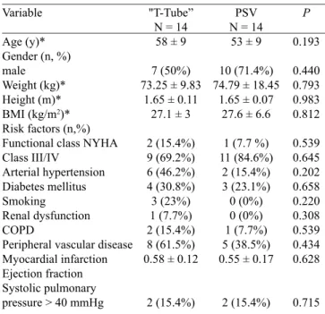
Table 1. Clinical characteristics. Variable
Age (y)* Gender (n, %) male
Weight (kg)* Height (m)*
BMI (kg/m2)*
Risk factors (n,%) Functional class NYHA Class III/IV Arterial hypertension Diabetes mellitus Smoking Renal dysfunction COPD
Peripheral vascular disease Myocardial infarction Ejection fraction Systolic pulmonary pressure > 40 mmHg
"T-Tube” N = 14
58 ± 9
7 (50%) 73.25 ± 9.83
1.65 ± 0.11 27.1 ± 3
2 (15.4%) 9 (69.2%) 6 (46.2%) 4 (30.8%) 3 (23%) 1 (7.7%) 2 (15.4%) 8 (61.5%) 0.58 ± 0.12
2 (15.4%)
PSV N = 14 53 ± 9
10 (71.4%) 74.79 ± 18.45
1.65 ± 0.07 27.6 ± 6.6
1 (7.7 %) 11 (84.6%) 2 (15.4%) 3 (23.1%) 0 (0%) 0 (0%) 1 (7.7%) 5 (38.5%) 0.55 ± 0.17
2 (15.4%) P 0.193 0.440 0.793 0.983 0.812 0.539 0.645 0.202 0.658 0.220 0.308 0.539 0.434 0.628 0.715
The mean aortic clamping time of the PSV group was 62 ± 35 minutes and 68 ± 36 minutes in the T-tube group (P=0.651, Mann-Whitney test). The mean cardiopulmonary bypass (CPB) times were 89 ± 44 minutes and 82 ± 42 minutes in the PSV and T-tube groups, respectively (P=0.75, Mann-Whitney test).
The mean Tobin index immediately before extubation in the PSV group was 51.1 ± 25 and 64.5 ± 23 in the T-tube group (P=0.153).
Clinical and radiologic postoperative evolution The mean duration of stay in the postoperative intensive care unit was 2.1 ± 0.36 days in the PSV group and 2.3 ± 0.61 days in the T-tube group (P=0.581, Mann-Whitney test). The mean duration of hospital stay for the PSV and T-tube groups, respectively, was 9.6 ± 4.83 days and 8.6 ± 2.8 days (P=0.829, Mann-Whitney test). There were no deaths and no patient required reintubation. One patient (T-tube group) experienced renal dysfunction, which was managed without dialysis (P=0.308). Forty-three percent of the patients had some degree of atelectasis. The mean atelectasis score in the T-tube group was 0.6 ± 0.8 and 0.5 ± 0.6 in the PSV group (P=0.979, Mann-Whitney test).
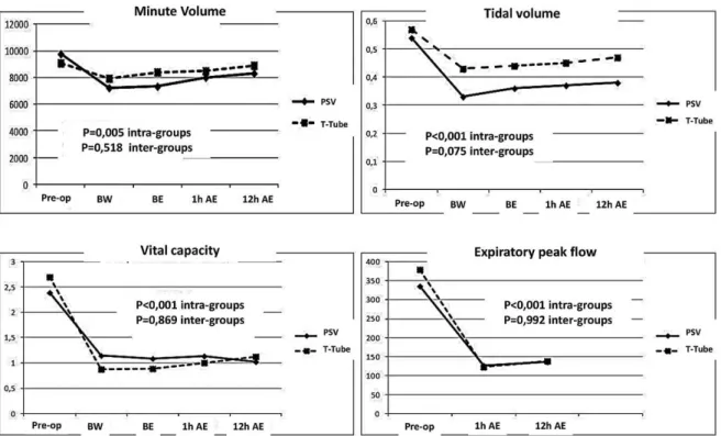
Postoperative ventilatory parameters
In both groups the minute volume, tidal volume, vital
capacity and peak expiratory low declined signiicantly
postoperatively, compared with the preoperative period (Figure 1).
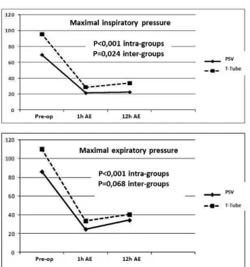
There was a signiicant decrease in both groups in the
inspiratory (IP) and expiratory (EP) maximal pressures after extubation compared with preoperative values (Figure
2). However, we found that the IP was signiicantly lower
in the PSV group (P=0.024). Notably, patients in this
group already had a signiicantly lower IP preoperatively
(P=0.028). As a result, the temporal pattern was similar in both groups, since there was no interaction between groups (P=0.150). Similar to the IP, the mean preoperative
maximum EP was signiicantly lower in the PSV group
(P=0.035). The differences between groups were not
statistically signiicant (P=0.068) and the temporal pattern was similar in both groups (P=0.133).
There were signiicant changes in the postoperatively
respiratory rate, heart rate, PaO2 and arterial oxygen saturation compared to the preoperative values, but the temporal patterns were similar in both groups and the differences were
not signiicant (Figure 3).
DISCUSSION
We found that there was no signiicant difference in
weaning from IMV using a trial period of either PSV or spontaneous breathing using a T-tube piece in low-risk patients who underwent valve and/or coronary artery bypass grafting surgery.
The main goal of a weaning trial is to identify patients who are able to breathe without a ventilator with the minimum risk of extubation failure and its potential complications [6]. Even though many institutions that perform cardiovascular surgery have routinely used pressure support as a weaning
trial before extubation, there is no consensus that a speciic
method of weaning is superior. The majority of patients can be successfully weaned from mechanical ventilation irrespective of whether this is executed by intermittent mandatory ventilation, pressure support, or a T-tube trial [7,8]; a spontaneous breathing trial using a T-tube is still routinely
performed in patients who fulill weaning criteria [9].
A study reported by IMV Esteban et al. [10], which compared four methods of weaning, found that once-daily trials of spontaneous breathing led to about three times more rapid extubation than intermittent mandatory ventilation and was about twice as rapid as PSV. There are hospitals
Fig. 2 - Perioperative evolution of the maximal inspiratory and expiratory pressures. Preo-op: preoperative; 1h AE: 1 hour after extubation; 12h AE: 12 h after extubation
Fig. 3 - Perioperative evolution of the respiratory rate, heart rate, PO2 and arterial oxygen saturation. Preo-op:
Author’s roles & responsibilities
ISL Author AMF Co-author SB Co-author AJR Co-author
in developing countries that may have limited resources and/or more simple mechanical ventilators. Therefore, in a resource limited setting more rapid weaning might result in
more eficient use of scarce ventilators and a shorter period
of intubation related discomfort.
PSV is a form of ventilatory support provided during IMV in which a predetermined, constant, positive inspiratory pressure is maintained by the ventilator, while the patient controls the respiratory cycle. In this ventilatory mode the
patient controls the respiratory rate, the inspiratory low, and
the inspiration/expiration ratio, thereby reducing the oxygen demand as a consequence of reduced respiratory muscle work. It also provides a better synchrony between patient and ventilator [11,12]. Although the use of pressure support has
been justiied by reducing the imposed work of the ventilator
circuit and the endotracheal tube [13], the use of even low levels of pressure support may lead to an underestimation of the risk of extubation failure [8]. Hence, a spontaneous breathing trial using a T-tube might be especially interesting in a population in which the risk of reintubation is particularly high [14].
The present study demonstrated that several parameters
of respiratory function were lower in the irst hours after
extubation than they were preoperatively. These declines resulted primarily from pain and changes related to the anesthesia, CPB and the use of mechanical ventilation, as observed by other investigators [15-18]. Thus, even though spontaneous breathing with a T-tube may be an adequate method for weaning from mechanical ventilation in the majority of the cases, the method might confer a higher risk for reintubation [19], especially in patients with less cardiorespiratory reserve.
Extubation failure seems to be determined more by the conditions inherent to the patient than by the method of weaning from the ventilator [9,20,21]. Pain, a major factor in the postoperative period [22], induces ventilation with smaller amplitude in an attempt to minimize discomfort. Moreover, the residual effect of anesthetic drugs and analgesics on the central nervous system also contributes to this breathing pattern. However, the respiratory parameters tend to improve gradually in the subsequent hours after surgery. The two methods for interruping IMV that we evaluated had no
inluence on the postoperative evolution of such parameters. The signiicant difference of maximal inspiratory
pressures that we observed between the groups was probably caused by the fact that patients in the T-tube group had higher ventilatory pressure preoperatively; however there was no apparent effect on the postoperative evolution in favor of
this group. Additionally, there was no signiicant difference
in the Tobin index [5] between the groups, ensuring a safe interruption of IMV; hence, the expected extubation success was similar for both groups.
The incidence of pulmonary complications in the
postoperative period of heart surgery depends on the diagnostic technique. Vargas et al. [23], in a prospective study using chest computed tomography scans, found that 86.7% of the patients who underwent CABG had some degree of pulmonary atelectasis in the second postoperative day. We believe that in our study the incidence of atelectasis was lower due to the lower sensitivity of chest radiographs to detect atelectasis. However, the extent of atelectasis, as
measured by atelectasis scores, did not differ signiicantly
between the methods used for interrupting the IMV, even though the PSV method had a greater theoretical potential to reduce the incidence and/or severity of atelectasis.
Postoperative pulmonary atelectasis is multifactorial: anesthesia, cardiopulmonary bypass, type of operation performed, preoperatively pulmonary function and mode and the duration of IMV play a role. It is unlikely that a short trial period of pressure support ventilation or spontaneous breathing without airway pressure before extubation would noticeably
inluence the incidence of postoperative pulmonary atelectasis
in patients with good cardiopulmonary functional reserve. Although we believe our study contributes to demonstrate the safety of using T-tube supported spontaneous breathing for weaning from IMV after cardiac surgery, it is not free of limitations. It is a study with a small sample of low-risk patients with good cardiopulmonary reserve, whose mean age was below 60 years old, and with uneventful operations. Because changing from mechanical to spontaneous ventilation increases preload and afterload [24] and because cardiac dysfunction is probably one of the most common causes of weaning failure [25,26], studies with larger numbers of patients at higher risk of cardiac and/or pulmonary dysfunction, including the elderly
(≥ 65 years), are required to evaluate the methods for weaning
from IMV in patients undergoing cardiovascular operations with greater external validity.
In conclusion, our results showed that in low-risk patients who underwent cardiac surgery with cardiopulmonary bypass the method used to interrupt invasive mechanical ventilation, a short trial of either spontaneous breathing through a T-tube
or pressure support ventilation, did not signiicantly affect
the postoperative course.
REFERENCES
2. Piotto RF, Maia LN, Machado MN, Orrico SP. Effects of the use of mechanical ventilation weaning protocol in the Coronary Care Unit: randomized study. Rev Bras Cir Cardiovasc. 2011;26(2):213-21.
3. Goldwasser R, Farias A, Freitas EE, Saddy F, Amado V, Okamoto V. III Consenso Brasileiro de Ventilação Mecânica. Desmame e interrupção da ventilação mecânica. J Bras Pneumol. 2007;33(Supl 2):S128-S36.
4. Gambaroto G. Fisioterapia respiratória em unidade de terapia intensiva. 1ª ed. São Paulo: Atheneu; 2006.
5. Tobin MJ. Predicting weaning outcome. Chest. 1988;94(2):227-8.
6. Frutos-Vivar F, Esteban A, Apezteguia C, González M, Arabi Y, Restrepo MI, et al. Outcome of reintubated patients after scheduled extubation. J Crit Care. 2011;26(5):502-9.
7. Esteban A, Aláa I, Gordo F, Fernández R, Solsona JF, Vallverdú I, et al. Extubation outcome after spontaneous breathing trials with T-tube or pressure support ventilation. The Spanish Lung Failure Collaborative Group. Am J Respir Crit Care Med. 1997;156(2 Pt 1):459-65.
8. Tobin MJ. Extubation and the myth of “minimal ventilator settings”. Am J Respir Crit Care Med. 2012;185(4):349-50.
9. Thille AW, Harrois A, Schortgen F, Brun-Buisson C, Brochard L. Outcomes of extubation failure in medical intensive care unit patients. Crit Care Med. 2011;39(12):2612-8.
10. Esteban A, Frutos F, Tobin MJ, Alía I, Solsona JF, Valverdú I, et al. A comparison of four methods of weaning patients from mechanical ventilation. Spanish Lung Failure Collaborative Group. N Engl J Med. 1995;332(6):345-50.
11. Annat G, Viale JP. Measuring the breathing workload in mechanically ventilated patients. Intensive Care Med. 1990;16(7):418-21.
12. Mancebo J, Amaro P, Mollo JL, Lorino H, Lemaire F, Brochard L. Comparison of the effects of pressure support ventilation delivered by three different ventilators during weaning from
mechanical ventilation. Intensive Care Med. 1995;21(11):913-9.
13. Brochard L, Rua F, Lorino H, Lemaire F, Harf A. Inspiratory pressure support compensates for the additional work of breathing caused by the endotracheal tube. Anesthesiology. 1991;75(5):739-45.
14. Thille AW, Cortés-Puch I, Esteban A. Weaning from the ventilator and extubation in ICU. Curr Opin Crit Care. 2013;19(1):57-64.
15. Giacomazzi MC, Lagni VB, Monteiro MB. A dor pós-operatória
como contribuinte o prejuízo na função pulmonar em pacientes submetidos à cirurgia cardíaca. Rev Bras Cir Cardiovasc. 2006;21(4):386-92.
16. Matte P, Jacquet L, Van Dyck M, Goenen M. Effects of conventional physiotherapy continuous positive airway pressure and non-invasive ventilatory support with bilevel positive airway pressure after coronary artery bypass grafting. Acta Anaesthesiol Scand. 2000;44(1):75-81.
17. Pasquina P, Merlani P, Granier JM, Ricou B. Continuous positive airway pressure versus noninvasive pressure support ventilation to treat atelectasis after cardiac surgery. Anesth Analg. 2004;99(4):1001-8.
18. Wynne R, Botti M. Postoperative pulmonary dysfunction in adults after cardiac surgery with cardiopulmonary bypass:
clinical signiicance and implications for practice. Am J Crit
Care. 2004;13(5):384-93.
19. Assunção MSC, Machado FR, Rosseti HB, Penna HG, Serrão CCA, Silva WG, et al. Evaluation of T tube trial as a strategy of weaning from mechanical ventilation. Rev Bras Ter Intensiva. 2006;18(2):121-5.
20. Epstein SK, Ciubotaru RL, Wong JB. Effect of failed extubation on the outcome of mechanical ventilation. Chest. 1997;112(1):186-92.
21. Mokhlesi B, Tulaimat A, Gluckman TJ, Wang Y, Evans AT, Corbridge TC. Predicting extubation failure after successful completion of a spontaneous breathing trial. Respir Care. 2007;52(12):1710-7.
22. Mueller XM, Tinguely F, Tevaearai HT, Revelly JP, Chioléro R, von Segesser LK. Pain location, distribution, and intensity after cardiac surgery. Chest. 2000;118(2):391-6.
23. Vargas FS, Uezumi KK, Jatene FB, Terra-Filho M, Hueb W, Cukier A, et al. Acute pleuropulmonary complications detected by computed tomography following myocardial revascularization. Rev Hosp Clin Fac Med S Paulo. 2002;57(4):135-42.
24. Buda AJ, Pinsky MR, Ingels NB Jr, Daughters GT 2nd, Stinson EB, Alderman EL. Effect of intrathoracic pressure on left ventricular performance. N Engl J Med. 1979;301(9):453-9.
25. Cabello B, Thille AW, Roche-Campo F, Brochard L, Gómez FJ, Mancebo J. Physiological comparison of three spontaneous
breathing trials in dificult-to-wean patients. Intensive Care Med.
2010;36(7):1171-9.