Orofacial myofunctional characterization in Parry-Romberg
syndrome
Caracterização miofuncional orofacial na síndrome de Parry-Romberg
Fernanda Chiarion Sassi1, Laura Davison Mangilli2, Bruna Rainho Rocha3, Claudia Regina Furquim de Andrade1
ABSTRACT
Purpose: To characterize the orofacial miofunctional aspects of patients with Parry-Romberg syndrome, using standardized clinical protocols and Surface Electromyography (sEMG) of the masticatory muscles.
Methods: Participants were four patients with Parry-Romberg syndro-me and four healthy individuals, paired by age and gender, divided in two groups: Study Group (SG) and Control Group (CG), respectively. The groups were compared regarding performance during clinical examination - Orofacial Myofunctional Evaluation with Expanded Scores (OMES-E) and record of facial anthropometric measures and of jaw movements, and during an instrumental evaluation - Surface Elec-tromyography (sEMG) of the masticatory muscles. Results: Statistically significant differences between the groups were observed for the follo-wing variables: 1) numerical: mandibular lateral movement to the right, OMES-E posture/position and total score, 2) categorical: palate, behavior of the tongue during swallowing and chewing function. At sEMG no statistically significant differences were observed. Conclusion: The pre-sent study is the first to evaluate the orofacial myofunctional system of individuals with Parry-Romberg syndrome using standardized protocols. Results indicate that these individuals present alterations specially when considering mandibular movements, tongue mobility during swallowing and chewing function.
Keywords: Speech, language and hearing sciences; Face; Facial hemia-trophy; Syndrome; Stomatognathic system
RESUMO
Objetivo: Caracterizar os aspectos miofuncionais orofaciais de pacientes acometidos pela síndrome de Parry-Romberg, por meio de protocolos clínicos padronizados e da Eletromiografia de Superfície (EMGs) dos músculos mastigatórios. Métodos: A amostra foi composta por quatro pacientes com síndrome de Parry-Romberg e quatro indivíduos normais, separados em Grupo Pesquisa (GP) e Grupo Controle (GC), respectiva-mente, pareados por gênero e idade. Os grupos foram comparados em relação ao desempenho na avaliação clínica fonoaudiológica - Protocolo de Avaliação Miofuncional Orofacial com Escores Expandido (AMIOFE-E), registro das medidas de antropometria facial e de movimentos mandibula-res e na avaliação instrumental - Eletromiografia de Superfície (EMGs) dos músculos mastigatórios. Para todas as comparações, foi utilizado o nível de significância de 5%. Resultados: Observou-se diferença significativa entre os grupos nas variáveis postura/posição e escore total do AMIOFE-E. Além disso, a análise das variáveis categóricas do AMIOFE-E indicou di-ferença significativa entre os grupos para palato - altura e largura - e com-portamento da língua na deglutição e função mastigatória. A análise das medidas antropométricas indicou diferença significativa entre os grupos somente para e lateralidade mandibular à direita. Não foram observadas diferenças para os dados eletromiográficos. Conclusão: O presente estudo é o primeiro a avaliar o Sistema Miofuncional Orofacial de indivíduos acometidos pela síndrome de Parry-Romberg, por meio de protocolos padronizados. Os resultados indicam que esses indivíduos apresentam alterações, principalmente quanto à mobilidade mandibular e mobilidade de língua, na deglutição e na função de mastigação.
Descritores: Fonoaudiologia; Face; Hemiatrofia facial; Síndrome; Sis-tema estomatognático
This work was developed at the Division of Speech-Language and Hearing Science, Central Institute of Hospital das Clínicas, School of Medicine, Universidade de São Paulo – USP – São Paulo (SP), Brazil.
(1) Department of Physiotheraphy, Speech-Language and Hearing Science, School of Medicine, Universidade de São Paulo – USP – São Paulo (SP), Brazil. (2) Collegiate of Speech-Language and Hearing Sciences, Faculty of Ceilândia, Universidade de Brasília – UnB – Brasília (DF), Brazil.
(3) Division of Speech-Language and Hearing Science, Central Institute of Hospital das Clínicas, School of Medicine, Universidade de São Paulo – USP – São Paulo (SP), Brazil.
Conflict of interests: No
Authors’ contribution: FCS performed data analysis and interpretation, wrote and organized the manuscript and was responsible for the submission of the article; LDM performed data analysis and interpretation, wrote and organized the manuscript; BRR performed the review of the literature and was responsible for data gathering and analysis; CRFA is the main researcher, responsible for the research design and final approval of the manuscript.
Endereço para correspondência: Claudia Regina Furquim de Andrade. R. Cipotânea, 51, Cidade Universitária, São Paulo (SP), Brazil, CEP: 05360-160. E-mail: [email protected]
INTRODUCTION
Parry-Romberg syndrome, described by Parry and Romberg in the nineteenth century, is also known as progressive facial hemiatrophy(1-10). This syndrome is usually unilateral(2-4,6,10,11),
affecting the skin(1,3,5,7-10,12), muscles(1,3,5-8,10,11), fatty tissue(4,6,8,12),
cartilage(4,7) and bones(1,3,4,6-12), and can also present as idiopathic
facial atrophy because its etiology remains unknown. The most important features of the syndrome are facial asymmetry(4,13), which can extend across the entire hemiface(13),
deviation of the mouth and nose to the affected side(6) and
uni-lateral exposure of teeth in the smile (3,4,12). When the forehead
is affected, the patient presents localized linear scleroderma, characterized by skin thickening due to excessive deposition of collagen fibers and known as en coup de sabre(8,12,13). Given
this clinical picture, patients often seek medical care with aesthetic complaints(3).
Parry-Romberg syndrome, which is more common in women(2,3,6), usually begins in the first two decades of life (2-4,6-8,10,11,13) and progresses slowly over a period of two to ten years(6)
until it stabilizes(3,6,7). Because it is a rare syndrome, the main
information on it is based on case studies or case series. Thus, considering that the syndrome affects the face as a whole, including both the soft and hard tissue, a literature se-arch on the orofacial myofunctional system (OMS) changes in patients affected by Parry-Romberg syndrome was conducted. However, no studies were found in the speech pathology area that answer the questions raised.
The OMS changes include conditions and specific behaviors that may negatively impact the oral posture and the stomatogna-thic functions – breathing, chewing, swallowing and speech(14)
– leading to vital and social problems. Speech therapists are trained to evaluate and treat OMS disorders, which include postural and morphological aspects, mobility, sensitivity and strength of the speech organs, in addition to oral functions(14).
The reasons why patients with Parry-Romberg syndrome seek care/treatment are primarily motivated by the aesthetic aspect and are not usually related to the functional needs. However, because this syndrome is characterized by facial atro-phy that can affect the structures from the skin to the bones, it is possible that these patients also have functional changes that they do not find relevant but that may lead to future problems if left untreated.
The aim of this study was to characterize the OMS aspects of patients affected by Parry-Romberg syndrome using standar-dized clinical protocols and surface electromyography (EMG) of masticatory muscles.
METHODS
This study was approved by the Ethics Committee for the Analysis of Projects and Research of Hospital das Clínicas da Faculdade de Medicina da Universidade de São Paulo
– HCFMUSP (CAPPesq 0373/09). The study procedures started only after the participants signed an Informed Consent Form.
Participants
The sample consisted of four patients affected by Parry-Romberg syndrome (22 ± 3.03 years old) and four normal sub-jects (24.5 ± 2.6 years old), divided into the Study Group (SG) and the Control Group (CG), respectively, matched for gender and age. Patients were diagnosed by the Craniomaxillofacial Surgery Team of the Plastic Surgery and Burns Division of the HCFMUSP and were then sent to the Speech Pathology Division of the same hospital for evaluation.
The SG patients were selected according to service demand, in a ten-month period, with no distinction of gender or socio-economic-cultural status. Individuals exhibiting dentofacial deformity(15) prior to the onset of the first symptoms and signs of
Parry-Romberg syndrome and those with a history of previous speech therapy, presence of speech comorbidities (i.e., com-plaints or deficits of communication and/or hearing), presence of neurological diseases, history of facial trauma, presence of cognitive impairments or impairments at the consciousness level that prevented understanding of the verbal information requested during the evaluation, according to medical records, were excluded.
The CG included adult volunteers with no changes in the orofacial myofunctional system and shoulder girdle region, accepting those with absence/extraction of the third molars, with Angle’s Class I molar relationship and absence of severe malocclusion(15), without the use of orthodontic appliances at
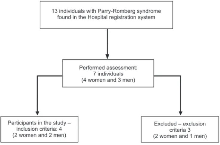
the moment of evaluation, and with no prior speech therapy. During the study period, 13 individuals with Parry-Romberg syndrome were evaluated. The selection flowchart of the SG participants is illustrated in Figure 1.
Procedures
Clinical evaluation
Participants underwent oral motor clinical evaluation while seated in a chair in a room with adequate lighting. The expan-ded protocol of the Orofacial Myofunctional Evaluation with Scores (OMES-expanded)(16) was used for this evaluation. This
protocol was developed based on previous evaluation models, with the addition of numerical scales that reflect the physical characteristics and orofacial behaviors of the individuals (who can obtain a maximum of 230 points).
The clinical protocol used for this study was one of the three protocols validated for orofacial myofunctional evalu-ation, published in the specialized literature(14). The protocol
tongue, mandible and cheeks. The stomatognathic system was also evaluated for mobility and performance in swallowing and chewing functions. Participants were individually evaluated by visual inspection, and the evaluation was subsequently complemented by the analysis of images recorded on a digital camera (Sony® DSC - W120).
To evaluate mobility, the participants performed separate movements of the lips, tongue, mandible and cheeks. In this analysis, the individual movements of each structure were consi-dered normal when they were performed accurately and without tremor. Dysfunction was considered to be present when there was a lack of precision in the movement, associated movements of other components (e.g., lips accompanying tongue movements) and an inability to perform the movement. The examiner scored mobility using a 6-point scale, as defined in the protocol: score 6 - normal; score 5 - insufficient ability; score 4 - insufficient abi-lity and associated movements; score 3 - insufficient abiabi-lity and tremors and/or deviation; score 2 - insufficient ability, tremors of associated movements and/or deviation; and score 1 - lack of ability, i.e., unable to perform the task.
To evaluate the oral phase of swallowing, the participants were asked to bring a room-temperature glass of water to their mouth. After putting the water in their mouth, they should lower the glass so that the entire face could be videotaped and should then swallow in their usual way. According to the methodology proposed by the OMES-expanded, a minimum of two and a maximum of four replicates were performed. Next, the participants were instructed to proceed as above, but the examiner would put their index finger under the participant’s chin and their thumb under the lower lip (mentalis muscle). At that moment, their lips would be separated after swallowing to visualize the teeth and tongue and to determine if there was tongue thrusting. The swallowing pattern was considered normal when the tongue remained contained in the oral cavity and contraction of the mandibular elevator muscles and sealing of the oral cavity occurred without effort.
As described in the protocol, the lip behavior during swallowing was considered normal if the lips were closed
with no apparent contraction, which was given a score of 6. The tongue was considered normal during swallowing when it stayed contained in the oral cavity, which was given a score of 4. The behaviors that changed during swallowing were sco-red as follows: 1 - if present or 2 - if absent. These behaviors included movements of the head, neck or other body parts during swallowing, uncoordinated mandibular movements, apparent tension of the facial muscles, food escape and noise during swallowing. Swallowing efficiency was also analyzed, considering the participant’s ability to propel the food bolus from the oral cavity into the oropharynx. The efficiency was evaluated with both solid foods (French bread) and liquid (wa-ter). A score of 3 was given when there was only one repetition of the swallowing of the food bolus, a score of 2 was given when there were two or three repetitions, and a score of 1 was given for multiple swallowing.
The OMES-expanded protocol recommends the use of the Bono cookie (Nestlé®) to evaluate chewing(16). However,
that cookie could not be used in this study due to the service’s inability to obtain it; thus, the cookie was replaced with French bread. All of the instructions indicated in the OMES-expanded protocol were also followed for the evaluation of chewing. Solid food intake was analyzed regarding the type of bite, using the following scores: 4 - bite with incisors; 3 - bite with canines; 2 - bite with molars and 1 - when the participant did not bite but broke the material into pieces with their hand and then took it into his/her mouth. Chewing was classified by observing the digital images (the chewing strokes), as follows: alternate bilateral - score 10; simultaneous bilateral - score 8; unilateral grade 1 (chewing cycles on the same side, 61%-77%) - score 6; unilateral grade 2 (chewing cycles on the same side, 78%-94%) - score 4; chronic unilateral (95%-100% on the same side) and when chewing occurred in the region of the incisors and/or canines - score 2; the participant received a score of 1 when he/she could not perform the function. As observed during swallowing, altered behaviors were also recorded, as previously described.
Two experienced speech therapists evaluated all partici-pants. The agreement between the evaluators was assessed using the Kappa coefficient. The speech therapists assigned the OMES-expanded scores and exhibited a high level of agreement (>0.83).
To analyze the facial anthropometry, parameters such as the ratio between the thirds and sides of the face, symmetries or asymmetries, and range of mandibular movements were obtained using a digital sliding caliper (Digimess, Pró-Fono®) and were directly on the faces and oral cavities of the partici-pants. Marking of the base points was performed individually and manually by the researcher with the help of an eye pencil, as previously described. The following static and dynamic measurements of the face were considered(14,17): 1) the upper
third – the height of the upper third of the face, marking the upper point on the face, was measured; 2) middle third – the Figure 1. Study sample
Participants in the study – inclusion criteria: 4 (2 women and 2 men)
Excluded – exclusion criteria 3 (2 women and 1 men) Performed assessment:
7 individuals (4 women and 3 men) 13 individuals with Parry-Romberg syndrome
height of the middle third of the face was measured; 3) lower third – the height of the lower third of the face was measured; 4) outer corner of the eye to the corner of the mouth – distances between the outer corner of the eye and labial commissure on both hemifaces were measured; 5) midline – with the teeth in occlusion, whether the lines between the maxillary and mandibular central incisors coincided was assessed; if they did not, the horizontal distance between the lines was measured; 6) maximum oral opening - the distance between the incisal surfaces of the maxillary and mandibular incisor teeth, plus the extent of the vertical overlap (overbite), was measured; 7) lateralization of the mandible to the right and then to the left - the horizontal distance of the line between the mandibular central incisors to the line between the maxillary central incisors was measured, after the lateral sliding of the mandible to either side; in case of midline deviation, the relevant adjustment was made; 8) mandi-bular protrusion - sum of the measure of the horizontal overlap (overjet) with the measure of the maximum horizontal sliding of the mandible was taken; 9) overjet - the distance between the occlusal surface of the maxillary central incisor and the distal face of the mandibular central incisor was measured in occlusion; and 10) overbite - the distance between the maxillary and mandibular incisors was measured in occlusion.
Instrumental evaluation – electromyography (EMG)
Electromyographic evaluation of the masticatory muscles of the participants was based on the methodology already pu-blished in the literature(18). The methodology followed referred
only to the collection and recording of data; for data analysis, the proposed methodology could not be employed because different electromyography devices were used.
The EMG was performed using an electromyography device: a Miotool 400 with 4 channels, calibrated at 500 mi-crovolts (µV), with a band-pass filter (20-500 Hz) and 100-fold gain, with a low noise level (<5 µV root mean square [RMS]). The software used to capture and process the EMG analysis was Miograph 2.0 (Miotec® Equipamentos Biomédicos, Brazil), which performs the acquisition, storage and on-line processing of the signals and runs under the Windows XP operating system. The signals for the electrical activity of the muscle movements were obtained using bipolar electrodes with Ag/AgCl surfaces (model SDS500 – disposable, double, fixed with transpore tape [3M®]).
A speech therapist with experience in the field performed all EMG tests under the same environmental conditions. The positioning of the electrodes followed the technique of placing the midpoint of the muscle belly in the longitudinal direction of the muscle bundle in the mesodistal position of the muscle(19),
where higher signal amplitude is observed for this type of electrode. To ensure correct positioning of the electrodes, the masseter and temporalis muscles were identified by palpation at rest and during maximal voluntary contraction – the maximum intercuspation was requested. After this step, muscle function
was tested to check for possible positioning errors, and elec-trodes were repositioned when necessary.
Simultaneous electrical activity of the temporal and masse-ter muscles was evaluated in both hemifaces for the following tasks(18):
- Rest;
- Maximum clenching with a cotton roller between the teeth (CT);
- Maximum clenching with maximum intercuspation (MIC). To collect the data, all participants were comfortably seated in a chair with their back supported, feet on the floor, hands resting on the lower limbs, head properly positioned (Frankfurt plane parallel to the ground), eyes open and looking at a pre-determined fixed point. All individuals received instructions about the test. The skin of the face was prepared using gauze soaked in 70% alcohol and was locally shaved for all partici-pants to ensure good impedance during the examination. The signals obtained were analyzed in RMS and are expressed in microvolts (µV). The reference (earth) cable was connected to the electrode and fixed on the right wrist.
First, data were collected for the masseter and temporal muscles at rest for 30 seconds. Three collections were perfor-med to obtain the mean electrical activity. Then, the participants were asked to remain at rest for 15 seconds without recording. After this command, a 10-mm cotton roller was placed bilate-rally between the first and second molars, and the participants were asked to bite the cotton with the maximum strength pos-sible for five seconds, three times in succession, at five-second intervals. The same procedure was performed to record the electrical activities of the masseter and temporal muscles in maximum intercuspation (maximum clenching without cotton).
The EMG may encounter interference from many factors, including the impedance in the skin; thus, this factor is con-sidered during the evaluation of patients with Parry-Romberg syndrome. Individuals with this syndrome may have facial atrophies; however, for the patients in this study, the evaluated areas showed no signs of atrophy and/or changes (most common in the buccinator muscles and frontal region).
Surface electromyography analysis
Data analysis
The asymmetry indices for the maximum intercuspation measurements with and without the cotton roller were cal-culated by dividing the side with less activation by the side with greater activation. These indices were calculated for each individual separately.
Data were statistically analyzed using SPSS software, version 22. Statistical analysis included the likelihood ratio test to compare the groups regarding the categorical data. The Mann-Whitney test was used to compare the groups regarding asymmetry measurements, and the Wilcoxon signed-rank test was used to compare the facial sides regarding the maximum intercuspation measurements. For all comparisons, a 5% sig-nificance level was used.
Reliability
Based on the literature, which indicates subjectivity in the reading of the EMG measurements, a reliability analysis was performed to determine the level of agreement between exami-ners and, thus, to ensure greater reliability of the measurements. To this end, ten EMG samples were randomly selected from
a total of 72. These samples were independently analyzed by two speech therapists experienced in the field and who were blind to the study. The correlation coefficient was high for all comparisons (95% confidence interval [CI] = 0.8745-0.9368), indicating high consistency among examiners.
RESULTS
Regarding the data in the clinical evaluation, according to the categories of the OMES-expanded protocol, the statistical analysis showed differences between the groups for the posture and position of the speech organs and for the total score of the OMES-expanded. Overall, the SG had lower scores for all categories, showing changes in posture and position, mobility and functions of the stomatognathic system (Table 1).
Concerning the comparative analysis of the items evaluated in each category of the OMES-expanded, there was a signi-ficant difference between groups for palate height and width, tongue mobility in swallowing and the chewing function. Again, the SG presented a worse score compared to the CG (Table 2).
Table 1. Between-group comparison for the overall results of OMES-E
Mean (SD) - mm
RG CG U p-value
Posture and position Mobility
Function
51.50 (2.02) 102.50 (4.77)
42.50 (3.28)
60.25 (1.44) 107.00 (1.73)
46.50 (1.50)
0.000 7.000 5.000
0.029* 0.886 0.486
Total 196.50 (7.58) 213.75 (0.85) 0.000 0.029*
*Significant values (p<0.05) – Mann-Whitney test
Note: OMES-E = Expanded Oral Myofunctional Assessment with Scores; mm = milimeters; SD = standard deviation; RG = research group; CG = control group; U = Mann-Whitney Test
Table 2. Between-group comparison for the categorical items of OMES-E
X2 DF p-value
OMES-E
Posture
Lips 6.086 3 0.107
Mandible 6.086 3 0.107
Cheeks 4.36 3 0.225
Face 5.545 3 0.136
Tongue 3.819 2 0.148
Palate 6.086 2 0.048*
Mentalis 1.726 2 0.422
Mobility
Lips 0 1 1
Tongue 5.545 5 0.353
Mandible 5.545 4 0.236
Cheeks 3.452 2 0.178
Function
Breathing 3.452 1 0.063
LBS 2.773 2 0.25
TBS 6.086 2 0.048*
OBS 4.36 2 0.113
SE 3.452 1 0.063
Mastication 6.592 2 0.037*
* Significant values (p<0.05) – Likelihood test
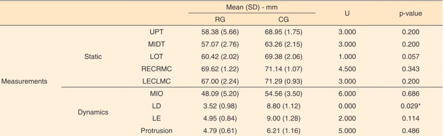
Table 3. Between-group comparisons for anthropometric and mandible amplitude measurements Mean (SD) - mm
U p-value
RG CG
Measurements
Static
UPT 58.38 (5.66) 68.95 (1.75) 3.000 0.200
MIDT 57.07 (2.76) 63.26 (2.15) 3.000 0.200
LOT 60.42 (2.02) 69.38 (2.06) 1.000 0.057
RECRMC 69.62 (1.22) 71.14 (1.07) 4.500 0.343
LECLMC 67.00 (2.24) 71.29 (0.93) 3.000 0.200
Dynamics
MIO 48.09 (5.20) 54.56 (3.50) 6.000 0.686
LD 3.52 (0.98) 8.80 (1.12) 0.000 0.029*
LE 4.95 (0.84) 9.00 (1.28) 2.000 0.114
Protrusion 4.79 (0.61) 6.21 (1.16) 5.000 0.486
*Significant values (p<0.05) – Mann-Whitney test
Note: mm = milimeters; SD = standard deviation; RG = research group; CG = control group; U = Mann-Whitney Test; UPT = upper third of the face; MIDT = middle third of the face; LOT = lower third of the face; RECRLC = right eye corner to right mouth corner; LECLMC = left eye corner to left mouth corner; MIO = maximal incisor opening; LD = lateralization to the right; LE = lateralization to the left
Table 4. Between group comparisons for the asymmetry indexes of the masseter and temporal muscles (µV)
Asymmetry index Muscle Group Median Interquartile range U Z p-value
1st quartil 3rd quartil
MTC
Temporal Research 0.64 0.34 0.82 8.0 0.000 1.000
Control 0.70 0.32 0.80
Masseter Research 0.55 0.23 0.93 5.0 -0.866 0.386
Control 0.78 0.57 0.81
MTCCR
Temporal Research 0.54 0.50 0.89 7.0 -0.290 0.772
Control 0.83 0.30 0.86
Masseter Research 0.57 0.37 0.87 5.0 -0.866 0.386
Control 0.78 0.65 0.81
Mann-Whitney test (p<0.05)
Note: µV = microvolts; U = Mann-Whitney test; Z = a value expressed as standard deviation; MTC = maximum voluntary teeth clenching; MTCCR = Maximum voluntary teeth clenching on cotton rolls
When comparing the groups for static and dynamic facial anthropometric measurements, there was significant difference between the groups only for lateralization of the mandible to the right. Considering the means obtained for the dynamic measurements (i.e., mandibular movement), the individuals of the SG showed a range of mandibular movements smaller than that observed for the CG (Table 3).
There was no significant difference between the groups when comparing the asymmetry indices in the EMG activity concerning the maximum intercuspation with and without the cotton roller, both for the temporal muscle and the masseter muscle. However, the descriptive analysis indicates that the SG mean values are lower than those of the CG (Table 4).
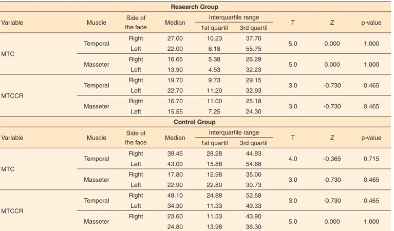
In the intragroup comparison regarding the electrical activities obtained in the different hemifaces, there were no significant differences when comparing the maximum inter-cuspations between the facial sides in both groups (Table 5).
DISCUSSION
Evidence-based practice requires that the relationships
between interventions and outcomes be well established. Given that this practice analyzes the results based on the evidence, it is necessary that health professionals identify, implement and organize rehabilitation processes to improve their effecti-veness(20,21). This practice is only possible if the initial health
status of the patients is properly mapped.
To date, this study is the first to conduct extensive clinical characterization, investigating the impact of Parry-Romberg syndrome on the orofacial myofunctional organs. Participants with this syndrome exhibited worse performance when compa-red to healthy participants with regard to the posture/position of the orofacial myofunctional organs, the functionality of these organs and mandibular mobility.
According to the literature, the association between cra-niofacial syndromes and facial asymmetries due to altered facial growth is common. In Parry-Romberg syndrome, the individuals show normal craniofacial growth until the onset of the first signs, which normally occurs in the first two decades of life(2-4,6,7,10,11,13). Normal craniofacial growth may explain
whose main characteristic is facial asymmetry(4,13).
The results of this study indicated the presence of a deficit regarding the mandibular movements. The groups differed sig-nificantly only when considering lateralization of the mandible to the right, with the group of individuals with Parry-Romberg syndrome showing more restricted movements. According to previous studies(22,23), the values expected for mandibular
move-ments in healthy individuals are maximum opening – between 40 and 60 mm; mandibular lateralization – between 7 and 11 mm (for each side); and mandibular protrusion – between 7 and 11 mm, without distinction between genders and age groups. In this study, the participants with Parry-Romberg syndrome exhibited greater restriction of all mandibular movements when compared with the values expected for healthy individuals.
The literature suggests that mandibular mobility is directly related to the functionality of the masticatory muscles and, consequently, with chewing efficiency(24-27). Previous studies
have shown that the lateral chewing preferences may be related to differences in the activation of the masticatory muscles and in the lateralization of the mandible(26-28). The worse outcome
observed during the overall chewing performance in patients with Parry-Romberg syndrome suggests that the impairment of the chewing function may be interfering with mandibular functionality. This finding should be considered in future stu-dies on the subject.
The mandibular function must adapt to a wide variety of factors that affect the stomatognathic system(29). Mandibular
movements are responsible for intraoral spatial modifications. These movements have a strong impact on chewing, swallo-wing and speech patterns because they are responsible for allowing appropriate movements of the tongue and other soft tissues (amplitude) within the oral cavity(30). This factor can
explain the difference between the groups for the movement of the tongue during chewing and swallowing. Once more, as expected, the group of individuals with the syndrome showed a worse performance.
Regarding the evaluation of the aspect/posture of the orofacial myofunctional organs, the individuals with Parry-Romberg syndrome presented lower scores compared to healthy individuals, indicating the presence of facial asymmetry in the syndromic individuals. Among these individuals, three of them were affected on the left side, and only one was affected on the right side. Additionally, according with the posture measurements, there was a statistically significant result for the variable palate. However, no descriptions of the change of this structure were available in the literature, and there are no other craniofacial alterations to justify separating groups by this criterion. Thus, the variability in the characterization of the palate may have been due to the features of this specific sample and is not necessarily related to the syndrome.
Table 5. Comparison between hemifaces for maximal voluntary teeth clenching (µV)
Research Group
Variable Muscle Side of
the face Median
Interquartile range
T Z p-value
1st quartil 3rd quartil
MTC
Temporal Right 27.00 10.23 37.70 5.0 0.000 1.000
Left 22.00 6.18 55.75
Masseter Right 16.65 5.38 26.28 5.0 0.000 1.000
Left 13.90 4.53 32.23
MTCCR
Temporal Right 19.70 9.73 29.15 3.0 -0.730 0.465
Left 22.70 11.20 32.93
Masseter Right 16.70 11.00 25.18 3.0 -0.730 0.465
Left 15.55 7.25 24.30
Control Group
Variable Muscle Side of
the face Median
Interquartile range
T Z p-value
1st quartil 3rd quartil
MTC
Temporal Right 39.45 28.28 44.93 4.0 -0.365 0.715
Left 43.00 15.88 54.68
Masseter Right 17.80 12.98 35.00 3.0 -0.730 0.465
Left 22.90 22.80 30.73
MTCCR
Temporal Right 48.10 24.88 52.58 3.0 -0.730 0.465
Left 34.30 11.33 49.33
Masseter Right 23.60 11.33 43.90 5.0 0.000 1.000
24.80 13.98 36.30
Wilcoxon signed-rank test (p<0.05)
Facial growth and facial structural development are promo-ted by functional, environmental and genetic factors. Therefore, the balance of morphological development with functional development is important for adequate growth. The atrophy of the muscles that make up the stomatognathic system in individuals with Parry-Romberg syndrome can have a direct effect on orofacial functions, especially in chewing because the mobility of the structures may be reduced, as observed in this study. Proper chewing stimulates the correct development of the jaws and related structures. Orofacial disorders may limit or even impair the physiological activities, which would justify the indication of speech therapy for these individuals(30).
Despite the differences in the groups concerning muscle atrophy and altered chewing, there were no significant di-fferences for the EMG measurements. These findings may be explained by the choice of the muscles evaluated in the examination (i.e., temporal and masseter), as they may or may not be affected by the syndrome. In the situations evaluated for this study, there was a higher occurrence of atrophy in the buccinator muscle region and no incidence of atrophy in the masseter muscle region. However, a previous study(6)
repor-ted the case of a patient with Parry-Romberg syndrome who presented with atrophy of the sternocleidomastoid, masseter and pterygoid muscles and of the subcutaneous soft tissues, though these findings were revealed via magnetic resonance imaging (MRI) rather than EMG. To validate these results, the test should be repeated, adding the buccinator muscle region to the data collection and analysis. Such a step would allow for evaluation of the main areas involved in chewing that may be affected by the syndrome.
When dealing with syndromic patients, it is always impor-tant to remember that the same syndrome can have different phenotypes, such as with Parry-Romberg syndrome. In some patients, the only sign of the syndrome is the scleroderma on the forehead - en coup de sabre(8,12,13), while in others, the facial
asymmetry may extend across the entire hemiface(13). Moreover,
as mentioned earlier, these signs may progress slowly over a period of two to ten years(6) until stabilization(3,6,7).
One limitation of this study was the sample size. Because this syndrome is rare, there is a natural lack of individuals to compose the study sample. The results are only applicable to the sample studied and cannot be generalized. However, the research methodology, which is based on the use of standar-dized and validated protocols, allows its replication in future studies. Therefore, further studies should be conducted in an attempt to increase the sample size and to ensure the validity of the results obtained.
CONCLUSION
The results of this study allowed the initial characterization of the OMS aspects of patients affected by Parry-Romberg syndro-me, demonstrating that participants with this syndrome exhibited
worse performance when compared to healthy participants regarding the posture/position of the orofacial myofunctional organs, the functionality of these organs and mandibular mobility.
REFERENCES
1. Hu J, Yin L, Tang X, Gui L, Zhang Z. Combined skeletal and soft tissue reconstruction for severe Parry-Romberg syndrome. J Craniofac Surg. 2011;22(3):937-41. http://dx.doi.org/10.1097/ SCS.0b013e31820fe27d
2. Miao J, Liu R, Lin H, Su C, Li H, Lei G et al. Severe bilateral pyramidal tract involvement in a patient with Parry-Romberg syndrome. Am J Med Sci. 2009;337(3):212-4. http://dx.doi. org/10.1097/MAJ.0b013e31818226f9
3. Pinheiro TPS, Silva CC, Silveira CSL, Botelho PCE, Pinheiro MGR, Pinheiro JJV. Progressive hemifacial atrophy: case report. Med Oral Patol Oral Cir Bucal. 2006;11(2):E112-E114.
4. O’Flynn S, Kinirons M. Parry-Romberg syndrome: a report of the dental findings in a child followed up for 9 years. Int J Paediatr Denti. 2006;16(4):297-301. http://dx.doi.org/10.1111/j.1365-263X.2006.00730.x
5. Guo ZN, Zhang HL, Zhou HW, Lan HJ, Wu J, Yang Y. Progressive facial hemiatrophy revisited: a role for sympathetic dysfunction. Arch Neurol. 2011;68(9):1195-7. http://dx.doi.org/10.1001/ archneurol.2011.190
6. Duymaz A, Karabekmez FE, Keskin M, Tosun Z. Parry-Romberg syndrome: facial atrophy and its relationship with other regions of the body. Ann Plast Surg. 2009;63(4):457-61. http://dx.doi. org/10.1097/SAP.0b013e31818bed6d.
7. Longo D, Paonessa A, Specchio N, Delfino LN, Claps D, Fusco L et al. Parry-Romberg syndrome and Rasmussen encephalitis: possible association. Clinical and neuroimaging features. J Neuroimaging. 2011;21(2):188-93. http://dx.doi.org/10.1111/j.1552-6569.2009.00398.x
8. Menascu S, Padeh S, Hoffman C, Ben-Zeev B. Parry-Romberg syndrome presenting as status migrainosus. Pediatr Neurol. 2009;40(4):321-3. http://dx.doi.org/10.1016/j. pediatrneurol.2008.11.007
9. Qureshi UA, Wani NA, Altaf U. Parry-Romberg syndrome associated with unusual intracranial vascular malformations and Phthisis bulbi. J Neurol Sci. 2010;291:107-9. http://dx.doi.org/10.1016/j. jns.2010.01.003
10. Haldar A, Mukherjee A. Parry Romberg’s disease with intractable partial epilepsy. Neurol Índia. 2007;55(2):160-2. http://dx.doi. org/10.4103/0028-3886.32791
11. Sommer A, Gambichler T, Buhles MB, Rothenburg T, Altmeyer P, Kreuter A. Clinical and serological characteristics of progressive facial hemiatrophy: a case series of 12 patients. J Am Acad Dermatol. 2006;54(2):227-33. http://dx.doi.org/10.1016/j. jaad.2005.10.020
13. Anderson PJ, Molony D, Haan E, David DJ. Familial Parry-Romberg disease. Int J Pediatr Otorhinolaryngol. 2005;69(5):705-8. http:// dx.doi.org/10.1016/j.ijporl.2004.12.004
14. Felício CM, Medeiros APM, Melchior MO. Validity of the ‘protocol of oro-facial myofunctional evaluation with scores’ for young and adult subjects. J Oral Rehabil. 2012;39(10):744-53. http://dx.doi. org/10.1111/j.1365-2842.2012.02336.x
15. Proffit WR, Fields HW, Moray LJ. Prevalence of malocclusion and orthodontic treatment need in the United States: estimates from the NHANES III survey. Int J Adult Orthodon Orthognath Surg. 1998;13(2):97-106.
16. Felício CM, Folha GA, Ferreira CL, Medeiros AP. Expanded protocolo f orofacial myofunctional evaluation with scores: validity and reability. Int J Pediatr Otorhinolaryngol. 2010;74(11):1230-9. http://dx.doi.org/10.1016/j.ijporl.2010.07.021
17. Cattoni DM. O uso do paquímetro na motricidade orofacial: procedimentos de avaliação. Barueri: Pró-Fono; 2006.
18. Sforza C, Peretta R, Grandi G, Ferronato G, Ferrario VF. Soft tissue facial planes and masticatory muscle function in skeletal Class III patients before and after orthognatic surgery treatment. J Oral Maxillofac Surg. 2008;66(4):691-8, 2008. http://dx.doi. org/10.1016/j.joms.2007.06.645
19. Soderberg GL, Cook MT. Electromyography in biomechanics. Phys Ther. 1984;64(12):1813-20.
20. HassanT, Naini FB, Gill DS. The effects of orthognathic surgery on speech: a review. J Oral Maxillofac Surg. 2007;65:2536-43. http:// dx.doi.org/10.1016/j.joms.2007.05.018
21. Weiner JB, Alexander JA, Shortell SM, Baker LC, Geppert JJ. Quality improvement implementation and hospital performance on quality indicators. Health Serv Res. 2006;41(2):307-34. http://dx.doi. org/10.1111/j.1475-6773.2005.00483.x
22. Celic R, Jerolimov V, Zlataric DK. Relationship of slightly limited mandibular movements to temporomandibular disorders. Braz Dent J. 2004;15(2):151-4. http://dx.doi.org/10.1590/S0103-64402004000200012
23. Buschang PH, Throckmorton GS, Travers KH, Hayasaki H. Incisor and mandibular condylar movements of young adult females during maximum protrusion and lateratrusion of the jaw. Arch Oral Biol. 2001;46(1):39-48. http://dx.doi.org/10.1016/S0003-9969(00)00096-0 24. Harper RP, Bruin H, Burcea I. Muscle activity during mandibular
movements in normal and mandibular retrognathic subjects. J Oral Maxillofac Surg. 1997;55(3):225-33. http://dx.doi.org/10.1016/ S0278-2391(97)90530-9
25. Sforza C, Ugolini A, Rocchetta D, Galante D, Mapelli A, Gianni AB. Mandibular kinematics after orthognathic surgical treatment: a pilot study. Br J Oral Maxillofac Surg. 2010;48(2):110-4. http://dx.doi. org/ 0.1016/j.bjoms.2008.09.002
26. Yazdani J, Ebrahimi H, Talesh KT, Khashabi E, Pourshahidi S, Tadbir AA. Comparing the effect of 3 orthognathic surgical methods on the mandibular range of movement. J Craniofac Surg. 2010;21(3):703-5. http://dx.doi.org/10.1097/SCS.0b013e3181d83fbc 27. Felício CM, Melchior MO, Silva MAMR. Effects of orofacial
myofunctional therapy on temporomandibular disorders. Cranio. 2010;28(4):249-59. http://dx.doi.org/10.1179/crn.2010.033 28. De Felício CM, Ferreira CLP, Medeiros APM, Silva MAMR,
Tartaglia GM, Sforza C. Electromyographic índices, orofacial myofunctional status and temporomandibular disorders severity: a correlation study. J Electromyogr Kinesiol. 2012;22(2):266-72. http://dx.doi.org/10.1016/j.jelekin.2011.11.013
29. Yamada R, Ogawa T, Koyano K. The effect of head posture on direction and stability of mandibular closing movement. J Oral Rehabil. 1999;26(6):511-20. http://dx.doi.org/ 0.1046/j.1365-2842.1999.00386.x