ISSN 1806-3713 © 2015 Sociedade Brasileira de Pneumologia e Tisiologia
http://dx.doi.org/10.1590/S1806-37132015000000095 J Bras Pneumol. 2015;41(5):482-483
Letter to the editor
482
To The ediTor:
Primary immunodeiciencies (PIDs) are characterized by impairment of one or more arms of the immune response, resulting in decreased defense, an increased number of infections, and, in certain cases, a higher incidence of autoimmune diseases and cancers.(1) Although PIDs are considered rare diseases, many of them are more common than are those currently diagnosed with the “heel prick” test. The manifestations of PIDs are heterogeneous and are usually caused by genetic defects of the immune system and its development.
Common variable immunodeiciency (CVI) is the most prevalent of the severe PIDs. A diagnosis of CVI is based on reduced levels of IgG, IgA, and (in some cases) IgM, together with reduced levels of speciic antibodies, after other causes of hypogammaglobulinemia have been excluded.(2) The incidence of CVI is similar in both genders, with either sporadic or familial distribution. Although it can manifest at any time in life, it is especially common in adolescence and young adulthood. The most striking characteristics of this disease include hypogammaglob
-ulinemia associated with frequent infections, especially with encapsulated bacteria, as well as a poor response to immunization protocols.(3)
A 27-year-old Black female, who worked as a maid, was admitted to the ER of a tertiary hospital with a 7-day history of productive cough, fever, and dyspnea. The patient reported a history of asthma, recurrent pneumonia, and some episodes of furunculosis. In addition, she reported that, one month prior, she had been hospitalized for ive days because of pneumonia and that the frequency of such infections had increased in the last ive years.
The initial examination revealed fever, an SpO2 of 84%
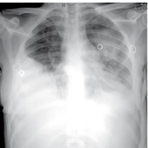
on room air (digital pulse oximetry), a heart rate of 120 bpm, and an arterial pressure of 90/60 mmHg. A chest X-ray of the chest showed right pleural effusion as well as pneumonic iniltrates in the middle third and lower lobe of the left lung (Figure 1). The patient was started on antibiotic therapy to treat the pulmonary focus and underwent thoracentesis followed by chest tube drainage, on the right side, because of empyema.
The patient was found to be antinuclear factor-neg
-ative. Serology for HIV, HTLV, hepatitis A, hepatitis B, hepatitis C, Epstein-Barr virus, rubella, toxoplasmosis, and cytomegalovirus was negative, as was the venereal disease research laboratory test (for syphilis). In addition, sputum smear microscopy and sputum cultures were negative for tuberculosis and fungi. No bacteria were isolated from blood or pleural luid culture. The results of
Bronchiectasis caused by common variable
immunodeiciency
Paulo Henrique do Amor Divino1, José Henrique de Carvalho Basilio1,
Renato Moraes Alves Fabbri1, Igor Polônio Bastos1, Wilma Carvalho Neves Forte2
thyroid function testing, anti-thyroglobulin testing, and anti-thyroid peroxidase testing were normal, ruling out the possibility of autoimmune thyroiditis. Quantiication of serum immunoglobulins revealed a persistent decrease in IgA, IgM, and IgG. Serology was negative for antibody to hepatitis B surface antigen (anti-HBs), although the patient had received three doses of the hepatitis B vaccine. Additional immunological assessment, which included determination of total complement activity (CH50), photoreduction of nitroblue tetrazolium, and measurement of phagocytosis by neutrophils and mononuclear phagocytes, yielded normal results.
On the basis of the clinical picture, indings from history taking, and laboratory test results, a diagnosis of CVI was made and the patient was started on human immunoglobulin replacement therapy (600 mg/kg), which resulted in rapid clinical and radiological improvement. Two months after admission, the patient was discharged to follow-up in the pulmonology and allergy/immunodeiciency outpatient clinics for continuation of her immunoglobulin replacement therapy (monthly administration).
Recurrent pneumonia can result in bronchiectasis and forms part of the core clinical picture of CVI. Among PIDs, CVI is the second most common, although it is believed to be underdiagnosed. Its incidence is reported as 1:10,000 among individuals of European descent, being rare among Asians, with a reported incidence of 1:2,000,000 individuals in Japan.(4) To date, there have been no studies investigating its incidence in the Black population.
Encapsulated bacteria, such as Streptococcus pneu-moniae and Haemophilus inluenzae, are combated by
IgG2 subclass antibodies to polysaccharide antigens. In the absence of such immunoglobulins, as in cases of CVI, patients experience sinopulmonary infections, especially pneumonia, bronchitis, sinusitis, and otitis.(1,2) Infections with atypical bacteria, such as some species of the genus Mycoplasma, have also been reported.(5) Often, patients experience tonsillitis, otitis, and giardiasis in childhood, all of which are facilitated by IgA deiciency, and, in adolescence or young adulthood, develop recurrent pneumonia, which is characteristic of IgG deiciency, suggesting that CVI is the result of progression of IgA deiciency.(6)
The patient in question had been immunized against hepatitis B, as recommended, having received all three doses of the vaccine. Nevertheless, she tested negative for anti-HBs, i.e., she had deicient production of speciic protein antibodies, which also occurs in CVI.(2) In this
Amor Divino PH, Basilio JHC, Fabbri RMA, Bstos IP, Forte WCN
PID, there can be deicient production of other speciic antibodies, especially those to polysaccharide antigens, and the pneumococcal vaccination status of the patient must therefore be taken into account.
Patients with CVI have normal or slightly reduced B lymphocyte counts. However, because of problems intrinsic to B lymphocytes, they can lack the ability to differentiate into antibody-producing plasma cells, might not function properly as antigen-presenting cells to T helper lymphocytes, or might not receive
suficient assistance from T helper lymphocytes, all of which can impair the response to immunizations and infections. (3-7) Such problems are probably due to disturbances in the expression of surface molecules on B lymphocytes or T helper lymphocytes, intracellular enzyme activity disturbances, or increased apoptosis.(8)
The prevalence of chronic pulmonary complications at CVI diagnosis is high (27.0-34.2%).(9) The most common such complication is bronchiectasis. Extensive pneumonia and the chronicity of infectious pulmonary episodes are responsible for the poor prognosis in CVI patients.(10) Bronchiectasis can accompany diseases and conditions other than CVI, including tuberculosis, aspergillosis, cystic ibrosis,alpha-1 antitrypsin dei
-ciency, AIDS, cancer, systemic lupus erythematosus, and rheumatoid arthritis. Therefore, it is necessary to make the differential diagnosis, whether it is based on indings from history taking or on ancillary test results.
The treatment for patients with CVI includes administering human immunoglobulin replacement therapy and combating infections. Immunoglobulin preparations contain neutralizing antibodies against a wide variety of bacteria and viruses, relecting the immunological memory of the donors, and should be administered every three or four weeks.
We believe that CVI should be borne in mind by health professionals who treat patients with recurrent pneumonia. Typically manifesting in adulthood, CVI is a PID that must be diagnosed early so that prompt treatment can be instituted, thereby lowering morbidity, improving quality of life, and, in many cases, making survival possible for these patients.
Figure 1. Chest X-ray showing right pleural effusion as
well as pneumonic iniltrates in the middle third and lower lobe of the left lung.
reFerences
1. Forte WC. Imunologia do Básico ao Aplicado. 3rd ed. São Paulo: Atheneu; 2015. p. 339.
2. European Society of Immunodeiciencies--Esid [homepage on the
Internet]. Geneva: Esid; c2015 [cited 2015 Mar 25]. Available from:
http://www.esid.org
3. Primary immunodeiciency diseases: report of a WHO scientiic
group. Clin Exp Immunol. 1997;109 Suppl 1:1-28.
4. Kokron CM, Errante PR, Barros MT, Baracho GV, Camargo MM, Kalil J, et al. Clinical and laboratory aspects of common variable
immunodeiciency. An Acad Bras Cienc. 2004;76(4):707-26. http://
dx.doi.org/10.1590/S0001-37652004000400007
5. Cunningham-Rundles C, Bodian C. Common variable
immunodeiciency: clinical and immunological features of 248 patients. Clin Immunol. 1999;92(1):34-48. http://dx.doi.org/10.1006/
clim.1999.4725
6. Carvalho Neves Forte W, Ferreira De Carvalho Júnior F, Damaceno
N, Vidal Perez F, Gonzales Lopes C, Mastroti RA. Evolution of
IgA deiciency to IgG subclass deiciency and common variable immunodeiciency. Alergol Immunopathol (Madr). 2000;28(1):18-20.
7. Conley ME, Notarangelo LD, Etzoni A. Diagnostic criteria for
primary immunodeiciencies. Representing PAGID (Pan-American Group for Immunodeiciency) and ESID (European Society for Immunodeiciencies). Clin Immunol. 1999;93(3):190-7. http://dx.doi.
org/10.1006/clim.1999.4799
8. Errante PR, Condino-Neto A. Imunodeiciência comum variável: revisão da literatura. Rev Bras Alerg Imunopatol. 2008;31(1):10-8.
9. Roxo Junior P. Primary immunodeiciency diseases: relevant aspects
for pulmonologists. J Bras Pneumol. 2009;35(10):1008-17.
10. Costa-Carvalho BT, Cocco RR, Rodrigues WM, Colla VA, Solé D, Carneiro-Sampaio MM. Pneumonias de repetição em paciente
com deiciência de anticorpos e imunoglobulinas normais. J Pneumol. 2002;28(3):155-8.
http://dx.doi.org/10.1590/S0102-35862002000300008
483