ORIGINAL
RES
EAR
CH
Correspondence to: Elaine Paulin – Centro de Ciências da Saúde e do Esporte (CEFID) – Universidade do Estado de Santa Catarina (UDESC) – Rua Pascoal Simone, 358 – CEP: 88080-350 – Florianópolis (SC), Brasil – E-mail: elaine.paulin@udesc.br
Presentation: may. 2013. Accepted for publication: nov. 2013 – Financing source: none – Conflict of interests: nothing to declare – Approval at the Ethics Committee n. 74/2011.
ABSTRACT | With the objective to evaluate possible dif-ferences in the values obtained in the thoracoabdominal cirtometry in orthostatism compared with the results in supine, 30 subjects with mean age 27.8±4.4 years were evaluated according to the following parameters: anthro-pometry, pulmonary function test and thocacoabdomi-nal cirtometry. Shapiro-Wilk test was used to verify data normality and the t test was performed in order to com-pare the thoracoabdominal cirtometry measurements in supine and in orthostatism positions. There were no sig-nificant differences in axillar and xiphoid mobility between measurements obtained in supine and orthostatism. The abdominal mobility measured in orthostatism (2.54±1.39 cm) was significantly lower (34.35%) when compared to the mobility obtained in supine (3.71±1.78 cm; p<0.001). The thoracic cirtometry can be performed in orthostatism as an alternative for the evaluation of patients with orthop-nea. The abdominal cirtometry can also be performed in this posture, with the expected one-third reduction in the abdominal mobility obtained in supine.
Keywords | Evaluation; Thorax; Supine Position.
RESUMO | Com o objetivo de avaliar possíveis diferenças nos valores obtidos na realização da cirtometria tóraco-ab-dominal em ortostatismo comparado com os resultados afe-ridos em decúbito dorsal, foram avaliados 30 participantes
Comparison between the measures of
thoracoabdominal cirtometry in supine and standing
Comparação entre as medidas de cirtometria tóraco-abdominal
realizadas em decúbito dorsal e em ortostatismo
Comparación entre las medidas de cirtometría tóraco-abdominal
realizadas en decúbito dorsal y en ortostatismo
Aline Pedrini1, Márcia Aparecida Gonçalves1, Bruna Estima Leal1, Wellington Pereira dos Santos Yamaguti2,
Elaine Paulin1
Study conducted at the Health and Sports Sciences Center at Universidade do Estado de Santa Catarina (UDESC) – Florianópolis (SC), Brazil. 1Graduate Program of Physical Therapy at UDESC – Florianópolis (SC), Brazil.
2Universidade de São Paulo (USP) – São Paulo (SP), Brazil.
com média de idade de 27,8±4,4 anos, por meio dos seguin-tes parâmetros: antropometria, prova de função pulmonar e mobilidade tóraco-abdominal pela cirtometria. O teste de Shapiro-Wilk foi utilizado para verificar a normalidade dos dados e o teste t pareado para a comparação entre as mensurações obtidas pela cirtometria tóraco-abdominal em decúbito dorsal e em ortostatismo. Não houve diferen-ças significativas na mobilidade axilar e xifoidea entre as medidas em decúbito dorsal e ortostatismo. A mobilidade abdominal mensurada em ortostatismo (2,54±1,39 cm) foi significativamente menor (34,35%) em comparação à mo-bilidade obtida em decúbito dorsal (3,71±1,78 cm; p<0,001). A cirtometria torácica pode ser realizada em ortostatismo como uma alternativa para a avaliação de pacientes que referem ortopnéia. A cirtometria abdominal também pode ser realizada nessa postura, com a ressalva de ser esperada uma redução em torno de um terço da mobilidade abdomi-nal obtida em decúbito dorsal.
Descritores | Avaliação; Tórax; Decúbito Dorsal.
RESUMEN | Con el objetivo de evaluar posibles
INTRODUCTION
Measuring thoracoabdominal mobility has been con-sidered as an important parameter to assess respiratory dysfunctions and to monitor training programs in dif-ferent populations1-3. Several instruments have been
used to analyze respiratory patterns4, among which are:
inductance plethysmography5, magnetometry6, laser
monitors7 and video image analysis systems8. Despite
being considered precise to assess the movements of the thoracic wall, these instruments are expensive, which makes their use in clinical practice limited9.
Cirtometry, also known as thoracoabdominal perim-etry, consists of a set of measurements of thoracic and abdominal circumferences during respiratory move-ments, and it aims at quantifying the thoracoabdomi-nal mobility in a simple manner, which is accessible and has low cost, therefore, only one metric tape is required for its performance10. Malaguti et al.11 conducted
thora-coabdominal cirtometry evaluations in 26 patients with Chronic Obstructive Pulmonary Disease (COPD) in 2 diferent days and with 2 independent observers, and they found high intra and interobserver reproducibility of measurements. he same result was described in the study by Caldeira et al.10, in which 2 independent
observ-ers performed 3 cirtometry measurements in 40 healthy individuals, and they also found high intra and interob-server reliability, which proves that cirtometry is a repro-ducible method to assess thoracoabdominal mobility.
Even though it is very common in clinical practice, cirtometry is still a method with little scientiic investi-gation; therefore, it is very questioned12, since there is no
standardization for its conduction. Most studies10,11,13
uses cirtometry with participants in the supine position, however, a technique with the participants in ortho-static position has recently been described and found good reproducibility among three diferent evaluators14.
However, the authors did not demonstrate if this form
of evaluation is diferent from that conducted with sub-jects in supine position.
he assessment of cirtometry in the orthostatic po-sition can facilitate the placement of the metric tape around the thorax and the abdomen, besides allowing the evaluation of patients submitted to thoracoabdomi-nal surgeries, obese patients and those with chronic pneumopathy and heart disease, who frequently present with orthopnea. herefore, it is important to investigate if there are diferences in values obtained by thoracoab-dominal cirtometry in diferent postures. he objective of this study was to compare the values obtained after the conduction of thoracoabdominal cirtometry in the orthostatic and the supine positions.
METHODOLOGY
his is a cross-sectional study. It was approved by the Human Research Ethics Committee (74/2011). All of the participants were previously enlightened as to the study and signed the informed consent form, as established in resolution 196/96 of the National Health Council.
In the laboratory of respiratory physical therapy (LAFIR) at Universidade do Estado de Santa Catarina
(UDESC), a convenience sample composed of 30 healthy volunteers was assessed. Participants should meet the following inclusion criteria: to present proof of normal pulmonary function and body mass index <30 kg/m2; not being smoker; not presenting
cardio-respiratory or neuromuscular conditions, or any other dysfunction that might interfere in the performance of the tests. Exclusion criteria were: inability to perform some of the proposed evaluation measurements for not understanding them or for not cooperating; request to be excluded from the study.
antropometría, prueba de función pulmonar y movilidad tóraco-abdominal por la cirtometría. El test de Shapiro-Wilk fue utilizado para verificar la normalidad de los datos y el test t pareado para la comparación entre las mediciones obtenidas por la cirtometría tóraco-abdominal en decúbito dorsal y en ortostatismo. No hubo diferencias significativas en la movilidad axilar y xifoidea entre las medidas en decúbito dorsal y ortostatismo. La movilidad abdomi-nal medida en ortostatismo (2,54±1,39 cm) fue significativamente
menor (34,35%) en comparación a la movilidad obtenida en decúbito dorsal (3,71±1,78 cm; p<0,001). La cirtometría torácica puede ser realizada en ortostatismo como una alternativa para la evaluación de pacientes que refieren ortopnea. La cirtometría abdominal también puede ser realizada en esa postura, con la salvedad de ser esperada una reducción en torno de un tercio de la movilidad abdominal obtenida en decúbito dorsal.
Participants were assessed only one by the same evaluator as to the parameters: anthropometry, proof of pulmonary function and thoracoabdominal cirtometry.
In order to measure body mass, a previously calibrat-ed scale was uscalibrat-ed. Participants were told to wear light clothes, to take of their shoes before standing on the scale and to remain erect, with their heads facing the front until the measured value was stable. A stadiome-ter was used to measure height, and participants should also be barefoot, with heels together. After anthropo-metric values were obtained (body mass and height), body mass index (BMI) was calculated by the following equation: body mass/height2 (kg/m2).
he proof of pulmonary function was conducted with a portable digital spirometer EasyOne (ndd Medical Technologies), previously calibrated, according to the methods and criteria recommended by the American horacic Society (ATS)15. he following parameters
were measured: Forced Vital Capacity (FVC), Forced Expiratory Volume in one second (FEV1) and the ra-tion FEV1/FVC. At least three acceptable and two reproducible maneuvers were performed. Spirometric variables were expressed in absolute values and in per-centages of the predicted normal values16.
he assessment of thoracoabdominal mobility was conducted by the cirtometry with a metric tape (Prim, Ind. Brasil. Korona), in two postures successively: (1) participant in the supine position; and (2) participant in orthostatic position. For the supine position, the par-ticipant was placed with 0º inclination, without a pil-low, with upper limbs along the body and uncovered thorax. After cirtometry was performed in this position, the participant was asked to stand up, with upper limbs along the body, and the examination was repeated.
In both postures, the circumferences of three ana-tomical points were measured in the following order: axillary fold, xiphoid process and umbilical line in two diferent moments: maximum inspiration and maxi-mum expiration. he diference between measurements obtained in maximum inspiration and expiration in each anatomic level was considered as the thoracoab-dominal mobility of each measured region. All of the measurements were conducted by the same evaluator, with experience to perform cirtometry, and each mea-surement was repeated twice in all of the anatomic lev-els, and the average between the two obtained values was considered.
Data were analyzed with the software SPSS for Windows, version 17.0 (IBM SPSS Statistics, IBM, Armonk, NY, USA) and treated with descriptive
analysis (mean and standard deviation) and inferen-tial analysis. he Shapiro-Wilk test was used to verify data normality and homogeneity of variance. In order to compare the measurements obtained by thoracoab-dominal cirtometry in supine and orthostatic positions, the paired t test was used. A 5% signiicance level was adopted (p<0.05).
Sample size was determined by means of a two-tailed test to calculate the diference of means, accord-ing to the followaccord-ing presuppositions obtained by the analysis of the 10 irst volunteers: diference between postures for axillary mobility of 0.75 cm; standard de-viation of 1.37 cm; test power of 80% and signiicance level of 5%, suggesting the sample size of 29 individuals.
RESULTS
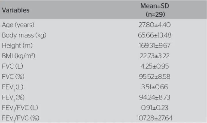
hirty participants were assessed (13 men and 17 women), with mean age of 27.8±4.4 years old. One participant was excluded from the study for present-ing altered proof of pulmonary function (FEV1<80%). he anthropometric characteristics and the pulmonary function of the participants are described in Table 1. Pulmonary function was within normality parameters (Table 1).
he values of thoracoabdominal mobility are de-scribed in Table 2. here was no signiicant diference of axillary (p=0.55) and xiphoid mobility (p=0.68) be-tween the measurements obtained in supine and ortho-static positions. he abdominal mobility measured in the orthostatic position (2.79±1.79 cm) was signiicant-ly lower (34.35%) in comparison to the mobility mea-sured in the supine position (4.25±2.08 cm) (p<0.001).
DISCUSSION
In this study, the thoracoabdominal mobility of healthy individuals was quantiied by the supine and orthostatic cirtometry. Results showed that thoracic cirtometry val-ues (axillary and xiphoid regions) were similar in both investigated postures. Considering abdominal cirtom-etry, a signiicant reduction of 34.35% was observed in the mobility obtained for the orthostatic position in re-lation to the one obtained in the supine position.
required for its performance10. Several studies have
used this resource to assess thoracoabdominal mobil-ity, and among the found studies, all of them describe the performance of cirtometry with the individuals placed in the supine position10,11,13.
Orthopnea is described as respiratory diiculty that occurs when the patient is in the supine position, being relieved when the person changes to the ortho-static position17. his symptom is frequently reported
by patients submitted to thoracic surgeries18, those
with heart diseases19, phrenic nerve palsy20, pulmonary
thromboembolism21, morbid obesity22, COPD23 and
other pictures of respiratory insuiciency24. In these
individuals, measuring cirtometry in the orthostatic position can be an option to enable the performance of the examination with less intolerance in comparison to the supine position. In our study, no signiicant difer-ences were found between cirtometry measurements conducted in supine and in orthostatic positions in the axillary (7.45%) and xiphoid (1.7%) regions. his re-sult may have rere-sulted from the structural architecture of the thoracic cage that sustained the thoracic wall in both analyzed postures, which made thoracic mo-bility similar. Some authors25,26 describe that, at rest,
in vertical positions (sitting down or orthostatic), the abdomen presents similar complacence to the one of the thoracic wall, and its elastic properties may be al-tered due to the position changing to more horizontal
levels. Concerning the mobility assessed in the ab-dominal region, there was signiicant diference when both postures were compared: the abdominal mobil-ity in the orthostatic position was lower (34.35%) in comparison to the mobility obtained in supine posi-tion. Our results corroborate those presented in previ-ous studies, using diferent measurement instruments to assess abdominal mobility27,28. hese studies report
that the adopted posture during evaluation signii-cantly inluenced the mobility of the abdominal wall, both for healthy individuals and those with neuromus-cular conditions.
Recently, Magalhães29 investigated the
thoracoab-dominal mobility by using the optoelectronic pleth-ysmography in 20 individuals (10 healthy ones and 10 with amyotrophic lateral sclerosis), and observed increased abdominal mobility in the supine position when compared to the sitting position, which corrob-orates the statement by Verschakelen and Demetes30. hey stated that, in supine position, people tend to present abdominal breathing. he increased abdomi-nal mobility in the supine position can be explained by the greater displacement of the diaphragm in the craniocaudal direction due to the stronger opposition generated by hydrostatic pressure of the abdomen in the dependent regions. In supine, the weight of the abdominal viscera dislocates the diaphragm in the ce-phalic direction, reducing the radius of curvature of the diaphragm and consequently generating more mo-bility due to the more favorable tension-length ratio31.
he supine posture has been more used by the re-searchers, however, facing the limitations to conduct the assessment in the supine position in individuals with orthopnea, there is the proposal to measure the cirtometry of these patients in the orthostatic posi-tion. herefore, the suggestion is that more studies are conducted with thoracoabdominal cirtometry being measured in the orthostatic position, consider-ing that its reproducibility has been conirmed in a previous study14.
he main limitation of this study was the absence of random postures to measure the thoracoabdominal cirtometry. he methodological design deined that individuals would have cirtometry assessed irstly in the supine position, and afterwards in the orthostatic position. herefore, the efect expected for the sec-ond assessed posture (orthostatic) would be the pos-sible optimization of results because of the learning efect, which would result in increased movement amplitude. However, it was observed that abdominal FVC: forced vital capacity; FEV1: forced expiratory volume in the first second; BMI: body mass
index; SD: standard deviation
Table 1. Anthropometric characteristics and pulmonar function of the participants
Variables Mean±SD
(n=29)
Age (years) 27.80±4.40
Body mass (kg) 65.66±13.48
Height (m) 169.31±9.67
BMI (kg/m²) 22.73±3.22
FVC (L) 4.25±0.95
FVC (%) 95.52±8.58
FEV1 (L) 3.51±0.66
FEV1 (%) 94.24±8.73
FEV1/FVC (L) 0.91±0.23
FEV1/FVC (%) 107.28±27.64
Table 2. Values of thoracoabdominal mobility of the participants assessed in supine and orthostatic position
Supine position
Orthostatic
position p-value
Axillary mobility (cm) 6.84±1.62 6.33±1.65 0.55
Xiphoid mobility (cm) 5.92±1.81 5.82±1.48 0.68
Abdominal mobility (cm) 4.25±2.08 2.79±1.79 <0.001*
mobility did not present reduction in this posture, which means that indeed the altered complacence of the abdominal section, due to the altered body posi-tion, was the main factor responsible for the variation of abdominal mobility.
CONCLUSION
It is possible to conclude that thoracic mobility values in the axillary and xiphoid regions were similar, both in the orthostatic and in the supine positions. herefore, the thoracoabdominal cirtometry can be performed in the orthostatic position as an alternative to assess pa-tients who report orthopnea. he abdominal cirtometry can also be conducted in this position, however, a reduc-tion of approximately one third of the abdominal mobil-ity obtained in supine position is expected.
REFERENCES
1. Paulin E, Brunetto AF, Carvalho CRF. Effects of a physical exercises program designed to increase thoracic mobility in patients with chronic obstructive pulmonary disease. J Bras Pneumol. 2003;29(5):287-94.
2. Ricieri DV, Rosário Filho NA. Effectiveness of a photogrammetric model for the analysis of thoracoabdominal respiratory mechanics in the assessment of isovolume maneuvers in children. J Bras Pneumol. 2009;35(2):144-50.
3. Yamaguti WP, Claudino RC, Neto AP, Chammas MC, Gomes AC, Salge JM, et al. Diaphragmatic breathing training program improves abdominal motion during natural breathing in patients with chronic obstructive pulmonary disease: a randomized controlled trial. Arch Phys Med Rehabil. 2012;93(4):571-7.
4. Cohn MA, Rao AS, Broudy M, Birch S, Watson H, Atkins N. The respiratory inductive plethysmograph: a new non-invasive monitor of respiration. Bull Eur Physiopathol Respir. 1982;18(4):643- 58.
5. Bloch KE, LI Y, Zhang J, Bingisser R, Kaplan V, Weder W, et al.
Effect of surgical lung volume reduction on breathing patterns in severe pulmonary emphysema. Am J Respir Crit Care Med. 1997;156(2Pt1):553-60.
6. Scano G. Normal thoracoabdominal motion. Monaldi Arch Chest Dis. 1999;54(3):287-8.
7. Kondo T, Uhlig T, Pemberton P, Sly PD. Laser monitoring of chest wall displacement. Eur Resp J. 1997;10(8):1865-9.
8. Aliverti A, Uva B, Laviola M, Bovio D, Lo Mauro A, Tarperi C, et al.
Concomitant ventilatory and circulatory functions of the diaphragm and abdominal muscles. J Appl Physiol. 2010;109(5):1432-40.
9. Ricieri DV. Validação de um protocolo de fotogrametria computadorizada e quantificação angular do movimento tóraco-abdominal durante a ventilação tranquila [dissertação]. Uberlândia (MG): Centro Universitário do Triângulo, 2000.
10. Caldeira VS, Starling CCD, Britto RR, Martins JA, Sampaio RF, Parreira VF. Precisão e acurácia da cirtometria em adultos saudáveis. J Bras Pneumol. 2007; 33(5):519-26.
11. Malaguti C, Rondelli RR, de Souza LM, Domingues M, Dal Corso S. Reliability of chest wall mobility and its correlation with pulmonary function in patients with chronic obstructive pulmonary disease. Respir Care. 2009;54(12):1703-11.
12. Tesch CB. Estudo da mobilidade tóraco-abdominal e da atividade muscular respiratória em diferentes posturas e em testes de função pulmonar [dissertação]. Piracicaba (SP): Universidade Metodista de Piracicaba, 2007.
13. Kakizaki F, Shibuya M, Yamazaki T, Yamada M, Suzuki H, Homma I. Preliminary report on the effects of respiratory muscle stretch gymnastics on chest wall mobility in patients with chronic obstructive pulmonary disease. Respir Care. 1999;44(4):409-14.
14. Borgui-Silva A, Mendes RG, Silva ES, Paulucci HL, Picchi PC, Di Lorenzo VAP. Medida da amplitude tóraco-abdominal como método de avaliação dos movimentos do tórax e abdômen em indivíduos jovens saudáveis. Fisioter Brasil. 2006;7(1):25-9.
15. Miller MR, Hankinson J, Brusasco V, Burgos F, Casaburi R, Coates A, et al. Standardisations of spirometry. Eur Respir J. 2005;26(2):319-38.
16. Pereira CAC, Sato, T, Rodrigues SC. Novos valores de referência para espirometria forçada em brasileiros adultos de raça branca. J Bras Pneumol. 2007;33(4):397-406.
17. Mukerji V. Dyspnea, Orthopnea, and Paroxysmal Nocturnal Dyspnea. In: Walker HK, Hall WD, Hurst JW, editors. Clinical Methods: The History, Physical, and Laboratory Examinations. 3rd edition. Boston: Butterworths; 1990. Chapter 11.
18. Foroulis CN, Zarogoulidis K, Papakonstantinou C. The role of surgery in the management of malignant pleural mesothelioma. J BUON. 2009;14(2):173-81.
19. Wexler RK, Elton T, Pleister A, Feldman D. Cardiomyopathy: an overview. Am Fam Physician. 2009;79(9):778-84.
20. Ikegami G, Abe T, Akasaka K, Kouyama A, Souma R, Matsuo T,
et al. Bilateral phrenic nerve paralysis manifested by orthopnea for 6 months in a patient with neuralgic amyotrophy. Intern Med. 2009;48(24):2123-7.
21. Stein PD, Sostman HD, Hull RD, Goodman LR, Leeper KV Jr, Gottschalk A, et al. Diagnosis of pulmonary embolism in the coronary care unit. Am J Cardiol. 2009;103(6):881-6.
22. Ferretti A, Giampiccolo P, Cavalli A, Milic-Emili J, Tantucci C. Expiratory flow limitation and orthopnea in massively obese subjects. Chest. 2001;119(5):1401-8.
23. Eltayara L, Ghezzo H, Milic-Emili J. Orthopnea and tidal expiratory flow limitation in patients with stable COPD. Chest. 2001;119(1):99-104.
24. Ursella S, Mazzone M, Portale G, Conti G, Antonelli M, Gentiloni Silveri N. The use of non-invasive ventilation in the treatment of acute cardiogenic pulmonary edema. Eur Rev Med Pharmacol Sci. 2007;11(3):193-205.
25. Agostoni E.; Rahn H. Abdominal and thoracic pressures at different lung volumes. J Appl Physiol, 1960;15:1087-92.
26. Wade OL. Movements of the toracic cage and diaphragm in respiration. J. Physiol. 1954;124(2):193-212.
27. Lo Mauro A, D’Angelo MG, Romei M, Motta F, Colombo D, Comi GP,
28. Romei M, Mauro AL, D’Angelo MG, Turcono AC, Bresolin N, Pedotti A, et al.
Efects of gender and posture on thoraco-abdominal kinematics during quiet breathing in healthy adults. Respir Physiol Neurobiol. 2010;172(3):184-91.
29. Magalhães CM. Análise da cinemática dos compartimentos da parede torácica nas posições supino e sentada de pacientes com esclerose lateral amiotrófica [dissertação]. Belo Horizonte (MG): Universidade Federal de Minas Gerais, 2011.
30. Verschakelen JA, Demedts MG. Normal thoracoabdominal motions. Influence of sex, age, posture and breath size. Am J Respir Crit Care Med. 1995;151(2Pt1):399-405.