ORIGIN
AL RESEAR
CH
Corresponding author: Rafaela Ferreira – Rua José de Campos Camargo, Piracicamirim – Piracicaba (SP), Brazil – CEP 13420-563 E-mail: [email protected] – Financing source: Nothing to declare – Conlict of interest: Nothing to declare – Presentation: Dec. 2015 – Accepted for publication: Aug. 2016 – Approved by by the Research Ethics Committee of the Universidade Federal de São Carlos, under approval protocol number 002/10.
1Masters in Physcal Therapy, Universidade Metodista de Piracicaba – Piracicaba (SP), Brazil.
2Professor, PhD in Physical Therapy, Department of Biomechanics, Faculdade de Medicina de Ribeirão Preto da Universidade de São
Paulo – Ribeirão Preto (SP), Brazil.
3Professor, PhD of the Graduate Program of the Universidade Metodista de Piracicaba – Piracicaba (SP), Brazil.
ABSTRACT | This study aims to evaluate the efect of cathodic high voltage electrical stimulation (HVES), associated with topical insulin, on rat integumentary lesions. For this purpose, 42 Wistar rats (240±30 g) were submitted to surgical removal of 1 cm2 of dorsal skin and divided
into six groups (n=7), treated for seven consecutive days: Control (C), placebo electrical stimulation (PES), cathodic electrical stimulation (ES), topical insulin (TI), placebo insulin (PI) and HVES associated with topical insulin (ES+I). HVES was administered 24 hours after surgery, 30 minutes per day, with a frequency of 100 Hz and a mean voltage of 60 V, maintained at the motor threshold. Lesion areas were recorded macroscopically on the irst, fourth and eighth day, submitted to histological treatment for inclusion in paraplast® and staining in Hematoxylin and Eosin. Epithelialization and the numerical proile of the cells were obtained by histometric analysis. The Shapiro-Wilk and Anova one-way test was followed by the Bonferroni (p<0.05). There was a signiicant reduction in the area of the lesion by the eighth day of treatment, both in the ES and ES+I groups, when compared with other groups. Reepithelialization did not difer between groups, but the distance between the edges of the lesion was lower in the ES and ES+I groups. These same groups showed a signiicant increase (p<0.05) in the number of ibroblasts and a decrease in leukocytes. Thus, we can conclude that cathodic HVES accelerated the lesion repair process, with the topical application of insulin showing no additional efect.
Keywords | Electric Stimulation; Insulin; Rats Wistar/ injuries.
352
RESUMO | O objetivo deste estudo foi avaliar o efeito da estimulação elétrica de alta voltagem (EEAV) catódica, associada à insulina tópica, em lesão tegumentar de ratos. Para tanto, foram utilizados 42 ratos Wistar (240±30 g), submetidos a retirada cirúrgica de 1 cm2 de pele do dorso
em 6 grupos (n=7), tratados por 7 dias consecutivos: Controle (C), estimulação elétrica placebo (EP), estimulação elétrica catódica (EE), insulina tópica (IT), insulina placebo (IP) e EEAV associada a insulina tópica (EE+I). A EEAV foi administrada 24 horas após a cirurgia, 30 minutos por dia, com frequência de 100 Hz e voltagem média de 60 V, mantida no limiar motor. Áreas das lesões foram registradas macroscopicamente no primeiro, quarto e oitavo dia, sendo submetidas a tratamento histológico para inclusão em paraplast® e coloração em Hematoxilina e Eosina. A epitelização e o peril numérico das células foram obtidos por análises histométricas. Utilizou-se o teste de Shapiro-Wilk e Anova one way seguida de Bonferroni (p<0,05). Observou-se redução signiicativa na área da lesão no 8º dia de tratamento, nos grupos EE e EE+I em relação aos demais grupos. A reepitelização não diferiu entre os grupos, mas a distância entre as bordas da lesão foi menor nos grupos EE e EE+I. Nestes mesmos grupos houve aumento signiicativo (p<0,05) no número de ibroblastos e diminuição de leucócitos. Pode-se concluir que a EEAV catódica acelerou o processo de reparação da lesão, não demonstrando efeito adicional com a aplicação da insulina tópica.
Descritores | Estimulação Elétrica; Insulina; Ratos Wistar/ lesões.
Efect of high-voltage electrical stimulation and
topical insulin on experimental cutaneous lesions
Efeito da estimulação elétrica de alta voltagem e insulina tópica em lesão cutânea
experimental
Resultados de la estimulación eléctrica por alta voltaje e insulina tópica en lesión cutánea
experimental
RESUMEN | El propósito de este estudio fue evaluar el resultado de la estimulación eléctrica por alta voltaje (EEAV) catódica, asociada a la insulina tópica, en lesión cutánea de ratas. Para ello, se utilizaron 42 ratas Wistar (240±30 g), les sometieron a cirugía de retirada de 1 cm2 de piel del dorso en 6 grupos (n=7), y
les trataron por siete días consecutivos: control (C), estimulación eléctrica placebo (EP), estimulación eléctrica catódica (EE), insulina tópica (IT), insulina placebo (IP) y EEAV asociada a la insulina tópica (EE+I). Se aplicó la EEAV 24 horas después de la cirugía, 30 minutos por día, con frecuencia de 100 Hz y voltaje de media tensión de 60 V, y la mantuvo en el umbral motor. Se registraron las áreas de las lesiones macroscópicamente en el primer, cuarto y octavo día, y las sometieron al tratamiento histológico para inclusión en paraplast® y tinción
hematoxilina-eosina. Se obtuvo la epitelización y el peril numérico de las células por análisis histométricos. Se empleó la prueba Shapiro-Wilk y ANOVA one way de Bonferroni (p<0,05). Se redujo signiicativamente el área de la lesión en el octavo día del tratamiento en los grupos EE y EE+I comparados a los demás grupos. La reepitalización no fue distinta entre los grupos, sin embargo, la distancia entre los bordes de la lesión fue menor en los grupos EE y EE+I. También en estos grupos aumentó signiicativamente (p<0,05) el número de ibroblastos y disminuyeron los leucocitos. Se concluye que la EEAV catódica aceleró el proceso de reparación de la lesión, pero no ocurrió resultado con la aplicación de la insulina tópica.
Palabras clave | Estimulación Eléctrica; Insulina; Ratas Wistar/ lesiones.
INTRODUCTION
Lesion repair is a combination of complex biological
and molecular events, with strong interference between
cells and their surrounding microenvironment. It has
several phases: coagulation, inlammation, migration,
proliferation and remodeling, which overlap in time
and space
1. hese events change the composition and
organization of the extracellular matrix and the local
expression of various growth factors
2.
Scientiic evidence has shown that high-voltage
electrical stimulation (HVES) has grown in both research
and clinical area as a prominent way to regenerate lesions
3-5.
Several studies have linked HVES with the healing of
chronic ulcers, though studies linking this type voltage use
with acute lesion regeneration are still scarce
3,6-9.
Several studies have shown success of the use
of insulin in the treatment of chronic lesions both
in diabetic humans
1,10,11and in rabbits
12. Liu et al.
1showed that topically applied insulin accelerated the
reepithelialization and maturation involved in lesion
repair, proving that it stimulated the migration and
diferentiation of keratinocytes.
Ideally, to treat a cutaneous lesion, one must institute
prophylactic measures. However, once a leasion appears,
early intervention is necessary to avoid or minimize
recurrent risks, as well as to facilitate the cicatrization
process.
Clinical use of resources that accelerate lesion
regeneration, such as HVES and insulin, has shown
positive cellular results with cutaneous lesions, even
when not linked to diabetes
13. his study’s hypothesis
is that association between electrical stimulation and
topical insulin has the potential to increase the inherent
efects of both resources.
he objective of this study was to evaluate the
efects of cathodic high-voltage electrical stimulation,
topical insulin, and the combination of both in the
integumentary regeneration of rat lesions.
METHODOLOGY
he study used 42 Wistar rats, aged from 3 to 4
months (240±30 g), randomly divided into six groups
(n=7): 1) Control (C) – untreated lesion; 2) Placebo
electrical stimulation (PES) – lesion treated with
HVES; 3) Eletrical stimulation (ES) – lesion treated
with cathodic HVES; 4) Insulin (I) – lesion treated
with topical insulin; 5) Placebo insulin (PI) – lesion
treated with dermatological cream without any active
ingredient; and 6) ES + insulin (ES+I) – lesion treated
with cathodic HVES + topical insulin.
Treatment occurred during a period of seven
consecutive days, and the animals were kept in individual
cages, equally supplied with ration and water
ad libitum
,
prior to the extraction of 1 cm² of skin, including the
hypodermis, achieved with the use of a graphite-cast
hollowed-out template and a scalpel 11.
Treatment occurred one day after surgery. he
application of HVES lasted 30 minutes, on seven
consecutive days, under anesthetic induction similar
to the one used during surgery. Frequency was of 100
Hz and voltage ranged from 20 to 100 V, using the
motor threshold of each rat as a parameter. Tension
was increased throughout the application time to avoid
current accomodation.
An active silicon-carbon composite electrode
measuring 2 cm² was placed on the lesion, over sterile
gauze moistened with saline solution, while the dispersive
electrode measuring 4 cm² was placed in the abdominal
region, over sterile gel. We used the Neurodyn High Volt
– ANVISA 10360310008 – IBRAMED® equipment.
PES group was exposed to the same procedures, though
the equipment was turned of.
Treatments using topical insulin and placebo insulin
used enough cream to cover the lesion area (0.7 grams
cream). he topical insulin (0.5 U/g) was supplied by the
Pharmacy of the University Hospital of UNICAMP,
under the patent: PI 0705370-3 UNICAMP. For the ES+I
group, insulin was applied only after electrical stimulation.
Standardized photographic record of the cutaneous
lesion was performed with a digital camera
(SONY-CYBERSHOT 8.1), positioned 40 cm from the
perpendicular surface of the lesion. A millimeter ruler
shared the same plane with the lesion. Records were
obtained on the irst day, after the surgery, on the fourth
and on the eighth day. Lesion was measured using the
Image Pro-plus 6.2 software (MediaCybernects). Each
image calibration used the ruler, with the margins
selected automatically. Lesion area was also calculated
automatically and expressed in cm².
Linear measures of reepithelialization were obtained
adding the edge of the lesion and the end of the
regenerating epithelium. Center lesion measurements
occurred between the extremities of the regenerated
epithelium, both in 15 non-serial cuts per animal, using
a Carl Zeiss millimeter eyepiece with a 4x objective.
hese measurements were adjusted according to the
micrometric coeicient of Mandarin-de-Lacerda et al.
14.
he sample was stained using Hematoxylin-Eosin
and to quantify ibroblasts and leukocytes, we used a
light microscope with a 100x objective, adapted with
a cross-linked eyepiece (Carl Zeiss, KF 10x/18) in 15
areas of 1000 μm
2each.
Lesion area was analyzed with the repeated measures
ANOVA test, followed by the Bonferroni
post-hoc
or
the Tamhane test (p<0.05). he Shapiro-Wilk test was
used to normalize the epithelization data and the cell
numerical proile, followed by the Anova one way test
and the Bonferroni
post-hoc
(p<0.05).
RESULTS
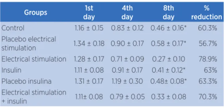
Table 1 shows that all experimental groups
experienced a reduction of the lesion area between the
irst and eighth day. On the other hand, the intergroup
analysis showed that the ES group saw a signiicantly
higher reduction percentage of the lesion area, when
compared with the other groups (p <0.05), except the
ES+I group.
Table 1. Mean ± Standard Deviation (in cm²) of the lesion area during diferent days (irst, fourth and eighth) and reduction percentage of the lesion area in the following groups: control – C, placebo electrical stimulation – PES, electrical stimulation – ES, insulin – I, placebo insulin – PI, electrical stimulation + insulin – ES+I
Groups 1st
day
4th day
8th day
% reduction
Control 1.16 ± 0.15 0.83 ± 0.12 0.46 ± 0.16* 60.3% Placebo electrical
stimulation 1.34 ± 0.18 0.90 ± 0.17 0.58 ± 0.17* 56.7% Electrical stimulation 1.28 ± 0.17 0.71 ± 0.09 0.27 ± 0.10 78.9% Insulin 1.11 ± 0.08 0.91 ± 0.17 0.41 ± 0.12* 63% Placebo insulina 1.31 ± 0.17 1.19 ± 0.30 0.48± 0.08* 63.3% Electrical stimulation
+ insulin 1.11± 0.08 0.79 ± 0.05 0.33 ± 0.08 70.3%
*p<0.05 versus ES
Table 2 shows the linear measure values of the
reepithelialization process, that is, the extension
between the edges of the epidermis and the free edge
at the center of the lesion, and also the central distance
of the lesion, that is, the distance between the growing
epithelia of both edges. his table shows similar values
for all experimental groups. he best lesion closure rate
occurred in the groups treated with HVES (ES and
ES+I), diferent from group I (p<0.05).
Table 2. Mean ± Standard Deviation (in µm) of reepithelialization (epithelial growth) and distance between lesion edges (lesion closure) of the following groups: control – C, placebo electrical stimulation – PES, electrical stimulation – ES, insulin – I, placebo insulin – PI, electrical stimulation + insulin – ES+I
Groups Reepithelialization Distance between edges
Control 1615.93 ± 1345.62 2159.51 ± 809.51 Placebo electrical
stimulation 1504.04 ± 1436.76 2156.60 ± 7470.80 Electrical stimulation 1437.70 ± 506.81 1120.40 ± 840.43* Insulin 1443.75 ± 228.82 3202.46 ± 1417.73 Placebo insulin 1845.30 ± 692.77 2683.02 ± 1162.61 Electrical stimulation
+ insulin 1936.76 ± 437.80 1108.38 ± 780.69*
*p<0.05 versus I
Table 3. Mean ± Standard Deviation of the number of ibroblasts and leukocytes per 1000 µm2 on the regenerating dermis of the
following groups: control – C, placebo electrical stimulation – PES, electrical stimulation – ES, insulin – I, placebo insulin – PI, electrical stimulation + insulin – ES+I
Groups Fibroblasts Leukocytes
Control 40.56 ± 7.4*$ 11.80 ± 5.47* Placebo electrical stimulation 39.8 ± 3.5*$ 10.37 ± 2.3*# Electrical stimulation 53.3 ± 5.21 5.00 ± 1.69# Insulin 46.9 ± 3.9*$ 10.46 ± 1.9*# Placebo insulin 43.18 ± 4.9*$ 17.18 ± 3.33* Electrical stimulation + insulin 55.71 ± 9.5 6.86 ± 2.01#
*p<0.05 versus ES; $ p<0.05 versus ES+I; #p<0.05 versus PI
DISCUSSION
Some factors interfere in the healing process, and
are classiied as local (infection, local tissue perfusion,
type of damaged tissue) and systemic inluences (age,
nutrition, tissue oxygen tension, diabetes, immunological
condition, associated diseases)
15. For more reliable
results, this study chose not to take these inluences
into consideration. herefore, all rats used were the
same age and race, had the same mechanism of injury,
same diet and were kept in a controlled environment.
We also considered the stress level, by using a group of
placebo animals, who underwent surgery, handling and
anesthesia daily, and provided the results presented by
the control group.
Electrical stimulation is believed to accelerate the
lesion reparation process, by imitating the electric
current that naturally occurs in the skin when damaged
16.
Our results show that all groups had their lesion
reduced during the acute phase, including the control
and placebo groups. The combined use of tratments
(ES+I) produced a high healing rate (70%). However, it
was the ES group that had the best reduction percentage
(78.9%), statistically standing out from the other groups
(p<0.05).
The application of diferent polarities can interfere
with tissue response, since diferent cells present in the
cicatrization process may migrate due to the charge
used, a fact identiied by Kloth and McCulloch
17, who
observed negative poles attracting cells like neutrophils
and macrophages, which would then sufer autolysis to
counteract the necrosis.
This study used a negative pole, without increase in
reepithelialization, in other words, epithelium growth.
However, the distance between the epithelial edges,
symbolizing lesion healing and its contraction, was
signiicantly better in HVES-treated groups, both by
itself (ES) and associated with topical insulin (ES+I).
The percentage of reduction in lesion areas was also
signiicantly higher in the ES group. These results
conirm the efects of cathodic HVES in the healing of
cutaneous lesions.
The dermis cells showed a signiicant decrease in
the number of leukocytes in the ES-treated groups and
a signiicant increase in ibroblasts in the ES and ES+I
groups, proving the beneits of the cathodic HVES. No
other groups showed similar results. The group treated
with placebo insulin had a signiicant increase in the
number of leukocytes, relecting the exacerbation of the
inlammatory process, caused by the lack of any active
therapeutic measure, such as electrical stimulation or
topical insulin.
The diference between the reepithelialization
measurements and the distance between the edges of
the lesion, both evaluated in this study, are justiied by
the action of myoibroblasts, cells responsible for the
contraction of the lesion. These cells align and attach
themselves to the thicker collagen ibers, pulling them
together and thus greatly contributing to the healing
process
18,19.
The number of ibroblasts conirms study data,
indicating that electrical stimulation improves lesion
regeneration, since it promotes cell migration, stimulates
ibroblasts and increases protein synthesis
17,20.
Isolated application of topical insulin showed
no signiicant diference in all analyzed variables,
though insulin is known to stimulate growth and the
development of diferent types of cells
21. It is also
important in the reepithelialization process, which
involves proliferation, migration and diferentiation of
Lima et al.
23used topical insulin with the same
formulation and provenance as our study and observed
accelerated healing of the cutaneous lesions of diabetic
rats. On the other hand, our study used non-diabetic rats
and the lesions were acute, which might explain the
diference in results. The distance between the edges
of the lesion in the group treated with topical insulin
did not difer from the control and placebo groups,
highlighting the electrical stimulation groups as the
ones with the best lesion closure.
Apikoglu-Rabus et al.
21studied the efect of topical
application of insulin on acute lesions in diabetic and
non-diabetic rats. Their study showed an accelerated repair
process with the use of insulin. The non-diabetic rats used
in this study failed to produce similar results. We believe
the diference lies with the experimental protocol, since
the authors of such study applied insulin twice a day.
In accordance with Sekhejane and Hourold
24, this
study proves that topical insulin is beneicial when
treating lesions, even if non-diabetes-related lesions, by
evidencing its role in the reduction of the inlammatory
reaction.
his study allows for future research, regarding the
combined use of high-voltage electrical stimulation and
topical insulin in an experimental model with diabetic
animals, with reduced growth factors, proliferation and
cell migration
25.
CONCLUSION
he cathodic HVES accelerated the lesion repair
process, with the addition of topical insulin showing no
additional efect.
REFERENCES
1. Liu Y, Petreaca M, Yao M, Martins-Green M. Cell and molecular mechanisms of keratinocyte function stimulated by insulin during wound healing. BMC Cell Biolog. 2009;10(1):1-15. 2. Yoon CS, Jung HS, Kwon MJ, Lee SH, Kim CW, Kim MK, et al.
Sonoporation of the minicircle-VEGF165 for wound healing of diabetic mice. Pharm Res. 2009;26(4):794-801.
3. Peters EJ, Lavery LA, Armstrong DG, Fleichili JG. Electric stimulation as an adjunct to heal diabetic foot ulcers: a randomized clinical trial. Arch Phys Med Rehabil. 2001;82(6):721-5.
4. Davini R, Nunes CV, Guirro ECO, Guirro RRJ. Tratamento de úlceras cutâneas crônicas por meio da estimulação elétrica de alta voltagem. Rev Ciênc Méd. PUCCAMP. 2005;14(3):249-58.
5. Tsai CH, Lin BJ, Chao PH. α2ß1 integrin and RhoA mediates electric ield-induced ligament ibroblast migration directionality. J Orthop Res. 2013;31(2):322-7.
6. Fitzgerald GK, Newsome D. Treatment of a large infected thoracic spine wound using high voltage pulsed monophasic current. Physical Therapy. 1993;73(6):355-6.
7. Griin JW, Tooms RE, Mendius RA, Clift JK, Zwaag RV, et al. Eicacy of high voltage pulsed current for healing of pressure ulcers in patients with spinal cord injury. Phys Ther. 1991;71(6):442-4.
8. Houghton PE, Kinkaid CB, Lovell M, Campbell KE, Keast DH, Woodbury MG, et al. Efect of electrical stimulation on chronic leg ulcer size and appearance. Phys Ther. 2003;83(1):17-28. 9. Davini R, Nunes CV, Guirro ECO, Guirro RRJ. Estimulação
elétrica de alta voltagem: uma opção de tratamento. Rev Bras Fisioter. 2005;9(3):249-56.
10. Hanam SR, Singleton CE, Rudek W. The efect of topical insulin on infected cutaneous ulcerations in diabetic and nondiabetic mice. J Foot Surg. 1983;22(4):298-301.
11. Pierre EJ, Barrow RE, Hawkins HK, Nguyen TT, Sakurai YMD, Desai M, et al. Efects of insulin on wound healing. Trauma. 1998;44(2):342-5.
12. Zhang XJ, Wu X, Wolf SE, Hawkins HK, Chinkes DL, Wolfe RR. Local insulin-zinc injection accelerates skin donor site wound healing. J Surg Res. 2007;142(1):90-6.
13. Madibally SV, Solomon V, Mitchell RN, Walter LV, Yarmush ML, Toner M. Inluence of insulin therapy on burn wound healigin rats. J Surg Res. 2003;109(2):92-100.
14. Mandarim-de-Lacerda CA, Fernandes-Santos C, Aguila MB. Image analysis and quantitative morphology. In: Hewitson TD, Darby JA (Editors), Histology protocols: methods in molecular biology. New Jersey: Humana Press; 2010;211-25. 15. Nuccitelli R, Nuccitelli P, Ramlatchan S, Sanger R, Smith PJS.
Imaging the electric ield associated with mouse and human skin wounds. Wound Rep Reg. 2008;16(3):432-41.
16. Thomaz JB, Herdy CDC, Oliveira JCP, Souza SR, Robadey
RA. Fundamentos da cicatrização das feridas. Arq Bras Med.
1996;70(2):65-72.
17. Kloth LC, McCulloch JM. Promotion of wound healing with electrical stimulation. Activ Wound Care. 1996;9(5):42-5. 18. Brown M, Gogia PP. Efects of high voltage stimulation
on cutaneous wound healing in rabbits. Phys Ther. 1987;67(5):662-7.
19. Zhao M, Bai H, Wang E, Forrester JV, McCaig CD. Electrical stimulation directly induces pre-angiogenic responses in vascular endothelial cells by signaling through VEGF receptors. J Cell Sci. 2004;117(Pt 3):397-405.
20. Isaac C, Ladeira PRS, Rego FMP, Aldunate JCB, Ferreira MC.
Processo de cura das feridas: cicatrização isiológica. Rev
Med. 2010;89(3/4):125-31.
21. Berry DP, Harding KG, Stanton MR, Jasani B, Ehrlich HP. Human wound contraction: collagen organization, ibroblasts and myoibroblasts. Plast Reconstr Surg. 1998;102(1):124-31.
23. Lima MH, Caricilli AM, Abreu LL, Araújo EP, Pelegrinelli FF, et al. Topical insulin accelerates wound healing in diabetes by enhancing the AKT and ERK pathways: a double-blind placebo-controlled clinical trial. PLoS One. 2012;7(5):e36974. 24. Sekhejane PR, Houreld NN, Abrahamse H. Irradiation at 636 nm positively afects diabetic wounded and hypoxic cells in vitro. Photomed Laser Surg. 2011;29(8):521-30.