Contemporary Series of Robotic-Assisted Distal Ureteral
Reconstruction Utilizing Side Docking Position
_______________________________________________
Rick C. Slater
1, Nicholas J. Farber
2, Julie M. Riley
3, Yaniv Shilo
1, Michael C. Ost
11 Department of Urology,University of Pittsburgh Medical Center, Pittsburgh, PA, USA; 2 Division of Urology, Rutgers Robert Wood Johnson Medical School, New Brunswick, NJ, USA; 3 Division of Urology, University of New Mexico, Albuquerque, NM, USA
ABSTRACT
ARTICLE
INFO
______________________________________________________________ ______________________
Purpose: The robot-assisted approach to distal ureteral reconstruction is increasingly utilized. Traditionally, the robot is docked between the legs in lithotomy position resul-ting in limited bladder access for stent placement. We examined the use of side docking of the daVinci robot® to perform distal ureteral reconstruction.
Materials and Methods: A retrospective review of distal ureteral reconstruction (ure-teral reimplantation and uretero-ureterostomy) executed robotically was performed at a single institution by a single surgeon. The daVinci robotic® Si surgical platform was positioned at the right side of the patient facing towards the head of the patient, i.e. side docking.
Results: A total of 14 cases were identified from 2011-2013. Nine patients underwent ureteral reimplantation for ureteral injury, two for vesicoureteral reflux, one for ure-teral stricture, and one for megaureter. One patient had an uretero-ureterostomy for a distal stricture. Three patients required a Boari flap due to extensive ureteral injury. Mean operative time was 286 minutes (189-364), mean estimated blood loss was 40cc (10-200), and mean length of stay was 2.3 days (1-4). Follow-up renal ultrasound was available for review in 10/14 patients and revealed no long-term complications in any patient. Mean follow-up was 20.7 months (0.1-59.3).
Conclusion: Robot-assisted laparoscopic distal ureteral reconstruction is safe and effec-tive. Side docking of the robot allows ready access to the perineum and acceptable placement of the robot to successfully complete ureteral repair.
Key words:
Video-Assisted Surgery; Ureter; Reconstructive Surgical Procedures
Int Braz J Urol. 2015; 41: 1154-9
_____________________
Submitted for publication: November 18, 2014 _____________________
Accepted after revision: August 20, 2015
INTRODUCTION
Laparoscopic techniques for ureteral reim-plantation and reconstruction continue to grow. The technique and efficacy of the laparoscopic ureteral reimplantation has been well described (1-3). However, creating a non-refluxing urete-ral reimplantation laparoscopically is technically very difficult, and has translated into poor adop-tion of the technique. With the introducadop-tion of
the daVinci Surgical System® (Intuitive Surgical, Sunnyvale, CA) minimally invasive surgery has now allowed surgeons to accomplish increasingly complex procedures with a shorter learning curve and better efficacy (4).
This position, however, results in limited access to the bladder for retrograde placement of a urete-ral stent. Previously, side docking of the daVinci® robot has been described for various gynecologic surgeries (5, 6) as well as for performing a radi-cal prostatectomy (7). We present an alternative docking position which simplifi es surgical set-up, allows ready access to the bladder for stent place-ment and may ultimately lead to shorter operating time without compromising surgical technique.
MATERIALS AND METHODS
Retrospective chart review was performed on all patients of the senior author’s who underwent robotic assisted laparoscopic ureteral reconstruction (i.e. ureteral reimplantation and uretero-ureteros-tomy) utilizing a side docking position.
Preoperatively, all patients were evaluated with retrograde pyelograms, except those with ve-sicoureteral refl ux (VUR) who were imaged with voiding cystourethrograms. Preoperative manage-ment included ureteral stenting or nephrostomy tube placement for patients with ureteral injury or stricture, and observation or defl ux in patients with VUR. Routine preoperative labs, including serum creatinine and urinalysis, were obtained in all patients.
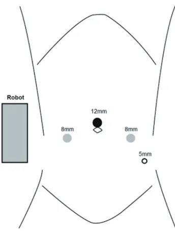
All operations employed the daVinci ro-botic Si surgical platform® (Intuitive Surgical, Sunnyvale, CA), with the robot docked on the patient’s right side parallel to the operative Ta-ble, e.g. “side docked” (Figures 1 and 2) (8). The patient was positioned in dorsal lithotomy posi-tion atop a memory foam pad to resist sliding, and legs were placed in yellow fi n stirrups. The patient was then placed in a Trendelenberg position. The trocar placement did not differ signifi cantly from traditional docking positions; we utilized an um-bilical port for camera placement and the robotic ports were placed 8 to 10cm apart and triangula-ted about the camera port with adjustments made to avoid the anterior superior iliac spine.
Ureteral reimplant performed for VUR uti-lized the non-refl uxing Lich Gregoire method (9). The ureter was identifi ed and dissected towards the bladder until its attachment to the bladder was visualized. The detrusor muscle was divided from
Figure 2 - Side docked position.
Figure 1 - daVinci robotic Si surgical platform port placement.
megaureter employed the Le Duc technique (10). The ureter was mobilized and introduced into the bladder through a short transmural channel in a nonrefluxing fashion. Distally, the ureteral end was widely spatulated and resulted in a distal ureteral plate that was fixed to the bladder mu-cosa, while the non-spatulated ureter remained unfixed. In all patients, ureteral stents and ure-thral Foley catheter were placed in a retrograde fashion during the procedure and Jackson-Pratt drains placed at the end of the operation.
Patients were postoperatively evaluated in the office setting approximately three to six weeks after the operation, with cystoscopy and stent re-moval. Additional follow-up with in-office renal ultrasound was scheduled at three months after surgery and yearly thereafter to assess the repair.
We collected the following demographic and procedural data from the electronic medical records of all patients: age, gender, body mass index (BMI), American Society of Anesthesio-logists (ASA) score, estimated blood loss (EBL), indication for surgery, operative time, time from ureteral injury, laterality of the operation, and intraoperative complications. Operative time was defined as time from start of incision to cessation of anesthesia.
Early postoperative outcomes were also extracted from the electronic medical record, in-cluding hospital length of stay (LOS). Postopera-tive complications were defined using the Clavien grading system (11). Office notes were reviewed for results of in-office renal ultrasound. We asses-sed change in renal function by comparing preo-perative and postopreo-perative serum creatinine (sCr) using a paired-sample Student t test. Statistical significance was defined as p<0.05.
RESULTS
From March 2011 to September 2013 a to-tal of 14 patients (13 female, 1 male) with a mean age of 39 years were identified and included in the study group. Indications for the procedure inclu-ded ureteral injury during primary hysterectomy in 9 patients, vesicoureteral reflux in 2 patients, congenital stricture in 2 patients and megaureter in 1 patient. Demographic data is listed in Table-1.
Operative and postoperative data are listed in Tables 1 and 2. Mean operative time was 286 minutes (189-364 minutes) and mean EBL was 40.0cc (10-200cc). All the procedures were com-pleted by the side-docking method without the need for re-docking. There was one intraoperati-ve complication: a contralateral ureter was erro-neously reimplanted and required reoperation and reimplantation of the correct ureter. All surgeries were completed without conversion to open or the need for re-docking. There was a single postope-rative Clavien grade I complication (postopepostope-rative fever).
Mean length of stay was 2.3 days (1-4 days). Creatinine was available for analysis in 12 of 14 patients. The difference between preoperati-ve and postoperatipreoperati-ve sCr was not statistically sig-nificant (p=68). Follow-up renal ultrasound was available for review in 10 out of 14 patients and demonstrated no evidence of complications in any patient.
DISCUSSION
Ureteral reconstruction can be accompli-shed using a variety of open procedures and has been described in the urologic literature with ex-cellent long-term outcomes. However, open sur-gery is associated with more blood loss, postope-rative pain, and longer lengths of hospital stay (2). The introduction of the daVinci robotic system® has changed the landscape of minimally invasive surgery. Despite the higher operating costs, longer setup, and loss of tactile feedback of the current robotic system, the benefits of a three-dimensio-nal field of vision, increased degrees of freedom of movement, tremor elimination, and motion sca-ling make robotic ureteral reconstruction advan-tageous (12, 13).
under-Table 1 - Patient Demographic Data and Operative Data.
Patient Age, years*
Side Indication Preop. Management
Procedure OT Postop. Complications
Postop. Imaging
FU
1 45 R HI NT UR 288 None N 4
2 34 R MU Observation UR, MT 322 None N 7
3 41 R HI Stent UR, UL 364 None Y 20
4 37 R HI NT UR, UL 236 None Y 6
5 22 L VUR Observation UR 241 None Y 59
6 21 R VUR Deflux UR 224 None Y 40
7 49 B HI NT UR, UL 362 None Y 24
8 61 R HI NT UR 350 None Y 44
9 47 R HI Stent UR, BF 366 None Y 26
10 28 L Stricture Stent UU 189 None Y 23
11 37 L HI Stent UR, UL, BF 328 Fever Y 12
12 64 L Stricture Stent UR, UL, BF 251 None Y 24
13 35 L HI NT UR 235 None N 0
14 25 R HI NT UR 254 None N 0
*Data include age (years); side of reconstruction (R = right; L = left; B = bilateral); indication for ureteral reconstruction (HI = Hysterectomy injury; MU = megaureter; VUR =
vesicoureteral reflux), preoperative management (NT = nephrostomy tube), operative procedure (UR = ureteral reimplantation; MR= megaureter tapering; UL = ureterolysis;
UU = ureteroureterostomy; BF = Boari Flap), operative time (OT = minutes), postoperative complications, postoperative imaging (Y = yes; N = No), and duration of follow-up (FU = months)
going robotic assisted ureteral reconstruction with a side docking position.
Our results suggest several important fin-dings. First, our case series demonstrates that side docking of the robot is comparable in operative time to other studies using the conventional do-cking approach (15, 16). Specifically, our mean operative time was 286.4 minutes which is similar to the 221 minutes reported in the largest case se-ries of conventionally docked robotic distal urete-ral reconstructions (16). Of note, our operative ti-mes include the entire duration of surgery, not just robotic console time, and may account for some of the disparity between our operative time and the literature time. In addition, many of our patients had additional concurrent procedures performed (e.g. ureterolysis and Boari flap) and nearly all had previously undergone abdominal surgery with subsequent formation of adhesions, both of which prolong operative times. Our mean length of stay of 2.3 days was also similar to the literature me-ans of 1.6 to 2.5 days using conventional docking, while our mean EBL of 40cc was also on par with
means of 50cc to 171cc quoted in the literature (16, 17). Our single postoperative complication a Clavien I postoperative fever–and the absence of any long-term complications (as assessed by ul-trasound and office evaluation) demonstrates the short and long-term safety of the repair. Additio-nally, the side-docking of the robot affords the sa-fety advantage of requiring less abduction of the patient’s legs, as the robot is no longer in that potential space. No patients in our series suffered from peroneal nerve injury or any musculoskeletal positioning complications. In patients with a his-tory of hip surgery or muscle contractures, side--docking of the robot is an excellent, safe alterna-tive to the traditional docking approach. Overall, our results suggest that the side-docking approach is safe, effective, and comparable to the conven-tional docking approach.
has been described as a complication (20). In our series, we had no stent migration, which may be attributed to the excellent visualization appre-ciated in typical ureteral stent placement. The direct access to the lithotomy position afforded by the side-docking position gives the assistant the opportunity to place a stent using a rigid rather than flexible cystoscope and therefore a better field of vision. The ability to easily place retrograde stents also allows for visual confir-mation of both the patency of the ureter and a good curl of the distal end of the stent. Ano-ther challenge to conventionally docked robo-tic ureteral reconstruction is that intraoperative stent placement may be cumbersome with the
robot blocking urethral access and requiring undocking of the robot or repositioning of the patient. Conversely, side docking of the robot allows easy cystoscopic access to the bladder for retrograde stent placement, especially when the ureter has been completely transected and the injury is managed via a nephrostomy tube.
There are several limitations to this stu-dy. First, its retrospective nature and relatively small sample size from a single surgeon intro-duce a possible selection bias. Nevertheless, this preliminary data may prompt other surgeons to adopt the side-docking approach to ureteral re-construction and generate additional, larger stu-dies of the approach. Second, we were unable to obtain the same surgeon’s data for compa-rison with the conventional docking approach. Third, postoperative imaging was unavailable in 4/10 patients, making it harder to determine the true success rate of the operation. However, two of those four patients did receive in-office follow-up at 4 and 7 months postoperatively and neither had evidence of complications on eva-luation. Moreover, 10/14 patients had extended follow-up with imaging and none had any long--term complications. Finally, this case series had a wrong site intraoperative complication. The error is attributed to the patient’s prior abdomi-nal surgery, which caused such extensive fibro-sis that the contralateral ureter was shifted to the intended side and was mistaken for the right ureter. The patient’s correct ureter was subse-quently reimplanted with no short or long-term postoperative complications.
CONCLUSIONS
Side docking of the robot during robotic assisted laparoscopic ureteral reconstruction of the distal ureter confers some benefit over the conventional docking approach, as the surgeon has unrestricted, ready access to the perineum for retrograde stent placement without undo-cking the robot or repositioning the patient. This approach is also safe and effective in terms of operative time, length of stay, and EBL compa-rable to literature values of the conventional do-cking approach.
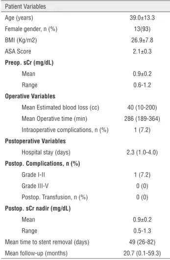
Table 2 - Preoperative, Operative and Postoperative Data.
Patient Variables
Age (years) 39.0±13.3
Female gender, n (%) 13(93)
BMI (Kg/m2) 26.9±7.8
ASA Score 2.1±0.3
Preop. sCr (mg/dL)
Mean 0.9±0.2
Range 0.6-1.2
Operative Variables
Mean Estimated blood loss (cc) 40 (10-200)
Mean Operative time (min) 286 (189-364)
Intraoperative complications, n (%) 1 (7.2)
Postoperative Variables
Hospital stay (days) 2.3 (1.0-4.0)
Postop. Complications, n (%)
Grade I-II 1 (7.2)
Grade III-V 0 (0)
Postop. Transfusion, n (%) 0 (0)
Postop. sCr nadir (mg/dL)
Mean 0.9±0.2
Range 0.5-1.3
Mean time to stent removal (days) 49 (26-82)
Mean follow-up (months) 20.7 (0.1-59.3)
CONFLICT OF INTEREST
None declared.
REFERENCES
1. Yohannes P, Chiou RK, Pelinkovic D. Rapid communication: pure robot-assisted laparoscopic ureteral reimplantation for ureteral stricture disease: case report. J Endourol. 2003; 17:891-3.
2. Rassweiler JJ, Gözen AS, Erdogru T, Sugiono M, Teber D. Ureteral reimplantation for management of ureteral strictures: a retrospective comparison of laparoscopic and open techniques. Eur Urol. 2007; 51:512-22.
3. Ogan K, Abbott JT, Wilmot C, Pattaras JG. Laparoscopic ureteral reimplant for distal ureteral strictures. JSLS. 2008; 12:13-7.
4. Rashid TG, Kini M, Ind TE. Comparing the learning curve for robotically assisted and straight stick laparoscopic procedures in surgical novices. Int J Med Robot. 2010; 6:306-10.
5. Einarsson JI, Hibner M, Advincula AP. Side docking: an alternative docking method for gynecologic robotic surgery. Rev Obstet Gynecol. 2011; 4:123-5.
6. Woods DL, Hou JY, Riemers L, Gupta D, Kuo DY. Side-docking in robotic-assisted gynaecologic cancer surgery. Int J Med Robot. 2011; 7:51-4.
7. Uffort EE, Jensen JC. Side docking the robot for robotic laparoscopic radical prostatectomy. JSLS. 2011; 15:200-2. 8. Chan ES, Yee CH, Lo KL, Chan CK, Hou SM, Ng CF.
Side-docking technique for robot-assisted urologic pelvic surgery. Urology. 2013; 82:1300-3.
9. Riedmiller H, Gerharz EW. Antireflux surgery: Lich-Gregoir extravesical ureteric tunnelling. BJU Int. 2008; 101:1467-82. 10. Le Duc A, Camey M, Teillac P. An original antireflux
ureteroileal implantation technique: long-term followup. J Urol. 1987; 137:1156-8.
11. Clavien PA, Barkun J, de Oliveira ML, Vauthey JN, Dindo D, Schulick RD, et al. The Clavien-Dindo classification of surgical complications: five-year experience. Ann Surg. 2009; 250:187-96.
12. Phillips EA, Wang DS. Current status of robot-assisted laparoscopic ureteral reimplantation and reconstruction. Curr Urol Rep. 2012; 13:190-4.
13. Patil NN, Mottrie A, Sundaram B, Patel VR. Robotic-assisted laparoscopic ureteral reimplantation with psoas hitch: a multi-institutional, multinational evaluation. Urology. 2008; 72:47-50.
14. Chan ES, Yee CH, Chiu PK, Chan CK, Hou SM, Ng CF. Robot-assisted radical cystectomy using a side-docking technique. J Laparoendosc Adv Surg Tech A. 2015; 25:207-11.
15. Stanasel I, Atala A, Hemal A. Robotic assisted ureteral reimplantation: current status. Curr Urol Rep. 2013; 14:32-6. 16. Fifer GL, Raynor MC, Selph P, Woods ME, Wallen EM,
Viprakasit DP, et al. Robotic ureteral reconstruction distal to the ureteropelvic junction: a large single institution clinical series with short-term follow up. J Endourol. 2014; 28:1424-8. 17. Baldie K, Angell J, Ogan K, Hood N, Pattaras JG. Robotic
management of benign mid and distal ureteral strictures and comparison with laparoscopic approaches at a single institution. Urology. 2012; 80:596-601.
18. Noh PH, Defoor WR, Reddy PP. Percutaneous antegrade ureteral stent placement during pediatric robot-assisted laparoscopic pyeloplasty. J Endourol. 2011; 25:1847-51. 19. Mufarrij PW, Rajamahanty S, Krane LS, Hemal AK.
Intracorporeal Double-J stent placement during robot-assisted urinary tract reconstruction: technical considerations. J Endourol. 2012; 26:1121-4.
20. Richter S, Ringel A, Shalev M, Nissenkorn I. The indwelling ureteric stent: a ‘friendly’ procedure with unfriendly high morbidity. BJU Int. 2000; 85:408-11.