Risk Factors for Bone Loss with Prostate Cancer in Korean Men
Not Receiving Androgen Deprivation Therapy
Sun-Ouck Kim, Taek Won Kang, Dongdeuk Kwon, Kwangsung Park, Soo Bang Ryu
Department of Urology, Chonnam National University Medical School, Gwangju, Korea
ABSTRACT
Purpose: Preexisting bone loss in men with prostate cancer is an important issue due to the accelerated bone loss during androgen deprivation therapy (ADT). In addition, a high prostate-speciic antigen (PSA) level has been reported to be re-lated to bone metabolism. This study assessed the factors associated with osteoporosis in Korean men with non-metastatic prostate cancer before undergoing ADT.
Materials and Methods: The study enrolled patients admitted for a prostate biopsy because of a high PSA or palpable nodule on a digital rectal examination. We divided the patients (n = 172) according to the results of the biopsy: group I, non-metastatic prostate cancer (n = 42) and group II, benign prostatic hypertrophy (BPH; n = 130). The lumbar bone mineral density (BMD) was evaluated using quantitative computed tomography. The demographic, health status, lifestyle, body mass index (BMI), serum testosterone concentration, and disease variables in prostate cancer (Gleason score, clinical stage, and PSA) were analyzed prospectively to determine their effect on the BMD.
Results: The estimated mean T-score was higher in group I than in group II (-1.96 ± 3.35 vs. -2.66 ± 3.20), but without statistic signiicance (p = 0.235). The signiicant factors correlated with BMD in group I were a high serum PSA (β = -0.346, p = 0.010) and low BMI (β = 0.345, p = 0.014) in the multiple linear regression model. Also old age (r = -0.481, p = 0.001), a high serum PSA (r = -0.571, p < 0.001), low BMI (r = 0.598, p < 0.001), and a high Gleason’s score (r = -0.319, p = 0.040) were the factors related to BMD in the correlation. The signiicant factors correlated with BMD in group II were old age (β = -0.324, p = 0.001) and BMI (β = 0.143, p = 0.014) in the multiple linear regression model.
Conclusions: The risk factors for osteoporosis in men with prostate cancer include a low BMI, and elevated serum PSA. Monitoring BMD from the outset of ADT is a logical irst step in the clinical strategy to avoid or minimize potential bone-related complications in these patients.
Key words: prostate neoplasm; osteoporosis; androgen deprivation therapy; prostate speciic antigen Int Braz J Urol. 2009; 35: 183-9
INTRODUCTION
In men, 36% of osteoporosis is due to low androgen levels, which can occur with congenital hypogonadism, the aging process, or androgen depri-vation therapy (ADT) for the treatment of advanced prostate cancer (1). Bone is the most common site of metastasis in many types of cancer, including
no data on this subject has been reported for Korean patients with prostate cancer.
Recently, the increased life expectancy,
advanced diagnostic techniques, and Westernized
eating habits have contributed to a high incidence of prostate cancer in Koreans and an increased mortal-ity rate due to co-morbidmortal-ity. Thus, predicting and preventing the progression of osteoporosis in patients with prostate cancer is of critical importance. Before initiating ADT, it is necessary to identify the causes of bone loss and related risk factors for osteoporosis. However, who should undergo bone mineral density (BMD) testing before ADT remains unclear. There is a major need to determine ways to treat patients with prostate cancer undergoing ADT without increasing the risk of osteoporosis. This study evaluated the fac-tors associated with osteoporosis in patients with non-metastatic prostate cancer before undergoing ADT as compared to those with benign prostate hypertrophy (BPH) alone.
MATERIALS AND METHODS
After informed consent was obtained from all patients, a prospective trial was initiated at Chonnam National University Hospital from January to
Decem-ber 2005. This study enrolled patients hospitalized for a prostate biopsy because of a high PSA or palpable
nodule on rectal examination. Based on previous med-ical history and physmed-ical examination, patients with thyroid or parathyroid disease, uncontrolled diabetes mellitus, cardiovascular disease, digestive disorders, and chronic steroid users were excluded, as well as
patients found to have bone metastasis on plain ilm
X-rays and a bone scan. Patient information on de-mographics, health status, lifestyle, tobacco use, and body mass index (BMI) were obtained. The patients were divided into two groups according to the result of the prostate biopsy: group I, patients with prostate cancer (n = 42), and group II, patients with BPH (n = 130). The general conditions of the patients assessed according to performance status were good and they reported light physical activity and moderate intakes of calcium, alcohol, and caffeine. We evaluated the relationship between the patient characteristics and
disease variables. This was analyzed prospectively us
-ing univariate and multivariate methods to determine their role in the BMD levels previously established using quantitative computed tomography (QCT) of the lumbar spine. The Institutional Review Board at our hospital approved the study.
Prostate Cancer Disease Variables
The patients’ charts were reviewed to obtain
information on clinical variables pertaining to
pros-tate cancer: clinical stage, Gleason score, and PSA. To measure PSA (Access Assay, Hybritech) and total testosterone (Immunoenzymatic assay, Beckman), serum was obtained at between 08:00 and 09:00 h.
Bone Mineral Density
The BMD in L1-4 was measured using
QCT. Using the World Health Organization Criteria, a normal BMD was deined as one greater than -1 standard deviation (SD) below the young adult mean
value (T-score), osteopenia as a T-score between -1
and -2.5 SDs, and osteoporosis as a T-score of -2.5
or less (6).
Statistical Analysis
Descriptive, comparative, univariate, and
multivariate analyses using the Statistical software package for the Social Sciences, version 12.0 (SPSS
Inc., Chicago, IL) were performed to describe BMD and the associations between it and the disease
vari-ables. Simple correlation analysis was performed using the nonparametric Spearman correlation coef
-icient. An independent samples t-test was used for comparison analysis. Variables statistically signiicant
in the univariate analysis were included in the multiple linear regression model with BMD of the lumbar spine as the dependent variable. Two-tailed tests were used for all correlation and comparison analyses. P values
of 0.05 or less were considered statistically signii -cant.
RESULTS
No differences were observed in the basic health characteristics between the two age-matched
groups (over 65 years old), except for PSA, as summa
and did not engage in physically demanding sports or recreational activity, but only in light exercise, such as short walks. Among the former and current smok-ers, the pack years ranged from 5 to 62. Additional information on the prostate cancer disease variables
for group I is summarized in Table-2.
The BMD between the Two Groups
No signiicant difference was detected in the
prevalence of bone loss between the two groups. In group I, 69.05% had osteopenia (16.67%) or
osteopo-rosis (52.38%) of the spine, while in group II, 55.38%
had osteopenia (9.23%) or osteoporosis (46.15%). The estimated mean T-score was higher in group I than in group II (-1.96 ± 3.35 vs. -2.66 ± 3.20), but the
dif-ference was not statistically signiicant (p = 0.235)
(Table-1). For all of the participants in this study, old
age (r = -0.371, p < 0.001), a high PSA (r = -0.209,
p = 0.006), and low BMI (r = 0.226, p = 0.003) were
signiicantly correlated with bone loss.
The BMD in Patients in Group-I
The mean patient age was 71.48 years. The
number (%) of participants by clinical stage T1, T2,
and T3 was 18 (42.8), 22 (25.4), and 2 (4.8) respec
-tively and by a Gleason’s score of 6, 7, 8, and 9 was 4 (9.5), 16 (38.1), 12 (28.6), and 10 (28.9), respectively.
Of those with prostate cancer, 69.05% had
osteope-nia (16.67%) or osteoporosis (52.38%) of the spine (mean T-score -2.66 ± 3.20). The signiicant factors
correlated with BMD in group I were a high serum
PSA (β = -0.346, p = 0.010) and low BMI (β = 0.345,
p = 0.014) in the multiple linear regression model
(Table-3). Also an old age (r = -0.481, p = 0.001), a high serum PSA (r = -0.571, p < 0.001), low BMI (r = 0.598, p < 0.001), and a high Gleason’s score (r =
-0.319, p = 0.040) were the factors related with BMD in the univariate analysis (Table-4).
The BMD in Patients in Group-II
The mean patient age was 70.7 years. Of
those with BPH, 55.38% had osteopenia (9.23%) or
osteoporosis (46.15%) of the spine (mean T-score
-1.96 ± 3.35). The signiicant factors correlated with BMD in group II were old age (β = -0.324, p = 0.001) and BMI (β = 0.143, p = 0.014). Smoking, serum testosterone and clinical stage were not signiicantly
correlated with BMD (Table-5).
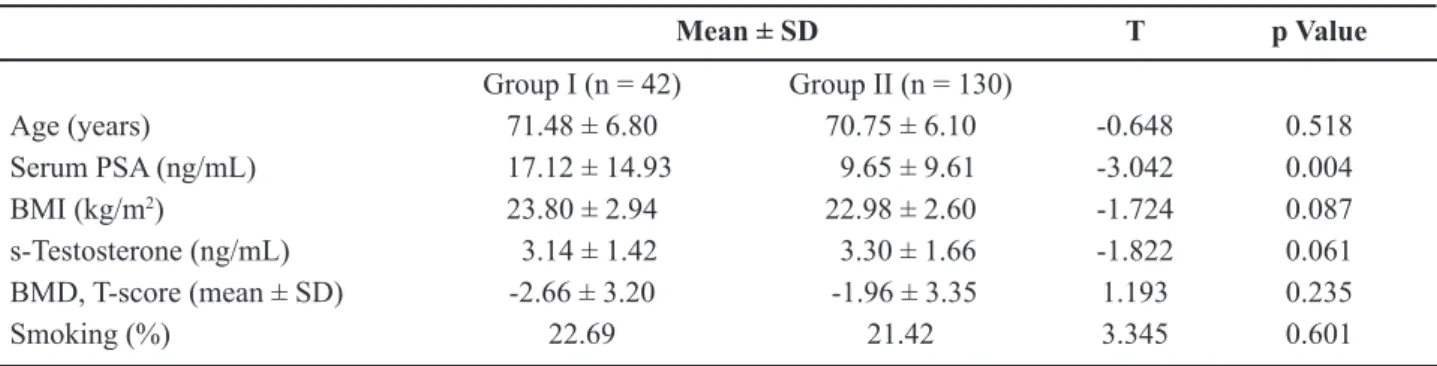
Table 1 – A comparison of the disease variables between the two groups.
Mean ± SD T p Value
Group I (n = 42) Group II (n = 130)
Age (years) 71.48 ± 6.80 70.75 ± 6.10 -0.648 0.518
Serum PSA (ng/mL) 17.12 ± 14.93 9.65 ± 9.61 -3.042 0.004
BMI (kg/m2) 23.80 ± 2.94 22.98 ± 2.60 -1.724 0.087
s-Testosterone (ng/mL) 3.14 ± 1.42 3.30 ± 1.66 -1.822 0.061
BMD, T-score (mean ± SD) -2.66 ± 3.20 -1.96 ± 3.35 1.193 0.235
Smoking (%) 22.69 21.42 3.345 0.601
Group I = prostate cancer group; Group II = benign prostate hyperplasia group; PSA = prostate speciic antigen; BMI = body mass index; BMD = bone mineral density; Dependent variables = T-score.
Table 2 – Basic characteristics of group I (prostate cancer
group).
Characteristics N (%)
Prostate cancer stage
cT1NxM0 18 (42.8)
cT2NxM0 22 (25.4)
cT3NxM0 2 (4.8)
Gleason’s score
3+3=6 4 (9.5)
7 (4+3, 3+4) 16 (38.1)
4+4=8 12 (28.6)
COMMENTS
As the prevalence of prostate cancer and osteoporosis increases with age, many patients may
already have osteoporosis when diagnosed with prostate cancer. Orchiectomy and the administration of a gonadotropin-releasing hormone agonist, which is the main treatment for metastatic prostate cancer,
Table 3 – The factor associated with bone loss in Group I (prostate cancer).
Standardized Coeficients T p Value*
Beta
Age (years) -0.228 -1.797 0.081
Smoking (%) -0.040 -0.334 0.741
Serum PSA (ng/mL) -0.346 -2.729 0.010
BMI (kg/m2) 0.345 2.587 0.014
Serum Testosterone (ng/mL) 0.095 0.711 0.482
Clinical stage 0.032 0.269 0.790
Gleason’s score -0.120 -0.991 0.328
Adjusted R2 = 0.479; Dependent variables = T-score; * calculated from multiple linear regression model.
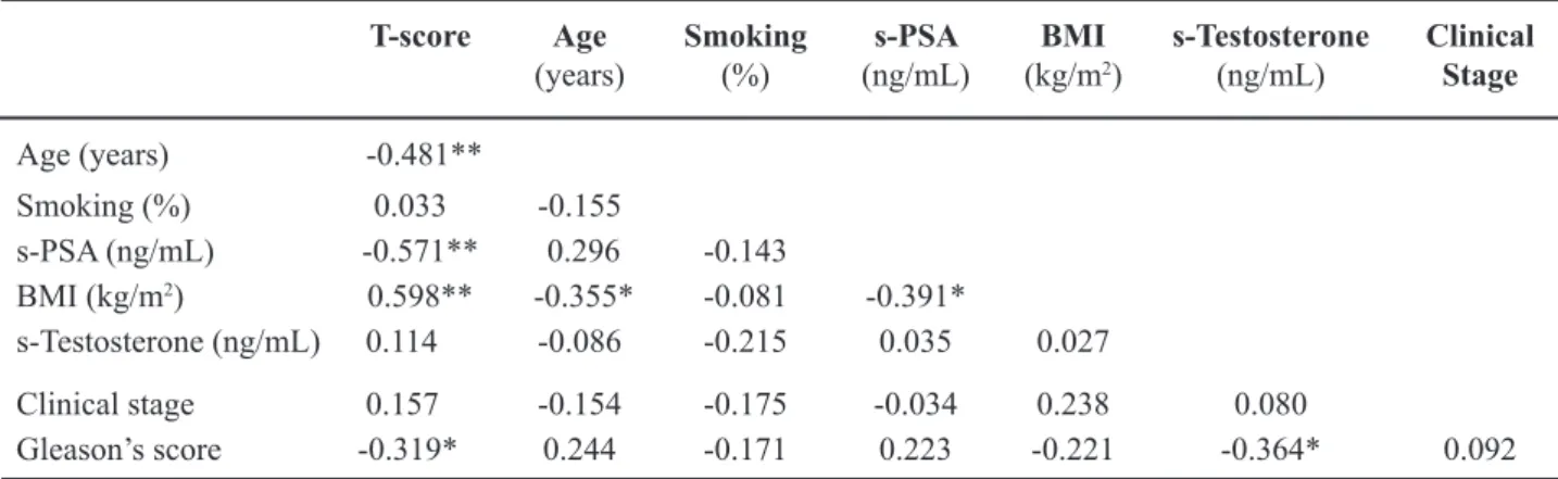
Table 4 – The factors associated with bone loss in Group I (prostate cancer group).
T-score Age
(years)
Smoking
(%)
s-PSA
(ng/mL) (kg/mBMI 2)
s-Testosterone
(ng/mL) Clinical Stage
Age (years) -0.481**
Smoking (%) 0.033 -0.155
s-PSA (ng/mL) -0.571** 0.296 -0.143
BMI (kg/m2) 0.598** -0.355* -0.081 -0.391*
s-Testosterone (ng/mL) 0.114 -0.086 -0.215 0.035 0.027
Clinical stage 0.157 -0.154 -0.175 -0.034 0.238 0.080
Gleason’s score -0.319* 0.244 -0.171 0.223 -0.221 -0.364* 0.092
**p < 0.01; *p < 0.05; calculated from Spearman’s correlation analysis; Dependent variables = T-score.
Table 5 – The factors associated with bone loss in Group II (benign prostatic hyperplasia group.
Standardized Coeficients T p Value*
Beta
Age (years) -0.324 -4.216 0.001
Serum PSA (ng/mL) -0.035 -0.475 0.635
BMI (kg/m2) 0.143 1.794 0.014
Smoking 0.113 1.496 0.075
Serum Testosterone (ng/mL) 0.021 0.274 0.784
have been reported to cause signiicant bone loss and
lead to bone fracture (7). This is of great concern for
men with prostate cancer who will receive ADT (8).
Therefore, osteoporosis should be prevented in men with prostate cancer who may require ADT.
Prostate cancer produces and secretes
abun-dant PSA, which is not synthesized in other tumors or tissues. PSA is an important, widely used sero -logic marker for prostate cancer, but its role in bone
metastases is still unclear. PSA, a serine protease,
and matrix metalloproteinases are involved in the breakdown of the extracellular matrix that promotes the invasion and metastasis of tumor cells in bone
(9). In addition, elevated serum PSA levels are as -sociated with advanced prostate cancer, and prostate cancer cells stimulate the release of various cytokines, which activate osteoclasts and bone resorption (10).
Prostate cancer preferably metastasizes to bone and
produces primarily osteoblastic phenotypes, unlike other cancers, which are associated with osteoclast formation. Among the known osteogenic factors pro-duced by prostate cells, bone morphogenic proteins, endothelin-1, insulin-like growth factors, parathyroid hormone-related peptide, transforming growth
factor-β, and PSA, the latter is uniquely produced by prostate
cancer cells (11-16). Men with prostate cancer with
poorly differentiated cells and a high Gleason’s score
have lower testosterone levels than those with well
differentiated cells and a low Gleason’s score (17).
Generally, poorly differentiated prostate cancer is very
progressive and metastasizes rapidly; the clinical stage
is already high at diagnosis.
Although aging and a low BMI are known risk
factors for osteoporosis, whether the serum PSA level or Gleason’s score are risk factors for osteoporosis
remains unclear in men with prostate cancer. In this
study sample, a low BMI and elevated serum PSA levels were signiicant factors of decreased BMD in
the multivariate analysis. Also, old age, a low BMI,
elevated serum PSA levels, and a high Gleason’s score were signiicantly associated with bone loss in
men with prostate cancer, in the univariate analysis. These results suggest that men with prostate cancer,
who are slender and have higher serum PSA levels,
are at increased risk of developing a decreased BMD after ADT. In this study, the total serum testosterone was not different between the two groups and was
not signiicantly correlated with BMD. In addition,
no correlation with bone loss was observed with the clinical stage of the disease. Although smoking causes osteoporosis, no correlation with bone loss was de-tected in our study sample, differing somewhat from our previous prediction. This may have been caused by the relatively small number of patients in this study sample.
To date, no convincing study on the status of BMD in non-metastatic prostate cancer prior to ADT had been conducted in Korea. In the present study, 69.05% of the patients with non-metastatic prostate cancer had osteopenia (16.67%) or osteoporosis
(52.38%) of the spine before ADT, which is similar to
another study in which 73.5% had osteopenia (55.9%)
or osteoporosis (17.6%) of the spine (8).
One of the limitations of this study is that the exact intake of calcium and vitamin D, as well as smoking status and the type of daily activities, which are other factors potentially affecting BMD, were not considered. For accuracy, a future study must include
all of these factors. In addition, the small size of the
non-metastatic prostate cancer group in this study is a limitation. Many studies have recommended that one should check the baseline BMD in all men before starting ADT when osteoporotic risk factors are found
(18,19). One should also consider performing BMD studies in older men who have a high serum PSA and
a slender stature before initiating ADT in prostate cancer.
CONCLUSIONS
The risk factors for osteoporosis in men with prostate cancer include old age, a low BMI, and
el-evated serum PSA. Consideration should be given to
performing BMD studies in these men before initiat-ing ADT in prostate cancer. Monitorinitiat-ing BMD from
the outset of ADT is a logical irst step in the clinical strategy to avoid or minimize potential bone-related
complications in these patients.
CONFLICT OF INTEREST
REFERENCES
1. Orwoll E, Ettinger M, Weiss S, Miller P, Kendler D, Graham J, et al.: Alendronate for the treatment of osteoporosis in men. N Engl J Med. 2000; 343: 604-10.
2. Daniell HW: Osteoporosis after orchiectomy for pros-tate cancer. J Urol. 1997; 157: 439-44.
3. Hatano T, Oishi Y, Furuta A, Iwamuro S, Tashiro K: Incidence of bone fracture in patients receiving lutein-izing hormone-releasing hormone agonists for prostate cancer. BJU Int. 2000; 86: 449-52.
4. Oefelein MG, Ricchuiti V, Conrad W, Seftel A, Bodner D, Goldman H, et al.: Skeletal fracture associated with androgen suppression induced osteoporosis: the clini-cal incidence and risk factors for patients with prostate cancer. J Urol. 2001; 166: 1724-8.
5. Mittan D, Lee S, Miller E, Perez RC, Basler JW, Bruder JM: Bone loss following hypogonadism in men with prostate cancer treated with GnRH analogs. J Clin Endocrinol Metab. 2002; 87: 3656-61.
6. Blake GM, Fogelman I: Principles of bone densitom-etry. In: Bilezikian JP, Raisz LG, and Rodan GA (ed.), Principles of Bone Biology. San Diego, Academic Press. 1996; pp. 1313-32.
7. McGrath SA, Diamond T: Osteoporosis as a compli -cation of orchiectomy in 2 elderly men with prostatic cancer. J Urol. 1995; 154: 535-6.
8. Conde FA, Sarna L, Oka RK, Vredevoe DL, Rettig MB, Aronson WJ: Age, body mass index, and serum prostate-specific antigen correlate with bone loss in men with prostate cancer not receiving androgen deprivation therapy. Urology. 2004; 64: 335-40. 9. Stetler-Stevenson WG, Aznavoorian S, Liotta LA:
Tumor cell interactions with the extracellular matrix
during invasion and metastasis. Annu Rev Cell Biol. 1993; 9: 541-73.
10. Ershler WB, Harman SM, Keller ET: Immunologic aspects of osteoporosis. Dev Comp Immunol. 1997; 21: 487-99.
11. Komori T, Yagi H, Nomura S, Yamaguchi A, Sasaki K, Deguchi K, et al.: Targeted disruption of Cbfa1 results in a complete lack of bone formation owing to maturational arrest of osteoblasts. Cell. 1997; 89: 755-64.
12. Yang J, Fizazi K, Peleg S, Sikes CR, Raymond AK, Jamal N, et al.: Prostate cancer cells induce osteoblast differentiation through a Cbfa1-dependent pathway. Cancer Res. 2001; 61: 5652-9.
13. Komori T: Runx2, a multifunctional transcription fac-tor in skeletal development. J Cell Biochem. 2002; 87: 1-8.
14. Karsenty G: The genetic transformation of bone biol-ogy. Genes Dev. 1999; 13: 3037-51.
15. Yingling JM, Blanchard KL, Sawyer JS: Development of TGF-beta signalling inhibitors for cancer therapy. Nat Rev Drug Discov. 2004; 3: 1011-22.
16. Bonewald LF, Mundy GR: Role of transforming growth factor-beta in bone remodeling. Clin Orthop Relat Res. 1990; 250: 261-76.
17. Eriksson A, Carlström K: Prognostic value of serum hormone concentrations in prostatic cancer. Prostate. 1988; 13: 249-56.
18. Higano CS: Management of bone loss in men with prostate cancer. J Urol. 2003; 170: S59-63; discussion S64.
19. Conde FA, Aronson WJ: Risk factors for male osteo-porosis. Urol Oncol. 2003; 21: 380-3.
Accepted after revision: December 3, 2008
Correspondence address:
Dr. Taek Won Kang Department of Urology
Chonnam National University Med. Sch. 8, Hak-dong, Dong-gu
Gwangju #501-757, Republic of Korea Fax: + 82 62 227-1643
EDITORIAL COMMENT
This is an interesting paper describing risk factors for osteopenia in men with prostate cancer and benign prostatic hyperplasia undergoing androgen deprivation therapy (ADT). The work describes high
prostate speciic antigen and low body mass index
as risk factors for men with prostate cancer about to undergo ADT. It is important to screen such men prior to ADT to determine if further steps are needed, such as vitamin D and calcium supplementation or bisphosphonate treatment.