ABSTRACT
Osteoporosis is defined as “a systemic skeletal disease characterized by low bone mass and microarchitectural deterioration of bone tissue with a consequent increase in bone fragility and susceptibility to fracture”. Approximately 40–50% of women sustain osteoporotic fractures in their lifetime; as such, it is appropriate that studies initially focused upon females. Despite an increased recognition of osteoporotic fractures in men, there continues to be neglect of this disease in males. This ongoing neglect is inappropriate as 25–33% of men in some populations will sus-tain osteoporotic fractures in their lifetime. Testosterone plays an impor-tant role in male skeletal health. However, recent data suggest that estrogen may in fact be the dominant hormone regulating skeletal sta-tus in both men and women. BMD measurement may be utilized for osteoporosis diagnosis and to assist with fracture risk prediction in men prior to their sustaining a fracture. Recognizing this need, the Internation-al Society for ClinicInternation-al Densitometry (ISCD) recommended and recently reaffirmed use of a BMD T-score of -2.5 or below be utilized to diagnose osteoporosis in men. Androgen therapy of hypogonadal men may be considered with the caveat that data do not exist to document that this treatment reduces fracture risk. At this time, the data is inadequate to support use of androgen treatment in eugonadal men with osteoporosis. Parathyroid hormone treatment does increase BMD; existing studies have not been of adequate size or duration to document fracture reduction efficacy. Bisphosphonate therapy increases BMD, reduces vertebral frac-ture risk and is considered the standard of care for osteoporotic men at this point in time. (Arq Bras Endocrinol Metab 2006;50/4:764-774)
Keywords:Osteoporosis; Androgens; Estrogens; Teriparatide; Bisphospho-nates
RESUMO
Osteoporose em Homens.
A osteoporose é definida como “uma doença esquelética sistêmica caracterizada por baixa massa óssea e deterioração da microarquite-tura do tecido ósseo com conseqüente aumento da susceptibilidade a fraturas”. Aproximadamente 40–50% das mulheres apresentarão uma fratura osteoporótica durante suas vidas, e por isso os estudos iniciais focalizaram o sexo feminino. A despeito do reconhecimento cada vez mais freqüente da ocorrência de fraturas osteoporóticas em homens, a doença continua sendo negligenciada no sexo masculino. Isto não é apropriado na medida em que 25–33% dos homens em algumas popu-lações apresentarão fraturas osteoporóticas durante suas vidas. A testosterona exerce um importante papel na integridade esquelética masculina, contudo dados recentes sugerem que os estrógenos são de fato os hormônios dominantes na regulação esquelética em ambos os sexos. A medida da densidade mineral óssea pode ser utilizada no dia-gnóstico e para avaliar o risco de fraturas, e a ISCD recomenda que o escore T de -2.5 ou menos seja usado como critério diagnóstico no homem. A terapia androgênica no hipogonadismo masculino deve ser
N ei l Bi nkley
Osteoporosis Clinical Center and R esearch Program and Institute on A ging, U niversity of
W isconsin, Madison, W I, U SA .
considerada, embora não existam dados sobre redução do risco de fraturas, e no momento não há dados que suportem o uso de andrógenos em homens eugonádicos com osteoporose. O trata-mento com paratormônio aumenta a massa óssea, porém os estudos existentes não apresentam amostra e duração suficientes para documentar redução no risco de fraturas. Os bisfosfonatos aumentam a massa óssea, reduzem o risco de fra-turas e são considerados como tratamento-padrão para o homem osteoporótico no presente momen-to. (Arq Bras Endocrinol Metab 2006;50/4:764-774)
Descritores: Osteoporose; Androgênios; Estrogênios; Teriparatida; Bisfosfonatos
DEFINITION AND EPIDEMIOLOGY
O
STEO PO RO SIS IS D EFIN ED AS “a systemic skeletaldisease characterized by low bone mass and microarchitectural deterioration of bone tissue with a consequent increase in bone fragility and susceptibility to fracture” (1). Approximately 40–50% of women sustain osteoporotic fractures in their lifetime (2); as such, it is appropriate that studies initially focused upon females. D espite an increased recognition of osteoporotic fractures in men, there continues to be neglect of this disease in males (3-5). This ongoing neglect is inappropriate as 25–33%% of men in some populations will sustain osteoporotic fractures in their lifetime (6). Furthermore, as is the case in women, osteoporotic fracture incidence rises exponentially in men with advancing age (though the rapid increase in fracture risk begins later, at approximately age 70, in men) (7). As such, males account for approximately 30% of hip and 20% of symptomatic vertebral fractures (8). Consistent with a major fracture burden among men, it is estimated that a 50-year-old male has a 17% chance of sustaining a hip fracture in his remaining lifetime (9). As such, it is clear that osteoporosis is a major health problem for both women and men. Moreover, due in large part to the increasing number of older adults, it is expected that the number of osteoporotic fractures in men will increase substantial-ly in the future.
It is widely appreciated that osteoporotic frac-tures cause substantial morbidity and mortality in women. Though not as well studied in men, similar adverse outcomes such as back pain, kyphosis and height loss occur following vertebral fractures in men. Additionally, other morbidities, including loss of ener-gy, impaired sleep and emotional difficulties, appear to be more common in men following vertebral fracture
(10). Similarly, hip fractures engender substantial morbidity; one year after hip fracture, over 50% of men require institutionalization and only ~20% return to their pre-fracture level of function (11). Furthermore, over 30 studies report higher mortality following osteoporotic fracture among men than observed in women. For example, a large five-year prospective study in Australia found the mortality ratio following hip fracture to be 3.2 for men and 2.2 for women (12). Even higher mortality was observed in a recent case-control study in the U K with only 37% of men surviving at two years post hip fracture (13). This increase in mortality among men is observed not only following hip, but also vertebral (14) and shoulder fracture (15). Potentially, the higher risk of death may reflect greater co-morbidities and suggest that men who sustain osteoporotic fractures are more frail than their female counterparts. Consistent with this, the recent U K report (13) found impaired pre-fracture function to be a major determinant of mortality. While further work to define the cause(s) of the greater mor-tality after hip fracture in men is warranted, it is cer-tainly reasonable for clinicians to assume that men who are frail and/ or have multiple co-morbidities be viewed as being at higher risk for adverse outcomes, notably infections such as pneumonia and septicemia (16), following osteoporotic fracture.
PATHOPHYSIOLOGY
decline in femoral neck BMD accelerates with age in older men (20). This bone loss with advancing age may be due to a multitude of factors; as is the case in women, the etiology of bone loss in men includes hypogonadism, excessive alcohol intake, glucocorti-coid excess, hyperparathyroidism, hyperthyroidism, hypercalciuria, antiepileptic drug usage, calcium/ vita-min D insufficiency and reduction in physical activi-ty/ immobilization, among others (21). H owever, hypogonadism, glucocorticoid excess and alcohol use are major factors and fell to contribute to osteoporosis in approximately 20% of men with this disease.
The development of estrogen deficiency at menopause has long been associated with rapid bone loss and fell to play a critical role in osteoporosis pathogenesis in women. As androgen levels decline slowly with advancing age in men, males do not expe-rience a similar mid-life phase of rapid bone loss. Absence of this rapid loss may explain the different his-tologic patterns observed with advancing age between men and women. Specifically, immediately following menopause, estrogen deficiency leads to elevated bone resorption, which causes trabecular perforation there-by weakening bone structure out of proportion to loss of density. Given the absence of this phase of enhanced osteoclast activity, trabecular architecture remains intact but progressively thins with age in men (22). H owever, in situations where androgen loss is abrupt, e.g., hypogonadism induced by surgery or androgen deprivation therapy as part of prostate can-cer treatment, rapid bone loss is observed (23) and fracture risk is increased (24). Importantly, testos-terone deficiency is associated with deterioration of trabecular architecture as determined by micro MRI (25). This microarchitectural change would be expect-ed to rexpect-educe the mechanical strength and increase frac-ture risk. Moreover, the possibility that testosterone treatment of hypogonadal men may improve trabecu-lar architecture was recently suggested (26).
It is clear from the above that testosterone plays an important role in male skeletal health. H owever, recent data suggest that estrogen may in fact be the dominant hormone regulating skeletal status in both men and women (27). This possibility was initially suggested by case reports of men with absent estrogen receptors or with aromatase deficiency in whom testosterone is unable to be converted to estrogen (28,29). In such men, estrogen treatment reduced bone turnover and increased bone mass (30,31). Sub-sequently, estrogen, not testosterone concentration, has been found to correlate with bone density in older men and appears to be the dominant hormone
regu-lating bone resorption (32). While these studies sug-gest that selective androgen receptor modulators could be a therapeutic approach for osteoporosis treat-ment in men, use of estrogen for this indication is inappropriate.
From the above, it is apparent that sex steroid inadequacy often contributes to the development of osteoporosis in men. H owever, as noted above, other diseases, so called “secondary causes” of bone loss, are found in 30–60% of osteoporotic men (33,34). As such, evaluation to detect, and if possible to correct, these conditions is appropriate in men with low bone mass or low-trauma fracture. The most common con-ditions to consider in such an evaluation are presented in table 1. H ypogonadism, corticosteroid use and alcohol abuse constitute the majority of secondary causes (21). Additionally, evaluation for hypercalciuria (≥ 4 mg/ kg/ day) is also worthy of consideration, as this condition may be present in up to 15% of osteo-porotic men (35,36).
When no clinically evident causes of osteoporosis are noted, and the laboratory evaluation is unrevealing, the diagnosis of idiopathic osteoporosis is appropriate. Idiopathic osteoporosis classically presents as vertebral fracture in relatively young men; the mean age in a recent series being 50.5 years (37). In such individuals, histomorphometric analysis of bone biopsies often reveals impairment in bone formation (38). Recent work suggests that this impairment reflects osteoblast dysfunction (39). Potential etiologies of idiopathic osteoporosis include altered estrogen status, low IGF-1 concentration and LRP5 gene mutations (40-42). As of this writing, none of these potential etiologies is amenable to specific corrective therapy.
In elderly men with no identifiable secondary cause, the diagnosis of age-related, rather than idio-pathic, osteoporosis seems appropriate. Some of this “age-related” osteoporosis likely reflects genetic acqui-sition of low peak bone and a family history of fracture should be sought as relatives of male osteoporotic patients have lower than average bone mass (43,44).
Table 1.Secondary causes of osteoporosis in men.
• Glucocorticoid excess (exogenous or endogenous) • Hypogonadism
• Other endocrine disease (hyperparathyroidism, hyper-thyroidism)
• Alcohol abuse • Tobacco use
• Gastrointestinal disease (malabsorption, post-gastrecto-my)
• Malignancy (notably multiple myeloma)
• Medications (heparin, excess thyroxine, antiepileptic drugs)
DIAGNOSIS OF OSTEOPOROSIS IN MEN
H istorically, osteoporosis was diagnosed only after the occurrence of a low-trauma fracture. In 1994, the World H ealth O rganization (WH O ) classification sys-tem was published allowing osteoporosis to be defined as a bone mineral density that is 2.5 or more standard deviations below that of a young normal adult i.e., a T-score of -2.5 or below (45). H owever, this classifi-cation system applied only to postmenopausal women and, until recently, no consensus densitometric defin-ition of osteoporosis in men existed (8). Given the high prevalence of osteoporotic fracture and projected increase in men, the ability to identify men at risk prior to their fracture is required. Fortunately, as is the case in women, low BMD in men is associated with an increase in fracture risk (46,47). As such, BMD mea-surement may be utilized for osteoporosis diagnosis and to assist with fracture risk prediction in men prior to their sustaining a fracture. Recognizing this need, the International Society for Clinical D ensitometry (ISCD ) recommended and recently reaffirmed use of a BMD T-score of -2.5 or below be utilized to diagnose osteoporosis in men (48,49).
When utilizing D XA in men, it must be recog-nized that the measured lumbar spine BMD will often be elevated by the presence of degenerative arthritis and/ or other calcifications (50). In fact, some studies do not demonstrate lower BMD with advancing age in men or a relationship between spine BMD and frac-ture risk (46). These observations almost certainly reflect elevation of the measured BMD by degenera-tive disease, an extremely common phenomenon among older men (50).
The normal population reference database to utilize for T-score derivation in men remains contro-versial. Briefly, data suggest that men and women with the same BMD are at the same risk for future fracture. As such, some experts recommend use of a female nor-mative database to derive T-scores in men (51,52). H owever, there is concern that use of a female norma-tive database will lead to the “underdiagnosis,” i.e., identification of too few men who will ultimately frac-ture, as having osteoporosis. Thus, the ISCD current-ly recommends use of a male normative database for T-score derivation in men, a position that will doubtlessly be revisited in the future (53). Interesting-ly, a recent small observational study found that changing from a male to female normative database for score derivation in men would only result in a T-score “improvement” of 0.3 at the lumbar spine and femur neck (54). Finally, it is important to recognize
that the WH O classification system (i.e., normal, osteopenia, osteoporosis) does not apply to healthy men under age 50. As such, the Z-score, not the T-score, should be reported if BMD measurement is per-formed in healthy men under age 50 (49).
Expert group recommendations for perfor-mance of BMD measurement in men have been pub-lished (8,52,55,56). A summary of these recommen-dations is presented in table 2. It is important to note that the cost-benefit relationship of performing “screening” bone mass measurement in men at a given age, as has been recommended for women in the U nited States, has not been determined.
As is the case among women (57), the majority of fragility fractures in men occur in individuals whose BMD T-score is not in the osteoporotic range (47). For example, in the Rotterdam study, 44% of non-ver-tebral fractures in women, and only 21% in men, occurred in individuals whose T-score was below -2.5. Such data indicate the need for development of more sensitive paradigms to identify individuals who are at higher risk for fracture. This need will soon be met by publication of a forthcoming WH O document that will utilize clinical factors to estimate absolute fracture probability over 10 years. Importantly, clinical risk fac-tors that increase the risk for future fracture in women, including prior fracture and glucocorticoid use, are similarly predictive for future fracture in men (58,59). Such an approach will allow treatment recommenda-tions to be based on this estimation of fracture risk, not simply on the BMD T-score (60). U se of fracture probability will allow thresholds to be defined at which osteoporosis treatment becomes cost effective. It is anticipated that this approach will be applied not only for women, but to men as well, thus allowing for treat-ment guidelines to be promulgated for men with osteoporosis. U sing this approach and data from Swe-den, it was recently found that intervention threshold are quite similar for men and women over age 60 (61). This is not surprising as the 10 year probability of osteoporotic fracture is quite similar for men and women with low BMD at many ages (59).
CLINICAL EVALUATION
multitude of other conditions that can cause bone loss and fractures (e.g. bone metastases, osteomalacia, multiple myeloma, etc.) must be considered. In this regard, the classical clinical history of osteoporotic fracture pain, i.e., relieved by laying down, worsened with activity, is reassuring, but does not obviate the need for radiographic or other imaging evaluation in men with low trauma fracture.
In men with osteoporosis diagnosed by BMD measurement or fracture, laboratory evaluation is indi-cated to evaluate for potential secondary causes of bone loss. A reasonable initial evaluation includes a complete blood count, serum calcium, creatinine, AST, TSH , total testosterone and 25O H D measure-ment (62). It could be argued that measuremeasure-ment of serum 25O H D is unnecessary if calcium and vitamin D supplementation is anticipated. H owever, given the widespread occurrence of vitamin D inadequacy (63), coupled with the modest increase in 25O H D that occurs with “routine” supplementation (64), which may not assure normalization of vitamin D status, rou-tine measurement of 25O H D in men with osteoporo-sis seems prudent.
Additional laboratory evaluations to consider in men with osteoporosis are noted in table 3 (65). Examples in which more extensive evaluation may be appropriate include, but are certainly not limited to, performance of serum and urine electrophoresis in younger men with vertebral fractures and measure-ment of prostate-specific antigen in men with bony sclerosis on D XA. Measurement of skeletal turnover markers, such as bone specific alkaline phosphatase, osteocalcin, n-telopeptide of type 1 collagen (N Tx), etc. may be considered based upon the clinician’s prac-tice. As is the case in women, elevated bone turnover is associated with increased fracture risk in men that is independent of BMD (66).
O ther laboratory considerations include evalua-tion of adrenal and parathyroid funcevalua-tion, estradiol, sex hormone-binding globulin, IGF-1 and bone biopsy. These more esoteric measures are not routine and their use should be individualized based upon the clin-ical presentation. O utside of patients with renal failure,
tetracycline labeling with subsequent biopsy for bone histomorphometric evaluation is not often required.
It is worthy of emphasis that age, in and of itself, should not preclude evaluation and treatment of men with osteoporosis. Though the average male life expectancy at birth in the U nited States is approxi-mately 75 years, a man at that age, on average, is expected to live an additional 10.5 years (67). Given the increased rate of bone loss observed and increasing fracture risk in men of this age, osteoporosis evaluation and treatment is warranted.
TREATMENT
The overall goal of osteoporosis treatment in men, as in women, is fracture prevention. A multifaceted approach, involving optimization of nutritional, phys-ical and pharmacologic factors, is ideal.
The classical osteoporosis nutritional approach focuses on attainment of adequate calcium and vitamin D . H owever, overall nutritional assessment is neces-sary as undernutrition, despite an epidemic of obesity, is extremely common among older adults. In fact, the Royal College of Physicians has emphasized the nutri-tional vulnerability of those over age 65 and estimated that 12% of community dwelling elders are at medium or high risk of malnutrition (68). Moreover, the preva-lence of undernutrition was estimated at 40% of those admitted to the hospital. In patients hospitalized with hip fracture, the simple provision of additional caloric supplementation improves outcomes (69).
After evaluating overall nutritional status, focus-ing upon calcium and vitamin D intake is reasonable as supplementation with these nutrients slows bone loss and reduces fracture risk in elderly women (70). Though less well studied in men, it seems probable that similar effects would be observed (71). Consistent with this, a recent two-year prospective study in 167 men of mean age 62 years found the daily provision of an additional 1,000 mg of calcium with 800 IU of
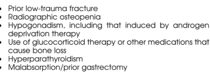
vit-Table 2.A summary of indications for bone mass measure-ment in men.
• Prior low-trauma fracture • Radiographic osteopenia
• Hypogonadism, including that induced by androgen deprivation therapy
• Use of glucocorticoid therapy or other medications that cause bone loss
• Hyperparathyroidism
• Malabsorption/prior gastrectomy
Table 3.Considerations in the laboratory evaluation of men with osteoporosis.
• CBC • ESR
• Serum calcium/phosphorus/creatinine and ALT or AST • TSH
• Free testosterone • 24 hour urine calcium • 25 hydroxyvitamin D • Serum/urine electrophoresis • PSA
• PTH
amin D3to suppress PTH and reduce bone loss (72).
As such, recommending a daily intake of approximate-ly 1,200 mg of elemental calcium through diet plus supplements, if necessary, for men with osteoporosis is appropriate (73).
Additionally, vitamin D supplementation is often necessary as even in locations with abundant sunshine, vitamin D inadequacy is common (74). Recent expert consensus suggests that the daily oral intake of vitamin D should be approximately 1,000 IU / day (75,76) with documentation of adequacy by measurement of serum 25O H D considered as noted above. A reasonable goal is to maintain the serum 25O H D above ~32 ng/ ml (70–80 nmol/ L) as values below this may be associated with secondary hyper-parathyroidism. Moreover, low vitamin D status is associated with muscle weakness and increased falls risk and simple provision of vitamin D reduces fracture risk (77). U sing the cutpoint of 32 ng/ ml, a recent U K report found that 54/ 56 men with osteoporosis attending a bone clinic had vitamin D inadequacy (78). As such, it is clear that vitamin D inadequacy is a common concern in men with osteoporosis.
Though calcium and vitamin D have received the most study, the possibility of phosphorus inade-quacy has recently been suggested as a contributor to osteoporosis therapy non-response in patients receiv-ing phosphate bindreceiv-ing calcium supplementation and pharmacologic osteoporosis therapy (79). In older, undernourished individuals, appreciation of this possi-bility and provision of calcium phosphate supplements seems prudent.
Physical measures, with a goal being reduction of falls risk, are often an important component of osteoporosis treatment (80). Simplistically, weight-bearing exercise is ideal and activities leading to spine flexion are to be avoided. H owever, evaluation by a physical therapist for provision of an exercise program, assessment of falls risk and evaluation for gait assistive devices (e.g., canes and walkers) may be indicated. The importance of falls assessment as part of a fracture risk reduction program cannot be overemphasized as more than one-third of people over age 65 fall annually (81) and approximately 5% of falls lead to fracture. Factors intrinsic to the patient, and those within the individu-al’s environment, should be identified and modified when feasible. A consensus approach to evaluating falls risk has been published by the American and British Geriatric Societies and includes evaluation of medica-tions, vision, neuromuscular function and gait/ bal-ance (82). In this regard, hip protectors would seem to be a logical approach to fracture reduction in selected
patients and some reports have documented efficacy in reduction of hip fracture in nursing home residents (83). H owever, the most recent Cochrane Review found only a marginally significant reduction in hip fracture incidence and thus “casts some doubt on the effectiveness” of hip protectors (84). Further work in this field is necessary as this simple, inexpensive option is clinically attractive. Moreover, focusing solely upon increasing bone mass seems unlikely to prevent hip fractures in individuals with recurrent falls.
this regard, a recent meta-analysis of placebo-con-trolled trials found the rates of prostate cancer, prostate specific antigen elevation and prostate biop-sies to be numerically, but not statistically, higher in men receiving testosterone (93).
To summarize, the role of testosterone replace-ment in aging men with osteoporosis remains inade-quately studied and definitive conclusions cannot be made. H owever, many older men with low total testosterone, often defined as below 250 ng/ ml, do have symptoms of hypogonadism which may be bene-fited by testosterone replacement. Concomitant low testosterone with clinical symptoms or signs of hypog-onadism such as reduced muscle mass/ strength, osteoporosis and increased body fat, is agreed upon as a situation in which testosterone therapy, with appro-priate monitoring, e.g., hematocrit, prostate specific antigen, etc., should be considered (94-97).
In contrast to the situation in women, where multiple large studies of osteoporosis therapies have been conducted, only a relatively small number of stud-ies utilizing anti-resorptive or anabolic osteoporosis treatment agents have been performed in men. More-over, as is the case for studies of testosterone therapy, the number of participants is much smaller than compa-rable studies in women. Though the available studies are relatively small, they do demonstrate similar effects as observed in the larger studies among women.
O f available osteoporosis therapies, the bispho-sphonate class has received the most study in men. For example, in a prospective randomized trial the impact of alendronate 10 mg daily versus placebo was evalu-ated in 241 men with osteoporosis, all of whom received 500 mg of calcium and 400 IU of vitamin D daily (98). After two years, lumbar spine BMD was 5.3% higher in the alendronate group and the inci-dence of radiographic vertebral deformities was reduced. Similarly, a small, open-label study in men with low BMD observed that daily alendronate over three years produced greater increases in spine BMD and significant reduction in radiographic vertebral fractures compared with alfacalcidol (99). U sing a very similar study design, in 316 osteoporotic men, rise-dronate 5 mg daily increased BMD to a greater extent than alfacalcidiol and reduced vertebral fracture risk (100). Moreover, daily risedronate increases BMD and reduces vertebral fracture risk in men receiving gluco-corticoid therapy (101). Importantly, intermittent bis-phosphonate therapy appears to be efficacious in men. Though the data are limited, a one-year study in which men with low BMD were randomly assigned to 70 mg of alendronate or placebo, demonstrated that weekly
alendronate administration significantly increased BMD at the spine and proximal femur (102). Finally, intravenous bisphosphonate therapy (zoledronic acid) increases BMD despite androgen deprivation therapy (103). It is apparent from the size and duration of the studies noted above that none of these were adequate-ly powered to detect an effect on non-vertebral frac-ture. A recent study of 280 men who had previously sustained a stroke found that daily risedronate did reduce hip fracture risk in comparison to place (104). Though encouraging, the number of hip fractures in this study (12) was small. D espite this limitation, experts in the field consider bisphosphonates to be the treatment of choice for osteoporotic men (105).
Parathyroid hormone has also been studied in osteoporotic men. A small study found that daily PTH injections increased lumbar spine BMD by ~13% over 18 months in the 10 men receiving therapy (106). Subsequently, in 151 men with a spine or hip T-score < -2.0, 11 months of daily PTH injections increased lumbar spine and hip BMD by a mean of 5.9 and 1.5% respectively (107). This study was terminated after a median duration of 11 months due to the develop-ment of osteosarcomas in rats. As such, whether this treatment reduces fracture risk in men remains to be determined.
At the time of this writing, no consensus state-ment or societal recommendation exists advising clini-cians which men to receive therapy. H owever, pub-lished expert opinion suggests that pharmacologic treatment at a T-score of ~-2.0 to -2.5 is indicated (108,109). The observation that the 10-year risk for fracture is virtually identical in men and women with a femur neck T-score of -2.5 supports use of similar treatment cutpoints regardless of gender. As noted above, it is expected that the forthcoming WH O absolute fracture risk paradigm will allow country-spe-cific treatment guidelines to be determined in men.
CONCLUSION
O steoporotic fractures become very common in men with advancing age. These fractures are associated with substantial morbidity and mortality. The number of men who sustain osteoporotic fractures will continue to increase for the foreseeable future. As such, osteo-porosis evaluation and treatment when indicated should be part of preventive care for older men. In men with low BMD or low-trauma fracture, this eval-uation should include laboratory assessment to exclude secondary causes. Androgen therapy of hypogonadal men may be considered with the caveat that data do not exist to document that this treatment reduces fracture risk. At this time, the data is inade-quate to support use of androgen treatment in eugo-nadal men with osteoporosis. Parathyroid hormone treatment does increase BMD ; existing studies have not been of adequate size or duration to document fracture reduction efficacy. Bisphosphonate therapy increases BMD , reduces vertebral fracture risk and is considered the standard of care for osteoporotic men at this point in time. The increasing number of men with osteoporosis, coupled with the availability of diagnostic recommendations and effective therapies demands that this disease no longer be neglected in men. It is hoped that the forthcoming WH O absolute fracture risk paradigm will enhance recognition and treatment of osteoporotic men.
REFERENCES
1. Anonymous. Consensus development conference: Pro-phylaxis and treatment of ssteoporosis. Am J Med 1990;90:107-10.
2. Chrischilles EA, Butler CD, Davis CS, Wallace RB. A model of lifetime osteoporosis impact. Arch Intern Med 1991;151:2026-32.
3. Gennari C, Seeman E. Introduction: The first internation-al conference on osteoporosis in men Siena, Itinternation-aly, Feb-ruary 23–25, 2001. Calcif Tissue Int 2001;69:177-8.
4. Kiebzak GM, Bienart GA, Perser K, Ambrose CG, Siff SJ, Heggeness MH. Undertreatment of osteoporosis in men with hip fracture. Arch Intern Med 2002;162:2217-22.
5. Feldstein AC, Nichols G, Orwoll E, Elmer PJ, Smith DH, Herson M, et al. The near absence of osteoporosis treat-ment in older men with fractures. Osteoporos Int 2005;16:953-62.
6. Nguyen TV, Eisman JA, Kelly PJ, Sambrook PN. Risk fac-tors for osteoporotic fractures in elderly men. Am J Epi-demiol 1996;144(3):255-63.
7. Cooper C, Melton LJ. Epidemiology of osteoporosis. Trends Endocrinol Metab 1992;3:224-9.
8. Eastell R, Boyle IT, Compston J, Cooper C, Fogelman I, Francis RM, et al. Management of male osteoporosis: report of the UK Consensus Group. QJM 1998;91:71-92.
9. Oden A, Dawson A, Dere W, Johnell O, Jonsson B, Kanis JA. Lifetime risk of hip fractures is underestimated. Osteoporos Int 1998;8:599-603.
10. Scane AC, Francis RM, Sutcliffe AM, Francis MJD, Rawl-ings DJ, Chapple CL. Case-control study of the patho-genesis and sequelae of symptomatic vertebral frac-tures in men. Osteoporos Int 1999;9:91-7.
11. Poor G, Atkinson EJ, O’Fallon WM, Melton LJI. Determi-nants of reduced survival following hip fractures in men. Clin Orthop 1995;319:260-5.
12. Center JR, Nguyen TV, Schneider D, Sambrook PN, Eis-man JA. Mortality after all major types of osteoporotic fracture in men and women: an observational study. Lancet 1999;353:878-82.
13. Pande I, Scott DL, O’Neill TW, Pritchard C, Woolf AD, Davis MJ. Quality of life, morbidity and mortality after low trauma hip fracture in men. Ann Rheum Dis 2006;65:87-92.
14. Kanis JA, Oden A, Johnell O, De Laet C, Jonsson B. Excess mortality after hospitalisation for vertebral frac-ture. Osteoporos Int 2004;15:108-12.
15. Johnell O, Kanis JA, Oden A, Sernbo I, Redlund-Johnell I, Petterson C, et al. Mortality after osteoporotic fractures. Osteoporos Int 2004;15:38-42.
16. Wehren LE, Hawkes WG, Orwig DL, Hebel JR, Zimmer-man SL, Magaziner J. Gender differences in mortality after hip fracture: The role of infection. J Bone Miner Res 2003;18:2231-7.
17. Orwoll ES. Osteoporosis in men. Endocr Rev 1995;16:87-116.
18. Riggs BL, Melton LJ 3rd, Robb RA, Camp JJ, Atkinson EJ, Oberg AL, et al. Population-based study of age and sex differences in bone volumetric density, size, geometry and structure at different skeletal sites. J Bone Miner Res 2004;19:1945-54.
19. Finkelstein JS. Overview of osteoporosis in men. Avail-able at: <http://www.uptodate.com/patient_info/topic-pages/topics/2092819.asp>. Accessed in January 15, 2002.
20. Burger H, de Laet CE, van Daele PL, Weel AE, Witteman JC, Hofman A, et al. Risk factors for increased bone loss in an elderly population. Am J Epidemiol 1998;147 (9):871-9.
21. Kamel HK. Male osteoporosis: New trends in diagnosis and therapy. Drugs Aging 2005;22:741-8.
22. Seeman E. The dilemma of osteoporosis in men. Am J Med 1995;98(suppl. 2A):76S-88S.
23. Stepan JJ, Lachman M, Zverina J, Pacovsky V, Baylink DJ. Castrated men exhibit bone loss: effect of calcitonin treatment on biochemical indices of bone remodeling. J Clin Endocrinol Metab 1989;69(3):523-7.
24. Shahinian VB, Kuo YF, Freeman JL, Goodwin JS. Risk of fracture after androgen deprivation for prostate can-cer. N Engl J Med 2005;352:154-64.
26. Benito M, Vasilic B, Wehrli FW, Bunker B, Wald M, Gomberg B, et al. Effect of testosterone replacement on trabecular architecture in hypogonadal men. J Bone Miner Res 2005;20:1785-91.
27. Khosla S, Melton LJI, Riggs BL. Estrogens and bone health in men. Calcif Tissue Int 2001;69:189-92.
28. Smith EP, Boyd J, Frank GR, Takahashi H, Cohen RM, Specker B, et al. Estrogen resistance caused by a muta-tion in the estrogen-receptor gene in a man. N Engl J Med 1994;331(16):1056-61.
29. Morishima A, Grumbach MM, Simpson ER, Fisher C, Qin K. Aromatase deficiency in male and female siblings caused by a novel mutation and the physiological role of estrogens. J Clin Endocrinol Metab 1995;80(12):3689-98.
30. Bilezikian JP, Morishima A, Bell J, Grumbach MM. Increased bone mass as a result of estrogen therapy in a man with aromatase deficiency. N Engl J Med 1998;339:599-603.
31. Rochira V, Faustini-Fustini M, Balestrieri A, Carani C. Estro-gen replacement therapy in a man with conEstro-genital aro-matase deficiency: effects of different doses of trans-dermal estradiol on bone mineral density and hormonal parameters. J Clin Endocrinol Metab 2000;35(5):1841-5.
32. Falahatik-Nini A, Riggs BL, Atkinson EJ, O’Fallon WM, East-ell R, Khosla S. Relative contributions of testosterone and estrogen in regulating bone resorption and formation in normal elderly men. J Clin Invest 2000;106:1553-60.
33. Kelepouris N, Harper KD, Gannon F, Kaplan FS, Haddad JG. Severe osteoporosis in men. Ann Intern Med 1995;123(6):452-60.
34. Compston J. Secondary causes of osteoporosis in men. Calcif Tissue Int 2001;69:193-5.
35. Khosla S, Lufkin EG, Hodgson SF, Fitzpatrick LA, Melton LJI. Epidemiology and clinical features of osteoporosis in young individuals. Bone 1994;15(5):551-5.
36. Vanderschueren D, Boonen S, Bouillon R. Osteoporosis and osteoporotic fractures in men: A clinical perspec-tive. Baillieres Clin Endocrinol Metab 2000;14:299-315.
37. Kurland ES, Rosen CJ, Cosman F, McMahon D, Chan F, Shane E, et al. Insulin-like growth factor-I in men with idiopathic osteoporosis. J Clin Endocrinol Metab 1997; 82(9):2799-805.
38. Ciria-Recasens M, Perez-Edo L, Blanch-Rubio J, Marinoso ML, Benito-Ruiz P, Serrano S, et al. Bone histomorphome-try in 22 male patients with normocalciuric idiopathic osteoporosis. Bone 2005;36:926-30.
39. Pernow Y, Granberg B, Saaf M, Weidenhielm L. Osteoblast dysfunction in male idiopathic osteoporosis. Calcif Tissue Int 2006;78:90-7.
40. Van Pottelbergh I, Goemaere S, Zmierczak H, Kaufman JM. Perturbed sex steroid status in men with idiopathic osteoporosis and their sons. J Clin Endocrinol Metab 2004;89:4949-53.
41. Patel MBR, Arden NK, Masterson LM, Phillips DI, Swami-nathan R, Syddall HE, et al. Investigating the role of the growth hormone-insulin-like growth factor (GH-IGF) axis as a determinant of male bone mineral density (BMD). Bone 2005;37:833-41.
42. Ferrari SL, Deutsch S, Baudoin C, Cohen-Solal M, Ostertag A, Antonarakis SE, et al. LRP5 gene polymor-phisms and idiopathic osteoporosis in men. Bone 2005;37(6):770-5.
43. Diaz MN, O’Neill TW, Silman AJ. The influence of family history of hip fracture on the risk of vertebral deformity in men and women: the European Vertebral Osteoporosis Study. Bone 1997;20(2):145-9.
44. Cohen-Solal ME, Baudoin C, Omouri M, Kuntz D, De Vernejoul MC. Bone mass in middle-aged osteoporotic men and their relatives: familial effect. J Bone Miner Res 1998;13(12):1909-14.
45. Kanis JA, Melton LJI, Christiansen C, Johnston CC, Khal-taev N. Perspective: The diagnosis of osteoporosis. J Bone Miner Res 1994;9(8):1137-41.
46. Melton LJI, Atkinson EJ, O’Connor MK, O’Fallon WM, Riggs BL. Bone density and fracture risk in men. J Bone Miner Res 1998;13(12):1915-23.
47. Szulc P, Munoz F, Duboeuf F, Marchand F, Delmas PD. Bone mineral density predicts osteoporotic fractures in elderly men: The MINOS study. Osteoporos Int 2005;16:1184-92.
48. Binkley N, Schmeer P, Wasnich R, Lenchik L. What are the criteria by which a densitometric diagnosis of osteo-porosis can be made in males and non-Caucasians? J Clin Densitom 2002;5(suppl.):s19-s27.
49. Leslie WD, Adler RA, El-Hajj Fuleihan G, Hodsman AB, Kendler DL, McClung M, et al. Application of the 1994 WHO Classification to Populations other than post-menopausal Caucasian women: The 2005 ISCD Official Positions. J Clin Densitom 2006;9(1):22-30.
50. Drinka PJ, DeSmet AA, Bauwens SF, Rogot A. The effect of overlying calcification on lumbar bone densitometry. Calcif Tissue Int 1992;50:507-10.
51. Kanis JA, Gluer C-C. An update on the diagnosis and assessment of osteoporosis with densitometry. Osteo-poros Int 2000;11:192-202.
52. Genant HK, Cooper C, Poor G, Reid I, Ehrlich G, Kanis J, et al. Interim report and recommendations of the World Health Organization task-force for osteoporosis. Osteo-poros Int 1999;10:259-64.
53. The Writing Group for the ISCD Position Development Conference. Diagnosis of osteoporosis in men, premenopausal women and children. J Clin Densitom 2004;7:17-26.
54. Wiemann L, Krueger D, Vallarta-Ast N, Binkley N. Effect of female database use for T-score derivation in men. 2006.
55. Leib ES, Lewiecki EM, Binkley N, Hamdy RC. Official posi-tions of the international society for clinical densitome-try. J Clin Densitom 2004;7:1-5.
56. Brown JP, Josse RG. 2002 clinical practice guidelines for the diagnosis and management of osteoporosis in Canada. Can Med Assoc J 2002;167(10 suppl):S1-S34.
58. Johnell O, De Laet C, Johansson H, et al. Oral corticos-teroids increase fracture risk independently of BMD. Osteoporos Int 2002;13(suppl):S14.
59. Kanis JA, Johnell O, Oden A, De Laet C, Mellstrom D. Diagnosis of osteoporosis and fracture threshold in men. Calcif Tissue Int 2001;69:218-21.
60. McClung MR. Do current management strategies and guidelines adequately address fracture risk? Bone 2006;38:S13-7.
61. Kanis JA, Johnell O, Oden A, Borgstrom F, Johansson H, De Laet C, et al. Intervention thresholds for osteoporosis in men and women: a study based on data from Swe-den. Osteoporos Int 2005;16:6-14.
62. Campion JM, Maricic MJ. Osteoporosis in men. Am Fam Physician 2003;67:1521-6.
63. Holick MF. Vitamin D: A millennium perspective. J Cell Biochem 2003;88:296-307.
64. Heaney RP, Davies KM, Chen TC, Holick MF, Barger-Lux MJ. Human serum 25-hydroxycholecalciferol response to extended oral dosing with cholecalciferol. Am J Clin Nutr 2003;77:204-10.
65. Bilezikian JP, Kurland ES, Rosen CJ. Male skeletal health and osteoporosis. Trends Endocrinol Metab 1999; 10(6):244-50.
66. Meier C, Nguyen TV, Center JR, Seibel MJ, Eisman JA. Bone resorption and osteoporotic fractures in elderly men: The Dubbo osteoporosis epidemiology study. J Bone Miner Res 2005;20:579-87.
67. Anonymous. Health, United States, 2005 with chartbook on trends in the health of Americans. Available at: <http://www.cdc.gov/nchs/faststats/lifexpec.htm>. Accessed in May 15, 2005.
68. Harris D, Haboubi N. Malnutrition screening in the elder-ly population. J R Soc Med 2005;98:411-4.
69. Delmi M, Rapin CH, Bengoa JM, Delmas PD, Vasey H, Bonjour JP. Dietary supplementation in elderly patients with fractured neck of the femur. Lancet 1990;335:1013-6.
70. Chapuy MC, Arlot ME, Duboeuf F, Brun J, Crouzet B, Arnaud S, et al. Vitamin D3 and calcium to prevent hip fractures in elderly women. N Engl J Med 1992;327:1637-42.
71. Francis RM. Male osteoporosis. Rheumatology (Oxford) 2000;39:1055-9.
72. Daly RM, Brown M, Bass S, Kukuljan S, Nowson C. Calci-um- and vitamin D3-fortified milk reduces bone loss at clinically relevant sites in older men: A 2-year random-ized controlled trial. J Bone Miner Res 2006;21:397-405.
73. Anonymous. Physician’s guide to prevention and treat-ment of osteoporosis. Available at: <http://www.nof.org/ physguide>. Accessed in April 20, 2003.
74. Levis S, Gomez A, Jimenez C, Veras L, Ma F, Lai S, et al. Vitamin D deficiency and seasonal variation in an adult south Florida population. J Clin Endocrinol Metab 2005;90:1557-62.
75. Heaney RP. Vitamin D: how much do we need, and how much is too much. Osteoporos Int 2000;11:553-5.
76. Dawson-Hughes B, Heaney RP, Holick MF, Lips P, Meunier PJ, Vieth R. Estimates of optimal vitamin D status. Osteo-poros Int 2005;16:713-6.
77. Trivedi DP, Doll R, Khaw KT. Effect of four monthly oral vit-amin D3 (cholecalciferol) supplementation on fractures and mortality in men and women living in the communi-ty: Randomized double blind controlled trial. Br Med J 2003;326:469-74.
78. Al-Oanzi ZH, Tuck SP, Raj N, Harrop JS, Summers GD, Cook DB, et al. Assessment of vitamin D status in male osteoporosis. Clin Chem 2006;52:248-54.
79. Heaney RP. Phosphorus nutrition and the treatment of osteoporosis. Mayo Clin Proc 2004;79:91-7.
80. Karlsson MK. Skeletal effects of exercise in men. Calcif Tissue Int 2001;69:196-9.
81. Tinetti ME. Preventing falls in elderly persons. N Engl J Med 2003;348:42-9.
82. Anonymous. The American Geriatrics Society, British Geriatrics Society and American Academy of Ortho-pedic Surgeons. 2001 Guideline for the prevention of falls in older persons. Annals of Long-Term Care 2001;11:42-54.
83. Lauritzen JB, Petersen MM, Lund B. Effect of external hip protectors on hip fractures. Lancet 1993;341:11-3.
84. Parker MJ, Gillespie WJ, Gillespie LD. Hip protectors for preventing hip fractures in older people. Cochrane Database Syst Rev 2005;20;(3):CD001255.
85. Allan CA, McLachlan RI. Age-related changes in testos-terone and the role of replacement therapy in older men. Clin Endocrinol (Oxf) 2004;60:653-70.
86. Hijazi RA, Cunningham GR. Andropause: Is androgen replacement therapy indicated for the aging male? Annu Rev Med 2005;56:117-37.
87. Harman SM, Metter EJ, Tobin JD. Longitudinal effects of aging on serum total and free T levels in healthy men. J Clin Endocrinol Metab 2001;86:724-31.
88. Anonymous. American association of clinical endocri-nologists medical guidelines for clinical practice for the evaluation and treatment of hypogonadism in adult male patients — 2002 update. Endocrine Practice 2002;8:439-56.
89. Snyder PJ, Peachey H, Hannoush P, Berlin JA, Loh L, Holmes JH, et al. Effect of testosterone treatment on bone mineral density in men over 65 years of age. J Clin Endocrinol Metab 1999;84(6):1966-72.
90. Isidori AM, Giannetta E, Greco EA, Gianfrilli D, Bonifacio V, Isidori A, et al. Effects of testosterone on body com-position, bone metabolism and serum lipid profile in mid-dle-aged men: A meta-analysis. Clin Endocrinol (Oxf) 2005;63:280-93.
91. Aminorroaya A, Kelleher S, Conway AJ, Ly LP, Han-delsman DJ. Adequacy of androgen replacement influences bone density response to testosterone in androgen-deficient men. Eur J Endocrinol 2005; 152: 881-6.
92. Francis RM. Androgen replacement in aging men. Cal-cif Tissue Int 2001;69:235-8.
94. Schlegel PN, Hardy MP. Androgen replacement overview and current guidelines. J Androl 2006;27:125.
95. Wald M, Meacham RB, Ross LS, Niederberger CS. Testos-terone replacement therapy for older men. J Androl 2006;27:126-32.
96. Jockenhovel F. Testosterone therapy — What, when and to whom? Aging Male 2004;7:319-24.
97. Rhoden EL, Morgentaler A. Risks of testosterone-replace-ment therapy and recommendations for monitoring. N Engl J Med 2004;350:482-92.
98. Orwoll E, Ettinger M, Weiss S, Miller P, Kendler D, Graham J, et al. Alendronate for the treatment of osteoporosis in men. N Engl J Med 2000;343:604-10.
99. Ringe JD, Dorst A, Faber H, Ibach K. Alendronate treat-ment of established primary osteoporosis in men: 3-year results of a prospective, comparative, two-arm study. Rheumatol Int 2004;24:110-3.
100.Ringe J, Faber H, Salem M, Grauer A, Moller G. Rise-dronate therapy reduces the risk of new vertebral frac-tures by 60% in osteoporotic men with osteoporosis with-in 1 year. J Bone Mwith-iner Res 2004;19(suppl. 1):s441.
101.Reid DM, Adami S, Devogelaer JP, Chines AA. Rise-dronate increases bone density and reduces vertebral fracture risk within one year in men on corticosteroid therapy. Calcif Tissue Int 2001;69:242-7.
102.Miller PD, Schnitzer T, Emkey R, et al. Weekly oral alen-dronic acid in male osteoporosis. Clin Drug Invest 2004;24:333-341.
103.Smith MR, Eastham J, Gleason DM, Shasha D, Tchekme-dyian S, Zinner N. Randomized controlled trial of zole-dronic acid to prevent bone loss in men receiving androgen deprivation therapy for nonmetastatic prostate cancer. J Urol 2003;169:2008-12.
104.Sato Y, Iwamoto J, Kanoko T, Satoh K. Risedronate sodi-um therapy for prevention of hip fracture in men 65 years or older after stroke. Arch Intern Med 2005;165:1743-8.
105.Bauer DC, Orwoll E. Quality indicators for management of osteoporosis. Ann Intern Med 2002;137:621-2.
106.Kurland ES, Cosman F, McMahon D, Rosen CJ, Lindsay R, Bilezikian JP. Parathyroid hormone as a therapy for idiopathic osteoporosis in men: effects on bone mineral density and bone markers. J Clin Endocrinol Metab 2000;85(9):3069-76.
107.Orwoll E, Scheele W, Paul S, Adami S, Syversen U, Diez-Perez A, et al. Brief therapy with recombinant human parathyroid hormone (1-34) increases lumbar spine bone mineral density in men with idiopathic or hypogo-nadal osteopenia or osteoporosis. J Bone Miner Res 2001;16(suppl. 1):S221.
108.Orwoll ES. Treatment of osteoporosis in men. Calcif Tis-sue Int 2004;72:114-9.
109.Colon-Emeric C, Yballe L, Sloane R, Pieper CF, Lyles KW. Expert physician recommendations and current prac-tice patterns for evaluating and treating men with osteoporotic hip fracture. J Am Geriatr Soc 2000;48:1261-3.
Address for correspondence:
Neil Binkley Suite 100