DISCLAIMER
This paper was submitted to the Bulletin of the World Health Organization and was posted to the Zika open site, according to the protocol for public health emergencies for international concern as described in Christopher Dye et al. (http://dx.doi.org/10.2471/BLT.16.170860).
The information herein is available for unrestricted use, distribution and reproduction in any medium, provided that the original work is properly cited as indicated by the Creative Commons Attribution 3.0 Intergovernmental Organizations licence (CC BY IGO 3.0).
RECOMMENDED CITATION
Rocha HAL, Correia LL, Leite AJM, Campos JS, Cavalcante e Silva A, Machado MMT, Rocha SGMO, Saraiva de Almeida NMG, Alves da Cunha AJL. Microcephaly: normality parameters and its determinants in northeastern Brazil: a multicentre prospective cohort study [Submitted]. Bull World Health Organ E-pub: 8 Feb 2016. doi:
http://dx.doi.org/10.2471/BLT.16.171215
Microcephaly: normality parameters and its determinants
in northeastern Brazil: a multicentre prospective cohort
study
Hermano Alexandre Lima Rocha,
aLuciano Lima Correia,
aÁlvaro Jorge
Madeiro Leite,
aJocileide Sales Campos,
bAnamaria Cavalcante e Silva,
bMárcia Maria Tavares Machado,
aSabrina Gabriele Maia Oliveira
Rocha,
aNádia Maria Girão Saraiva de Almeida
a& Antonio José Ledo
Alves da Cunha
ca Federal University of Ceará, Rua Prof. Costa Mendes 1608, Fortaleza,CE 60430140, Brazil.
b Unichristus, Fortaleza, Brazil.
c Federal University of Rio de Janeiro, Rio de Janeiro, Brazil.
Correspondence to Hermano Alexandre Lima Rocha (email: [email protected]).
Abstract
Brazil was marked by a serious epidemic of microcephaly due to still obscure causes.
According to the Ministry of Health, the number of suspected cases of microcephaly reached 2401. However, there is controversy in Brazil regarding the most appropriate cut-off point and diagnosis criteria. The present study aimed to measure the prevalence of microcephaly and identify its determinants, in a prospective cohort from a multicentre hospital-based study. The sample comprised 3623 live births admitted to the NICU, the dependent variable being cephalic perimeter. Bootstrap and logistic regression models were used to analyse data. 32 cm corresponded to tenth percentile in this population, with a confidence interval between 31.82 and 32.18. Intrauterine growth restriction, pregnancies resulting in congenital anomalies, oligohydramnios and the maternal use of steroids were associated with microcephaly. This work raises discussion on the cut-off point used for the diagnosis of microcephaly at a population level in a country with an outbreak as well as factors that can be associated with the outcome presented.
Introduction
The year 2015 in Brazil was marked by a serious epidemic of microcephaly due to still obscure causes. According to the Ministry of Health of Brazil, the number of suspected cases of microcephaly reached 2401 in 19 states and the Federal District, with 29 cases evolving to death. The states with the highest number of cases were in the Northeast region of Brazil, including all of its nine states (1).
The initial diagnosis of microcephaly is performed in the maternity ward by measuring the cephalic perimeter at birth. However, there is controversy in Brazil regarding the most appropriate cut-off point (2, 3). After the identification of an excessive number of suspected cases, the Ministry of Health of Brazil issued a statement reducing the cephalic perimeter representative of the suspicion of microcephaly from 33 cm to 32 cm (4).
The volumes of three main components (brain matter, cerebrospinal fluid and blood) determine the size of the skull during childhood. Microcephaly is defined as an occipitofrontal circumference measurement of less than the third percentile or below two standard deviations of the mean measurement for age, gender and ethnicity (5).
Microcephaly can be primary when the brain is small because it does not undergo proper embryonic development due to genetic factors and malformations. Microcephaly can also be secondary, when the brain completes normal embryonic development but suffers damage that alters its further development, including damage caused by vascular processes, perinatal diseases and antenatal systemic postnatal diseases. A study in Germany found that 41% of children diagnosed with microcephaly did not have a definitive diagnosis (3).
Approximately 90% of microcephaly cases are associated with mental retardation, except in those with familiar origin, which may exhibit normal cognitive development. The type and the severity level of sequelae may vary in each case. Treatment in the early years of life enhance development and the quality of life (6). The later recovery of the cephalic perimeter can guarantee normal cognitive development (7).
The pathogenesis of microcephaly is heterogeneous, ranging from genetic causes and intrauterine growth restriction to environmental factors, which can influence the development and size of the brain (6, 8). Studies found an association between microcephaly and diarrhoea and malnutrition (9). Infections during pregnancy can also cause microcephaly, including cytomegalovirus (10), toxoplasmosis (11) and rubella virus infection (12, 13).
two cases evaluated; however, this case also suffered from intrauterine growth restriction (14).
The present study aimed to measure the prevalence of microcephaly and identify its determinants in the nine states in the Northeast region of Brazil, which presents the highest number of cases in the country, using data relating to a period prior to the current outbreak. We intend to contribute to the scientific discussion of the problem by providing baseline information of relevance.
Methods
Study Design
This study utilized a prospective cohort from a multicentre hospital-based study of live births at gestational age from 230/7 weeks, carried out by RENOSPE (Perinatal Health Network of Northeast, Brazil). The purpose of this network was to enhance the competence of the public health system in areas such as management, assistance, education and perinatal research through the articulation of a network of neonatal units from public secondary and tertiary maternity wards located in states in the north and north-east of Brazil (15).
Setting
The study was conducted in high-risk neonatal units of public maternity wards located in capitals of nine north-eastern states, namely Maranhão, Piauí, Ceará, Rio Grande do Norte, Paraíba, Pernambuco, Alagoas, Sergipe and Bahia. Together, the north-eastern region comprises a population of 53 million people, most of it living in a semiarid climate.
During the period from July to December 2007, 37 health establishments participated in the study: 4 (10.8%) in Maranhão, 2 (5.4%) in Piauí, 7 (18.9%) in Ceará, 3 (8.2%) in Rio Grande do Norte, 7 (18.9%) in Paraíba, 6 (16.2%) in Pernambuco, 2 (5.4%) in Alagoas, 2 (5.4%) in Sergipe and 4 (10.8%) in Bahia.
The collection of the data in each unit was carried out prospectively from the time of admission to the time of discharge or death by a field researcher (doctor or nurse) trained by a RENOSPE coordinator.
Participants
The study sample included new-borns hospitalized after birth in the Neonatal Intensive Care Unit (NICU) whose record in the original database contained the discharge from the NICU and entrance to the NICU that did not include transfer from another health facility.
Exclusion criteria were infants admitted to the NICU with a transfer, congenital malformations and death that occurred in the delivery room. Children with a lack of information on these fields were also excluded.
Study Size
The sample comprised 3623 live births admitted to the NICU members of RENOSPE. Of these, 654 (18.1%) evolved to death during the neonatal period (at the NICU due to death) and 2969 (81.9%) who survived were admitted to the intensive care unit. Only the new-born infants with a gestational time between 37 and 41 weeks were included in the analysis, yielding 1314 individuals. This sample size conferred a power of approximately 80% for a type I error of 0.05 to identify risk differences as small as 2 with an estimated prevalence of 5% (exact number necessary for this power is 1418).
Variables
The dependent variable was cephalic perimeter, which was expressed as a continuous variable and was also categorized into positive or negative for microcephaly according to the definition provided by OMS growth chart curves (3).
Block I (distal variables): social, economic and demographic characteristics: Race, mother’s years of study, mother’s occupation.
Block II (intermediate variables): maternal characteristics:
Maternal age (in years), number of pregnancies (including current), number of abortions, number of stillborn children.
Block II (proximal variables): prenatal care and childbirth:
Number of prenatal visits, type of birth, length of pregnancy in weeks, use of antenatal corticosteroids (among new-borns with birth weight less than 1,500 g), complications in pregnancy (urinary infection, chorioamnionitis, acute foetal distress, premature rupture of the membranes, intrauterine growth retardation, premature labour, premature separation of placenta, hypertension, preeclampsia, eclampsia, oligohydramnios, gestational diabetes, previous diabetes, positive HBsAg antigen, HIV/hepatitis B, syphilis, toxoplasmosis).
Statistical methods
The public domain software EPI INFO 6.04 D was used for data file creation and processing.
The general description of the sample was presented using counts and percentages for categorical variables and measures of central tendency for numeric variables. The normality of the distribution of numeric variables was tested with the Kolmogorov-Smirnov test.
Central tendency analyses were performed to identify normal population and percentile values with the bootstrap technique using 1000 samples for each analysis. Descriptive box plots were built with subgroups. To identify statistical associations, regressive logistic bivariate models were used. Variables identified with p-values less than 0.1 were considered for the construction of a hierarchical regressive logistic multivariate model. The gross risk rates adjusted with their respective 95% confidence intervals were tabulated and analysed. Analyses were performed with SPSS for Windows software, v 17 (SPSS Inc.).
Ethical statements
The references of bioethics, recommended in resolution 196/96 of the National Health Council (CNS), were considered in this study. The project was submitted and approved by the Research Ethics Committee of the Federal University of Ceará under Protocol 31/2007.
Results
A total of 1006 children participated in this analysis, geographically distributed as follows: the State of Maranhão (295), State of Ceará (257), State of Bahia (181), State of Alagoas (139), State of Paraíba (134), State of Sergipe (79), State of Piaui (60), and the State of Rio Grande do Norte (44). The State of Pernambuco did not contribute to the sample due to the absence of data collection in this state by the field researchers.
The following results present a descriptive analysis of the normal patterns of the cephalic perimeter of children, followed by multivariate analysis of risk factors associated with microcephaly in this population.
The average cephalic perimeter of this population of children in north-eastern Brazil was 34.29 cm, with a standard deviation of 2.50 cm.
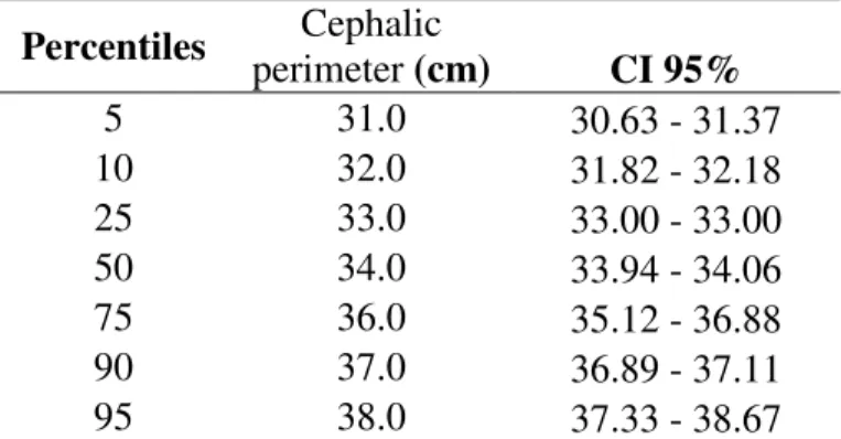
Table 1. Percentiles of cephalic perimeter measured at birth in states of the northeast region of Brazil, 2007
Percentiles Cephalic
perimeter (cm) CI 95%
5 31.0 30.63 - 31.37
10 32.0 31.82 - 32.18
25 33.0 33.00 - 33.00
50 34.0 33.94 - 34.06
75 36.0 35.12 - 36.88
90 37.0 36.89 - 37.11
95 38.0 37.33 - 38.67
The average cephalic perimeter was 34 cm, and the point of 32 cm for the diagnosis of microcephaly corresponded to P10 in this population, with a confidence interval between 31.82 and 32.18. The cephalic perimeter of 33 cm, considered earlier in Brazil, corresponded to the first quartile.
Table 2. Incidence of microcephaly1 within six months in different states of the northeast Region of Brazil, 2007.
Estados
Microcefalia
Não Sim
N % N %
Sergipe 53 93.00% 4 7.00%
Ceará 213 93.80% 14 6.20%
Paraíba 114 95.80% 5 4.20%
Piauí 54 96.40% 2 3.60%
Bahia 149 96.80% 5 3.20%
Maranhão 246 97.20% 7 2.80%
Alagoas 106 99.10% 1 0.90%
Rio Grande do Norte 23 100.00% 0 0.00% Região Nordeste 958 96.2% 38 2.8%
1
Defined as cephalic perimeter less than 2 standard deviations.
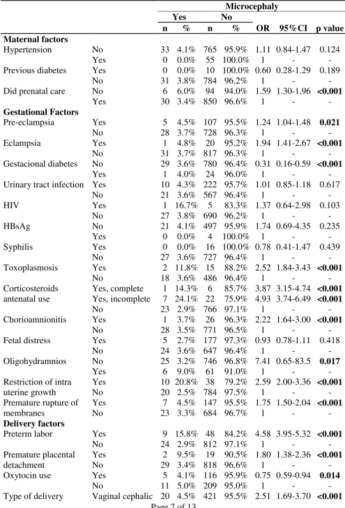
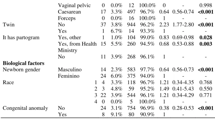
Table 3 lists the main risk factors for microcephaly between children born between 37 to 41 weeks.
Table 3. Bivariate analysis of associated factors of microcephaly in new-borns within 37 a 41 weeks of gestation in the northeast region of Brazil, 2007
Microcephaly
Yes No
n % n % OR 95%CI p value
Maternal factors
Hypertension No 33 4.1% 765 95.9% 1.11 0.84-1.47 0.124
Yes 0 0.0% 55 100.0% 1 - -
Previous diabetes Yes 0 0.0% 10 100.0% 0.60 0.28-1.29 0.189
No 31 3.8% 784 96.2% 1 - -
Did prenatal care No 6 6.0% 94 94.0% 1.59 1.30-1.96 <0.001
Yes 30 3.4% 850 96.6% 1 - -
Gestational Factors
Pre-eclampsia Yes 5 4.5% 107 95.5% 1.24 1.04-1.48 0.021
No 28 3.7% 728 96.3% 1 - -
Eclampsia Yes 1 4.8% 20 95.2% 1.94 1.41-2.67 <0.001
No 31 3.7% 817 96.3% 1 - -
Gestacional diabetes No 29 3.6% 780 96.4% 0.31 0.16-0.59 <0.001
Yes 1 4.0% 24 96.0% 1 - -
Urinary tract infection Yes 10 4.3% 222 95.7% 1.01 0.85-1.18 0.617
No 21 3.6% 567 96.4% 1 - -
HIV Yes 1 16.7% 5 83.3% 1.37 0.64-2.98 0.103
No 27 3.8% 690 96.2% 1 - -
HBsAg No 21 4.1% 497 95.9% 1.74 0.69-4.35 0.235
Yes 0 0.0% 4 100.0% 1 - -
Syphilis Yes 0 0.0% 16 100.0% 0.78 0.41-1.47 0.439
No 27 3.6% 727 96.4% 1 - -
Toxoplasmosis Yes 2 11.8% 15 88.2% 2.52 1.84-3.43 <0.001
No 18 3.6% 486 96.4% 1 - -
Corticosteroids antenatal use
Yes, complete 1 14.3% 6 85.7% 3.87 3.15-4.74 <0.001 Yes, incomplete 7 24.1% 22 75.9% 4.93 3.74-6.49 <0.001
No 23 2.9% 766 97.1% 1 - -
Chorioamnionitis Yes 1 3.7% 26 96.3% 2.22 1.64-3.00 <0.001
No 28 3.5% 771 96.5% 1 - -
Fetal distress Yes 5 2.7% 177 97.3% 0.93 0.78-1.11 0.418
No 24 3.6% 647 96.4% 1 - -
Oligohydramnios No 25 3.2% 746 96.8% 7.41 0.65-83.5 0.017
Yes 6 9.0% 61 91.0% 1 - -
Restriction of intra uterine growth
Yes 10 20.8% 38 79.2% 2.59 2.00-3.36 <0.001
No 20 2.5% 784 97.5% 1 - -
Premature rupture of membranes
Yes 7 4.5% 147 95.5% 1.75 1.50-2.04 <0.001
No 23 3.3% 684 96.7% 1 - -
Delivery factors
Preterm labor Yes 9 15.8% 48 84.2% 4.58 3.95-5.32 <0.001
No 24 2.9% 812 97.1% 1 - -
Premature placental detachment
Yes 2 9.5% 19 90.5% 1.80 1.38-2.36 <0.001
No 29 3.4% 818 96.6% 1 - -
Oxytocin use Yes 5 4.1% 116 95.9% 0.75 0.59-0.94 0.014
No 11 5.0% 209 95.0% 1 - -
Vaginal pelvic 0 0.0% 12 100.0% 0 - 0.998 Caesarean 17 3.3% 497 96.7% 0.64 0.56-0.74 <0.001
Forceps 0 0.0% 16 100.0% 1 - -
Twin No 37 3.8% 944 96.2% 2.23 1.77-2.80 <0.001
Yes 1 6.7% 14 93.3% 1 - -
It has partogram Yes, other 1 1.0% 104 99.0% 0.83 0.69-0.98 0.028 Yes, from Health
Ministry
15 5.5% 260 94.5% 0.68 0.53-0.88 0.003
No 11 3.9% 268 96.1% 1 - -
Biological factors
Newborn gender Masculino 14 2.3% 583 97.7% 0.64 0.56-0.73 <0.001
Feminino 24 6.0% 375 94.0% 1 - -
Race 1 4 3.3% 118 96.7% 1.21 0.34-4.35 0.768
2 3 4.8% 59 95.2% 1.49 0.41-5.43 0.550 3 22 3.9% 544 96.1% 1.21 0.34-4.29 0.771
4 0 0.0% 5 100.0% 1 - -
Congenital anomaly No 24 3.1% 754 96.9% 0.38 0.28-0.53 <0.001
Yes 8 9.1% 80 90.9% 1 - -
Bivariate analysis showed an association with microcephaly in the following factors: oligohydramnios (OR = 7.41; 95% CI 0.65-83.5, p = 0.017), premature labour (OR = 4.58; 95%CI 3.95-5.32), use of corticosteroid ante-natal ultrasound (OR = 3.87; 95%CI 3.15-4.74), intrauterine growth restriction (OR = 2.59; 95%CI 2.00-3.36), positive test for toxoplasmosis (OR = 2.52; 95% CI 1.84-3.43), new-born of the female gender (OR = 1.56; 95% CI 1.37-1.78), and congenital anomalies (OR = 0.38; 95% CI 0.28-0.53).
Maternal infections with syphilis, hepatitis B and HIV showed no statistically significant risk for microcephaly, only toxoplasmosis.
Table 4. Average and standard deviation of the selected maternal parameters, according to the diagnosis of microcephaly in new-borns at 37 to 41 weeks of gestation in the north-eastern region of Brazil, 2007
Microcefalia
Sim Não
Média SD Média SD p
Maternal age 25 7 25 7 0.809
Maternal educacional years 6 3 7 4 0.259
Number of pregnancies 2 2 2 2 0.689
Number of abortations 0 1 0 1 0.591
Number of children born alive 1 2 1 1 0.750
Number of stillbirths 0 0 0 0 0.787
Number of prenatal visits 5 2 6 2 0.278
The average and standard deviation of factors such as maternal age and education,
reproductive background and prenatal care attendance were compared among infants with and without microcephaly but did not show any statistically significant association.
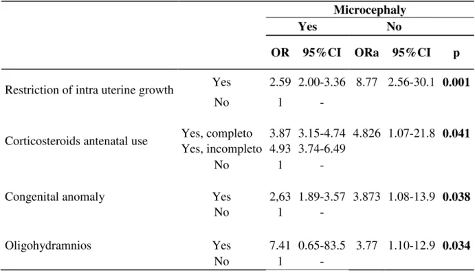
Table 5. Adjusted analysis of the factors associated with microcephaly in new-borns at 37 to 41 weeks of gestation in the north-eastern region of Brazil, 2007
Microcephaly
Yes No
OR 95%CI ORa 95%CI p
Restriction of intra uterine growth Yes 2.59 2.00-3.36 8.77 2.56-30.1 0.001
No 1 -
Corticosteroids antenatal use Yes, completo 3.87 3.15-4.74 4.826 1.07-21.8 0.041 Yes, incompleto 4.93 3.74-6.49
No 1 -
Congenital anomaly Yes 2,63 1.89-3.57 3.873 1.08-13.9 0.038
No 1 -
Oligohydramnios Yes 7.41 0.65-83.5 3.77 1.10-12.9 0.034
No 1 -
1
Controlled by: premature labour, congenital anomalies, gender of the new-born, meconium in the amniotic fluid, positive maternal HIV tests.
Discussion
This study suggests measures to be used as cut-offs that could be considered normal for the cephalic perimeter as well as factors that may contribute to the manifestation of microcephaly in new-borns.
Brazil has experienced an outbreak of cases of children identified with microcephaly (1), with more than 2000 cases already registered. Microcephaly is a rare condition, with an estimated prevalence of 1.9% (16). The incidence during the most current outbreak in one of the states of Brazil, Pernambuco, was approximately 4%. The first step for public strategies to confront this pathology is proper diagnosis. The Brazilian government is still working on the definition of the ideal cut-off point; the initial use of 33 cm as the cut-off cephalic perimeter for new-borns of gestational age between 37 and 41 weeks has been modified, establishing a new less sensitive cut-off point of 32 cm (4).
Considering that the diagnosis of microcephaly depends on a cephalic perimeter less than the second standard deviation of the normal curve of the population (17), it is reasonable to use the 5% percentile as the cut-off point for diagnosis. The percentile of 5% found in the sample studied in this work, with more than 1000 individuals, was 31 cm, with a confidence interval between 30.6 and 31.4 and a small variation between the different states.
Microcephaly can represent the structural deficiency of cerebral neurons or dendrites, and depending on its development, it can be primary or secondary, changing its prognosis (5). Most patients identified as carriers of microcephaly do not have a confirmed aetiologic diagnosis (3).
Recently, genetic causes and mutations have been associated with cases of microcephaly as their main determining factors (3, 18-20). This work identified that congenital anomalies were independently associated with microcephaly, but they did not constitute the most important factor. Intrauterine growth restriction has increased the risk of development of microcephaly by about eight times. This association was found in a follow-up study conducted in Canada (7), and it was identified that when both conditions are present and there is no recovery of the cephalic perimeter, the prognosis for a child's development is reserved. Oligohydramnios is a sign associated with a wide range of genetic diseases (21) and may be caused by many genetic anomalies.
Infectious causes have not been independently associated with the diagnosis of microcephaly in this work, unlike other studies, in which cytomegalovirus infection was significantly associated (10). However, the identification of an association in bivariate analysis with toxoplasmosis and the tendency towards association with HIN infection can justify further studies in this field.
The use of steroids during pregnancy, especially in early stages of pregnancy, appears to have effects on the foetus and on dose dependence (22). Animal models indicate that the use of corticosteroids during pregnancy are associated with cognitive and behavioural changes (23). In addition, an Australian cohort study identified that the use of antenatal corticosteroids may reduce the cephalic perimeter by up to 4% (24). A meta-analysis in 2015 suggested that use may lead to damage in the development of the new-born (25). This study adds to this body of knowledge, favouring the association suggested, which may serve as a basis for future work. It is noteworthy that while the first three factors, intrauterine growth restriction, congenital anomalies and oligohydramnios, are pathological conditions of pregnancy intrinsically correlated to the development of the braincase and its content, resulting in a predictably high level of risk, the fourth factor, the use of steroids during pregnancy, presents itself as an external condition, which suggests serious side effects of therapy with pharmacological mechanisms that merits investigation.
References
1. Brasil MdSd. Informe Epidemiológico nº 03/2015 – Semana Epidemiológica 48 (29/11
A 04/01/2016) Monitoramento dos casos de microcefalia no Brasil. 2015.
2. Vannucci RC. Barron TF. Vannucci SJ. Craniometric measures of microcephaly using
MRI. Early human development. 2012;88(3):135-40.
3. von der Hagen M. Pivarcsi M. Liebe J. von Bernuth H. Didonato N. Hennermann JB. et
al. Diagnostic approach to microcephaly in childhood: a two-center study and review of the literature. Developmental medicine and child neurology. 2014;56(8):732-41.
4. Brasil MdSd. Nota sobre medida do perímetro cefálico para diagnóstico de
microcefalia Brasil2015 [cited 2015 12/08/2015].
http://portalsaude.saude.gov.br/index.php/o-ministerio/principal/secretarias/svs/noticias- svs/21109-nota-sobre-medida-do-perimetro-cefalico-para-diagnostico-de-bebes-com-microcefalia-relacionada-ao-virus-zika].
5. Woods CG. Human microcephaly. Current opinion in neurobiology. 2004;14(1):112-7.
6. Leroy JG. Frías JL. Nonsyndromic microcephaly: An overview. Advances in pediatrics. 2005;52:261-93.
7. Amin H. Singhal N. Sauve RS. Impact of intrauterine growth restriction on
neurodevelopmental and growth outcomes in very low birthweight infants. Acta Pædiatrica. 1997;86(3):306-14.
8. Abuelo D. editor Microcephaly syndromes. Seminars in pediatric neurology; 2007:
Elsevier.
9. Skull SA. Ruben AR. Walker AC. Malnutrition and microcephaly in Australian
aboriginal children. The Medical journal of Australia. 1997;166(8):412-4.
10. Yamamoto AY. Figueiredo LT. MussiPinhata MM. Prevalência e aspectos clínicos da
infecção congênita por citomegalovírus. J Pediatr (Rio J). 1999;75:23-8.
11. Ozeki Y. Shimada Y. Tanikawa A. Horiguchi M. Takeuchi M. Yamazaki T. Congenital
toxoplasmosis mimicking microcephaly-lymphedema-chorioretinal dysplasia. Japanese journal of ophthalmology. 2010;54(6):626-8.
12. Tokugawa K. Ueda K. Fukushige J. Koyanagi T. Hisanaga S. Congenital rubella
syndrome and physical growth: a 17-year. prospective. longitudinal follow-up in the Ryukyu Islands. Reviews of infectious diseases. 1986;8(6):874-83.
13. Macfarlane DW. Boyd RD. Dodrill CB. Tufts E. Intrauterine rubella. head size. and intellect. Pediatrics. 1975;55(6):797-801.
14. Besnard M. Lastère S. Teissier A. Cao-Lormeau V. Musso D. Evidence of perinatal transmission of Zika virus. French Polynesia. December 2013 and February 2014. Euro Surveill. 2014;19(14):1-5.
15. Leite AJM. Mortalidade Neonatal: situação atual e perspectivas futuras. Rio de Janeiro: Artmed; 2008.
16. Sells CJ. Microcephaly in a normal school population. Pediatrics. 1977;59(2):262-5. 17. Opitz JM. Holt MC. Microcephaly: general considerations and aids to nosology. J Craniofac Genet Dev Biol. 1990;10(2):175-204.
18. Najm J. Horn D. Wimplinger I. Golden JA. Chizhikov VV. Sudi J. et al. Mutations of CASK cause an X-linked brain malformation phenotype with microcephaly and hypoplasia of the brainstem and cerebellum. Nature genetics. 2008;40(9):1065-7.
19. Shen J. Gilmore EC. Marshall CA. Haddadin M. Reynolds JJ. Eyaid W. et al. Mutations in PNKP cause microcephaly. seizures and defects in DNA repair. Nature genetics.
20. Dahlgren L. Wilson RD. Prenatally Diagnosed Microcephaly: A Review of Etiologies. Fetal Diagnosis and Therapy. 2001;16(6):323-6.
21. Stoll C. Alembik Y. Roth M. Dott B. Study of 224 cases of oligohydramnios and
congenital malformations in a series of 225.669 consecutive births. Public Health Genomics. 1998;1(2):71-7.
22. French NP. Hagan R. Evans SF. Mullan A. Newnham JP. Repeated antenatal
corticosteroids: Effects on cerebral palsy and childhood behavior. American Journal of Obstetrics and Gynecology. 2004;190(3):588-95.
23. Nulman I. Laslo D. Fried S. Uleryk E. Lishner M. Koren G. Neurodevelopment of children exposed in utero to treatment of maternal malignancy. British Journal of Cancer. 2001;85(11):1611-8.
24. French NP. Hagan R. Evans SF. Godfrey M. Newnham JP. Repeated antenatal
corticosteroids: Size at birth and subsequent development. American Journal of Obstetrics and Gynecology. 1999;180(1):114-21.
25. Sotiriadis A. Tsiami A. Papatheodorou S. Baschat AA. Sarafidis K. Makrydimas G. Neurodevelopmental outcome after a single course of antenatal steroids in children born preterm: A systematic review and meta-analysis. Obstetrics and Gynecology.