Computational analysis suggests that virulence of
Chromobacterium
violaceum
might be linked to biofilm formation and poly-NAG biosynthesis
Sidnei Becker, Cíntia Soares and Luismar Marques Porto
Laboratório de Tecnologias Integradas, Departamento de Engenharia Química e de Alimentos,
Universidade Federal de Santa Catarina, Florianópolis, SC, Brazil.
Abstract
Groups of genes that produce exopolysaccharide with aN-acetyl-D-glucosamine monomer are in the genome of sev-eral pathogenic bacteria. Chromobacterium violaceum, an opportunistic pathogen, has the operon hmsHFR-CV2940, whose proteins can synthesize such polysaccharide. In this work, multiple alignments among proteins from bacteria that synthesize such polysaccharide were used to verify the existence of amino acids that might be critical for pathogen activity. Three-dimensional models were generated for spatial visualization of these amino acid resi-dues. The analysis carried out showed that the protein HmsR preserves the amino acids D135, D228, Q264 and R267, considered critical for the formation of biofilms and, furthermore, that these amino acids are close to each other. The protein HmsF ofC. violaceum preserves the residues D86, D87, H156 and W115. It was also shown that these residues are also close to each other in their spatial arrangement. For the proteins HmsH and CV2940 there is evidence of conservation of the residues R104 and W94, respectively. Conservation and favorable spatial location of those critical amino acids that constitute the proteins of the operon indicates that they preserve the same enzymatic function in biofilm synthesis. This is an indicator that the operon hmsHFR-CV2940 is a possible target inC. violaceum pathogenicity.
Key words:biofilms, exopolysaccharide,Chromobacterium violaceumpathogenicity, comparative genomics.
Received: November 4, 2008; Accepted: May 25, 2009.
Introduction
Some microorganisms develop cooperative strategies in the formation of biofilms. Biofilm can be defined as in-terdependent communities of microorganisms, usually connected with a surface presenting high resistance to envi-ronmental stress. It consists of a complex symbiotic system of great importance to biotechnological and medical appli-cations, since it is correlated with bacterial resistance to an-tibiotics and promotion of lethal infections.
The formation of biofilms in several microorganisms involves the presence of a complex matrix where the poly-mer poly(beta-1,6-N-acetyl-D-glucosamine), poly-NAG, can play an important role (Itohet al., 2005), possibly re-lated to resistance to antibiotics and promotion of bacterial infections. Poly-NAG has been implicated in the formation of biofilms in several pathogen bacteria. The polymer, which is involved in cell adhesion in Staphylococcus epidermidis(Macket al., 1996), is also involved in abiotic
surface binding, and in intercellular adhesion formation of biofilm inEscherichia coli(Itohet al., 2005).
ThehmsHFRS operon has been involved in the for-mation of biofilmsin vitroinYersinia pestis. This operon was related to the blockage of the digestive system of fleas, and it is associated with the transmission of Y. pestis to mammals (Jarrettet al., 2004). The bacteriumBordetella pertussis has the operon bpsABCD related to a polymer that contributes to the formation of biofilms in the respira-tory tract of rats (Sloanet al., 2007). An understanding of the relationship between the operons of those bacteria in the formation of biofilms could potentially lead to the develop-ment of a vaccine applicable to different bacterial species. Recently, the sequencing of the complete genome of the Chromobacterium violaceum, strain ATCC 12472 (Brazilian National Genome Project Consortium, 2003; www.brgene.lncc.br; www.ncbi.nlm.nih.gov: access num-ber NC_005085) was carried out to promote domestic ge-nome projects. The investment was justified due to the great biotechnological and pharmaceutical potential of this microorganism. Chromobacterium violaceum is consid-ered a possible pathogen for humans and animals, with sev-eral reported cases of infection in humans and animals in tropical and subtropical areas, where it is normally found.
Send correspondence to Sidnei Becker. I Laboratório de Tecnolo-gias Integradas, Departamento de Engenharia Química e de Ali-mentos, Universidade Federal de Santa Catarina, Caixa Postal 476, 88040-900 Florianópolis, SC, Brazil. E-mail: [email protected].
Its potential pathogenicity in humans was first described in 1927 in Malaysia (Sneathet al., 1953). Although cases of infection withC. violaceumare rare, it has a mortality rate of more than 57%, as reported in a case of chronic granu-lomatous disease in children (Macher et al., 1982). C. violaceumreveals some potentially pathogenic genes (Bra-zilian National Genome Project Consortium, 2003); how-ever, their extent is not yet known or how these genes are organized in their genome.
This work aims to link, through comparative analysis, the geneshmsH,hmsF,hmsR and the ORF CV2940 ofC. violaceumwith the genes of organisms known and related to the formation of biofilm in an effort to shed light on the pathogenic potential ofC. violaceum. The analysis of theC. violaceumgenome reveals the presence of possible genes involved in the formation of biofilms, and the sequence of the group of geneshmsHFR and CV2940 are potentially functional for it. Initially, the relationship among the pro-teins is carried out by the alignment of amino acid se-quences and it is complemented by structural modeling in order to identify the importance of the amino acid through spatial viewing.
Methods
Comparisons were carried out using the amino acid sequences of the organisms listed in Table 1 and obtained from GenBank. These organisms with their genomic re-gions were selected for this study because of their con-firmed connection with biofilm formation or production of polysaccharides such as poly(beta-1,6-N-acetyl-D -gluco-samine).
An initial approach among groups of proteins (Ta-ble 1) was performed using the Clustal W server (Jean-mouginet al., 1998) through multiple alignments in order to identify possible amino acids conserved among the pro-teins. The structural modeling was performed by 3D-JIGSAW (Bateset al., 2001) and PHYRE servers (Kelley
et al., 2000) with the models with higher identity chosen as the template. The ProCheck program (Laskowski et al., 1996) was used to check the quality of the three-dimensional model generated. The quality of the model was analyzed through the Ramachandran diagram (Ramachan-dran et al., 1963). Analyses were carried out using the Verify3D program (Eisenberget al., 1997) in order to en-sure a more solid and comprehensive quality of the model.
Results
In a multiple alignment comparison generated by the Clustal W server with other glycosyltransferase proteins, the critical amino acids related to formation of biofilms are preserved also in the HmsR protein ofC. violaceum (Figu-re 1). Conserved amino acids that a(Figu-re critical for function of HmsR ofY. pestis(D176, D269, Q305 and R308) corre-spond to residues D135, D228, Q264 and R267 in C. violaceum,respectively.
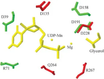
The structural model generated (Figure 2) comprises the critical amino acids for the function of this protein in the formation of biofilms arranged in a three dimensional con-formation with the possibility of fitting a substrate. The res-idue D228 is in exactly the same position as D191 (SpsA) with other conserved residues (D135, Q264 and R267) also positioned on the active site.
The multiple alignment among organisms in this study shows that the polypeptide HmsF ofC. violaceum re-tains the amino acids that are crucial to the formation of extracellular matrix in biofilms (Figure 3). The D114 and D115 aspartate amino acids of the protein HmsF of Y. pestis, which have HmsF deacetylase activity functionality, are conserved on all related organisms in this study, and in
C. violaceumcorrespond to aspartate D86 and D87. The important amino acids in the synthesis of extracellular ma-trix inY. pestis(W143 and H184), which correspond inC. violaceumto W115 and H156, are also retained.
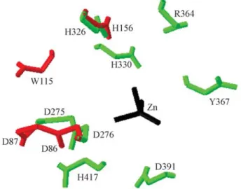
In the three-dimensional model of HmsF of C. violaceum obtained (Figure 4), the 3D-JIGSAW server made use of the proteinSpPgdA as a template (PDB code: 2c1g) (Blairet al., 2005), which is aN-acetyl-glucosamine deacetylase. The model generated for HmsF of C. violaceumpresenting the spatial location of critical resi-dues in the biofilm formation toY. pestisshows that the res-idues D86, D87, W115 and H156 are closely located in space (Figure 4). These residues are situated around the ac-tive site.
In the multiple alignment of HmsH protein, it was ob-served that the residue arginine R113, which had a poor performance in the formation of biofilms inY. pestis
(For-Table 1- Bacteria with their genomic regions used in the comparative study.
Organism and GenBank access number
GenBank access number Gene/ORF
manet al., 2006), and that inC. violaceumis the residue R104, was conserved in almost all organisms studied, but it was not observed inS. epidermidis, a Gram-positive bacte-ria (not shown). The multiple alignment performed among bacteria of this study for ORF CV2940 shows that the criti-cal residue tryptophan W80 ofY. pestisis also conserved in Gram-negative bacteria E. coli and C. violaceum, corre-sponding to the residue W94 inC. violaceum.
The analysis of the three-dimensional model gener-ated for HmsR ofC. violaceumby the Ramachandran dia-gram shows that the model holds 99.1% of the residues in allowed regions, demonstrating a good quality. The analy-sis of the environment of each amino acid residue carried out by the Verify3D program presented two regions (resi-dues 175 to 187 and 230 to 237) in the HmsR with a nega-tive 3D-1D score, indicating a probable incorrect folding in this region. However, the critical residues for HmsR in the formation of biofilm do not appear in this region. The
struc-Figure 2- Superimposed proteins closing up the active site of SpsA pro-tein bound to Mn-UDP with the built model of HmsR propro-tein C. violaceum. The residues of SpsA are green with HmsR residues in red and the UDP-Mn, Mg and Glycerol in yellow.
Figure 1- Partial multiple alignment of HmsR fromC. violaceumcompared with HmsR ofY. pestis; PgaC ofE. coli; IcaA ofS. epidermidisand HmsR of
B. pertussis. The critical residues aspartate (D), glutamine (Q) and arginine (R) are demarcated.
tural model of HmsF protein generated also presents a good quality, with a Ramachandran plot showing 98.5% of the residues in allowed regions. The model also shows a good quality for HmsF when compared with the results obtained in the Verify3D program, providing a great number of amino acids in a positive 3D-1D score.
Discussion
The production of biofilms by several organisms sug-gests the conservation of proteins or a set of proteins with similar function for its synthesis.
The HmsR protein, an important glycosyltransferase in polysaccharide synthesis, maintains the critical residues for its function also inC. violaceum. Critical residues in the formation of biofilms in HmsR protein ofY. pestis(Forman
et al., 2006), which match in C. violaceum to aspartate D135 and D228, arginine R267 and glutamine Q264, are retained in all the bacteria shown in this study, indicating functional similarity for this protein. The spatial location of the critical residues for biofilm formation in an active site strongly emphasizes the enzymatic role that the HmsR pro-tein ofC. violaceumcan perform in the synthesis of polysa-ccharide.
The HmsF protein of C. violaceum retains all the amino acids that are critical to the formation of biofilms. The generated model with the spatial location of critical residues in the formation of biofilms inY. pestisshows that they are close to each other. The use of a deacetylase as template was important due to the role that this enzyme plays in the formation of biofilms. The deacetylation in-volves mainly aspartate (D) and histidine (H) residues (Blairet al., 2005), and in this work these residues are lo-cated in a favorable spatial condition. Although actual evi-dence can be obtained from additional techniques such as substrate-enzyme reactions, it can be assumed that this model has a deacetylase catalytic region. The protein
HmsH preserves the residue arginine, which shows a mod-erate effect on the formation of biofilms inY. pestisamong all bacteria of this study, except forS. epidermidiswhich is a Gram-positive bacterium. In the three-dimensional mo-del, the choice of the template of the OGT enzyme (PDB accession code 1w3b) was very useful, since this enzyme has anN-acetylglucosamine (GlcNac) additive activity (Ji-nek et al., 2004) that is the basic unit of the polymer poly(beta-1,6-N-acetyl-D-glucosamine).
Conservation and favorable spatial arrangement of those critical amino acids that constitute the proteins en-coded by the elucidated operon indicate that they may ex-press the necessary enzymatic function for resistant biofilm formation. Therefore the conclusion is that the operon
hmsHFR-CV2940 might be linked toC. violaceum patho-genicity.
References
Bates PA, Kelley LA, Maccallum RM and Sternberg MJ (2001) Enhancement of protein modeling by human intervention in applying the automatic programs JIGSAW and 3D-PSSM. Proteins 5:39-46.
Blair DE, Schuttelkopf AW, Macrae JI and Van Aalten DM (2005) Structure and metal-dependent mechanism of pepti-doglycan deacetylase, a streptococcal virulence factor. Proc Natl Acad Sci USA 102:15429-15434.
Brazilian National Genome Project Consortium (2003) The com-plete genome sequence ofChromobacterium violaceum re-veals remarkable and exploitable bacterial adaptability. Proc Natl Acad Sci USA 100:11660-11665.
Eisenberg D, Luthy R and Bowie JU (1997) VERIFY3D: Assess-ment of protein models with three-dimensional profiles. Meth Enzymol 277:396-404.
Forman S, Bobrov AG, Kirillina O, Craig SK, Abney J, Fet-therston JD and Perry RD (2006) Identification of critical amino acid residues in the plague biofilm Hms proteins. Mi-crobiology 152:3399-3410.
Itoh Y, Wang X, Hinnebusch BJ, Preston JF and Romeo T (2005) Depolymerization of beta-1,6-N-acetyl-D-glucosamine dis-rupts the integrity of diverse bacterial biofilms. J Bacteriol 187:382-387.
Jarrett CO, Deak E, Isherwood KE, Oyston PC, Fischer ER, Whit-ney AR, Kobayashi SD, Deleo FR and Hinnebusch BJ (2004) Transmission of Yersinia pestisfrom an infectious biofilm in the flea vector. J Infect Dis 190:783-792. Jeanmougin F, Thompson JD, Gouy M, Higgins DG and Gibson
TJ (1998) Multiple sequence alignment with Clustal X. Trends Biochem Sci 23:403-405.
Jinek M, Rehwinkel J, Lazarus BD, Izaurralde E, Hanover JA and Conti E (2004) The superhelical TPR-repeat domain of O-linked GlcNAc transferase exhibits structural similarities to importin alpha. Nat Struct Mol Biol 11:1001-1007. Kelley LA, Maccallum RM and Sternberg MJ (2000) Enhanced
genome annotation using structural profiles in the program 3D-PSSM. J Mol Biol 299:499-520.
Laskowski RA, Rullmann JA, Macarthur MW, Kaptein R and Thornton JM (1996) AQUA and PROCHECK-NMR: Pro-grams for checking the quality of protein structures solved by NMR. J Biomol NMR 8:477-486.
Figure 4- Superimposed proteinsSpPgdA and HmsF. The residues of
Macher AM, Casale TB and Fauci AS (1982) Chronic granu-lomatous disease of childhood and Chromobacterium violaceuminfections in the southeastern United States. Ann Intern Med 97:51-55.
Mack D, Fischer W, Krokotsch A, Leopold K, Hartmann R, Egge H and Laufs R (1996) The intercellular adhesin involved in biofilm accumulation of Staphylococcus epidermidis is a linear beta-1,6-linked glucosaminoglycan: Purification and structural analysis. J Bacteriol 178:175-183.
Ramachandran GN, Ramakrishnan C and Sasisekharan V (1963) Stereochemistry of polypeptide chain configurations. J Mol Biol 7:95-99.
Sloan GP, Love CF, Sukumar N, Mishra M and Deora R (2007) TheBordetellaBps polysaccharide is critical for biofilm de-velopment in the mouse respiratory tract. J Bacteriol 189:8270-8276.
Sneath PH, Whelan JP, Bhagwan SR and Edwards D (1953) Fatal infection by Chromobacterium violaceum. Lancet 265:276-277.
Guest Editor: José Carlos Merino Mombach