Bo ne marro w fibro blasts in patie nts
with advance d lung cance r
1Research Member of the Consejo Nacional de Investigaciones Científicas y
Técnicas de la República Argentina (CO NICET), Buenos Aires, Argentina
2Instituto de Biología y Medicina Experimental, Buenos Aires, Argentina 3Departamento de O ncologia, I. Iriarte Hospital, Buenos Aires, Argentina 4Departamento de Hematologia e Transplante de Médula Ó ssea,
Hospital Británico, Buenos Aires, Argentina N.A. Chasseing1,2,
E. Hofer2,
R.H. Bordenave3,
C. Shanley4 and
L.S. Rumi1,2
Abstract
In a previous study we demonstrated that the incidence of fibroblast colony-forming units (CFU-F) was very low in bone marrow primary cultures from the majority of untreated advanced non-small lung cancer patients (LCP) compared to normal controls (NC). For this reason, we studied the ability of bone marrow stromal cells to achieve confluence in primary cultures and their proliferative capacity follow-ing four continuous subcultures in consecutive untreated LCP and NC. We also evaluated the production of interleukin-1ß (IL-1ß) and prostaglandin E2 (PGE2) by pure fibroblasts. Bone marrow was obtained from 20 LCP and 20 NC. A CFU-F assay was used to investigate the proliferative and confluence capacity. Levels of IL-1ß and PGE2 in conditioned medium (CM) of pure fibroblast cultures were measured with an ELISA kit and RIA kit, respectively. Only fibroblasts from 6/13 (46%) LCP confluent primary cultures had the capacity to proliferate following four subcultures (NC = 100%). Levels of spontaneously released IL-1ß were below 10 pg/ml in the CM of LCP, while NC had a mean value of 1,217 ± 74 pg/ml. In contrast, levels of PGE2 in these CM of LCP were higher (77.5 ± 23.6 pg/ml) compared to NC (18.5 ± 0.9 pg/ml). In conclusion, bone marrow fibroblasts from LCP presented a defective proliferative and confluence capacity, and this deficiency may be associated with the alteration of IL-1ß and PGE2 production.
Co rre spo nde nce
N.A. Chasseing Instituto de Biología y Medicina Experimental Vuelta de O bligado, 2490 1428 Buenos Aires Argentina
Fax: + 54-11-4786-2564 E-mail: [email protected]
Some of these data were presented at the XLIII Reunión Anual de la Sociedad Argentina de Investigación Clínica (SAIC), Mar del Plata, Argentina, November 26-29, 1998.
Research supported by CO NICET (PEI No. 0098/97) and the Roemmers Foundation.
Received May 24, 2001 Accepted August 27, 2001
Ke y words
·Fibroblasts
·Interleukin-1
·Prostaglandin E2
·Cancer
Intro ductio n
Several in vitro studies of the
hematopoi-etic microenvironment using long-term bone marrow and fibroblast colony-forming units (CFU-F) culture systems have proved that most (60-90%) adherent stromal cells are fibroblasts (1-3). Reports also indicate that normal human bone marrow fibroblasts
primary bone marrow cultures from the ma-jority of untreated advanced non-small lung cancer patients (LCP) (7). This reduction in the number of CFU-F may be related to an increase in the levels of inhibitor factors and/or to a decrease of the levels of stimulant mediators such as interleukin-1 (1), IL-17, platelet derived growth factor, transform-ing growth factor-ß1 (TGF-ß1), epidermal growth factor, tumor necrosis factor recep-tor, collagen, fibronectin, IL-2, interferon-g, and prostaglandin E2 (PGE2) (8-13). For this reason, we have evaluated the ability of bone marrow stromal cells (mostly fibro-blasts) to achieve confluence in primary cul-tures and their proliferative capacity follow-ing four continuous subcultures in consecu-tive untreated LCP and normal controls (NC). At the same time, we studied the release of IL-1ß and PGE2 into CM of pure fibroblast cultures treated or not with muramyl-dipep-tide (MDP).
Mate rial and Me thods
Patie nts
Bone marrow samples were obtained from 20 NC and 20 consecutive untreated patients with non-small lung epidermoid carcinoma (stage III A, III B and IV). We used a UICC TNM classification. All LCP and NC were age and sex matched. The age intervals were: LCP = 40 to 60 and NC = 45 to 67 years. All individuals gave consent to participate in these studies, which were performed in ac-cordance with the principles of the Declara-tion of Helsinki. Patient bone marrow aspi-rates were provided by Dr. R.H. Bordenave, Department of Oncology, I. Iriarte Hospital, Buenos Aires, Argentina. Healthy control bone marrow aspirates were provided by Dr. C. Shanley, Department of Hematology and Bone Marrow Transplantation, Británico Hospital, Buenos Aires, Argentina. NC were healthy donors for bone marrow transplantation.
The present investigation was approved
by the Británico Hospital and I. Iriarte Hos-pital Ethics Committees.
Bone marrow micrometastases
Bone marrow infiltration with neoplastic cells was detected by immunocytochemistry staining (biotin-streptavidin-peroxidase, Uni-versal Dako LSAB System, Carpinteria, CA, USA) and an analysis of cell morphology was done by the Pappenheim technique. Bone marrow samples were stained with mono-clonal antibodies to cytokeratin AE1-AE3 (Dako), cytokeratin 7 (CK7, Dako) and cyto-keratin 20 (CK20, Dako). Patients were con-sidered positive for micrometastasis only if cells expressed cytokeratin AE1-AE3, CK7 and/or CK20 and if these were morphologi-cally malignant.
Colle ction and pre paration of bone
marrow ce lls
Bone marrow samples were collected under local anesthesia from the posterior iliac crest into heparinized saline without preservatives (25 units/ml, Gibco, Grand Is-land, NY, USA). Aspirates were diluted 1/2 with PBS, pH 7.5, and were layered onto Histopaque (density = 1,075 g/cm3; Sigma,
St. Louis, MO, USA). After being centri-fuged for 25 min at 1,500 rpm, mononuclear cells were harvested from the interface, washed twice in PBS and resuspended in a -medium (Gibco) containing 100 IU/ml peni-cillin (Gibco), 100 µg/ml streptomycin (Gibco) and 25 µg/ml of amphotericin B (Gibco). The cell suspension was counted with 3% acetic acid solution and cell viabil-ity was determined by Trypan blue exclu-sion.
Fibroblast colony-forming units assay
Viable, light density mononuclear cells (5 x 10⁶) were placed in 25-cm2 tissue
of supplemented previously described a -me-dium and 20% heat-inactivated FBS (Gibco cat No. 16,000-044) (1). The cells were in-cubated at 37ºC in a 5% CO2 humidified
environment for 7 days. After this period, the non-adherent cells were removed and the medium was renewed. Then the primary cul-tures were returned to incubation for an ad-ditional 7 days. At the end of this period, the medium was discarded and the adherent cells were washed twice with PBS, fixed with 100% methanol, and stained with Giemsa. Clones of >50 cells were scored as CFU-F under a binocular microscope.
Conflue nt primary culture formation
Viable, light density mononuclear cells (5 x 10⁶)of each sample were placed in
25-cm2 tissue culture flasks (Corning) which
contained 10 ml of supplemented previously described a-medium and 20% FBS. This supplemented medium is known to be selec-tive for fibroblast progenitor proliferation (1). The cells were incubated at 37ºC in a 5% CO2 humidified environment for 7 days. After
this period, the non-adherent cells were re-moved and the medium was renewed. This period of time was selected in order to allow the maximum release of growth factors, which are necessary for fibroblast progeni-tor proliferation (1). The primary cultures were returned to incubation for further days until confluence. From the initiation of the experiment until day 60, or until the cells reached confluence, the medium was changed every 7 days. At the end, the medium was discarded and the confluent adherent cells were washed twice with PBS and then trypsinized with a solution of trypsin-EDTA (0.05-0.02% in PBS, respectively; Gibco). Finally, trypsin-sensitive adherent cells were further induced to proliferate following four continuous subcultures. The adherent cells were subcultured only after confluence in each case. The number of days the adherent cells (most of them fibroblasts) took to
achieve confluence in primary cultures was studied, as well as the number of patients with fibroblast proliferative capacity when cells from confluent primary cultures were further induced to proliferate following four subcultures.
The fibroblastic nature of the adherent cells that composed the cultures was demon-strated by immunofluorescent staining with monoclonal antibodies against human fibronectin (a gift from Dr. A. Kornbliht, Facultad de Ciencias Exactas y Naturales, Universidad de Buenos Aires, Argentina) and against the human ß subunit of prolyl-4-hydroxylase (Dako). We also used cyto-chemical analyses with alkaline phosphatase stain. Adherent cells were fixed with 50% methanol.
Pre paration of CM from pure fibroblasts
Bone marrow fibroblasts isolated after four continuous confluent subcultures were adjusted to 5 x 10⁴ viable cells/ml in fresh a
-medium containing 20% FBS and 1% antibi-otic-antimycotic agent. The fibroblasts were allowed to adhere to plastic tissue culture plates (6 wells/plate, Falcon) for at least 24 h after the fourth subculture in order to obtain confluence before the experiment began. After this period, fibroblasts were incubated in the presence and absence of MDP (1 µg/ ml, Sigma) for an additional 72 h at 37ºC and 5% CO2 (14). The CM were obtained by
centrifugation at 1,000 rpm for 10 min and frozen at -20ºC before use for IL-1ß assay and at -70ºC for PGE2.
De te rmination of IL-1ß and PGE2 in the CM
of pure fibroblast culture s
levels between 10 and 7,400 pg/ml and the PGE2 assay detects levels between 2.5 and 250 pg/ml.
Simultaneously we evaluated IL-1ß and PGE2 levels in samples of supplemented a -medium after 7 days of incubation, and ob-served that the concentration for both soluble factors was below the minimum detectable dose. All samples and standards were ana-lyzed in duplicate.
Statistical analysis
Statistical analyses were performed us-ing parametric and nonparametric tests
de-pending on the data studied (P<0.05).
Re sults
Bone marrow micrometastases
Using the Pappenheim and immunocy-tochemistry techniques, morphological evi-dence of bone marrow infiltration with neo-plastic cells was not observed in cancer pa-tients.
Evaluation of prolife rative and conflue nce
capacity of the adhe re nt ce lls in bone
marrow primary cultures and four
continuous subculture s
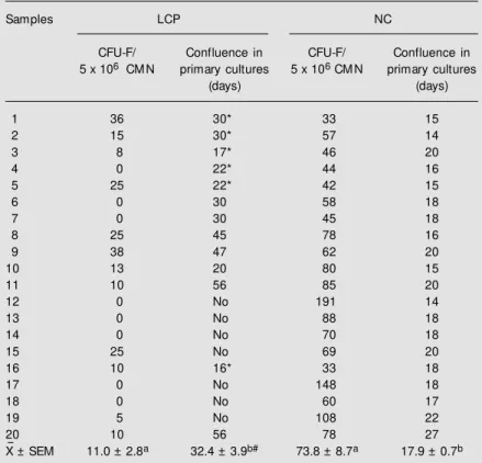
Table 1 shows that only 5/20 (25%) bone marrow cultures from LCP had adherent cells which were able to achieve confluence in primary cultures within the normal range (14 to 27 days). Furthermore, in the other 15/ 20 (75%) cultures from LCP we observed that there was lack of confluence (35% of the total) or that more days were needed for the stromal layers to obtain confluence (40% of the total). The values (mean ± SEM) were: LCP = 32.4 ± 3.9 days and NC = 17.9 ± 0.7 days (P<0.0004, nonparametric Mann-Whitney test).
In addition, after 14 days of incubation of bone marrow primary cultures, the number of CFU-F was very low (2/7) or zero (5/7) in the LCP samples with a lack of confluence. Moreover, all the cultures of adherent cells from LCP samples which were able to achieve confluence in primary cultures showed a significantly decreased number of CFU-F compared to the values for NC. This last observation was independent of whether or not the cultures were able to achieve conflu-ence within the normal range (Table 1).
On the other hand, when adherent cells from confluent primary cultures of LCP (13/ 20) were further induced to proliferate fol-lowing four continuous subcultures, we ob-served that the growth had diminished or
Table 1. Evaluation of fibroblast colony-forming units and the ability of the stromal cells to achieve confluence in bone marrow primary cultures.
Samples LCP NC
CFU-F/ Confluence in CFU-F/ Confluence in 5 x 10⁶ CM N primary cultures 5 x 10⁶ CM N primary cultures
(days) (days)
1 36 30* 33 15
2 15 30* 57 14
3 8 17* 46 20
4 0 22* 44 16
5 25 22* 42 15
6 0 30 58 18
7 0 30 45 18
8 25 45 78 16
9 38 47 62 20
10 13 20 80 15
11 10 56 85 20
12 0 No 191 14
13 0 No 88 18
14 0 No 70 18
15 25 No 69 20
16 10 16* 33 18
17 0 No 148 18
18 0 No 60 17
19 5 No 108 22
20 10 56 78 27
X- ± SEM 11.0 ± 2.8a 32.4 ± 3.9b# 73.8 ± 8.7a 17.9 ± 0.7b
Data are reported as means of duplicate individual values. #This mean w as obtained from the patients w ho show ed confluent adherent cells up to 60 days after the beginning of incubation. LCP = lung cancer patients; NC = normal controls; CM N = light density mononuclear cells; CFU-F = fibroblast colony-forming units; No = did not reach confluence w ithin the first 60 days.
* Six LCP confluent primary cultures w hose adherent cells could proliferate in the four subsequent subcultures compared to the 100% of NC.
stopped between the second and third sub-culture in seven LCP sub-cultures. Moreover, a high percentage of these seven LCP cultures showed the longest times to achieve conflu-ence in bone marrow primary cultures (Table 1). Regarding, the six LCP cultures that were able to proliferate and to reach confluence in each of the four continuous subcultures, the number of days they needed to achieve con-fluence in each of the four individual subcul-tures was always longer than for the NC subcultures, although we incubated the same amount of adherent cells in both groups. In contrast, all NC cultures presented full con-fluent stromal layers in primary cultures and had the capacity to proliferate following four continuous subcultures.
In terms of lineage markers, up to 90% of the non-hematopoietic adherent cells from patients and NC primary cultures expressed three fibroblast markers (fibronectin, prolyl-4-hydroxylase and alkaline phosphatase). Moreover, the majority of adherent cells in primary cultures presented a fusiform shape in both groups.
Regarding the number of trypsin-EDTA (0.05-0.02%)-sensitive adherent cells (pure fibroblasts) in the fourth confluent subcul-ture, LCP values did not differ from NC values [LCP = (0.805 ± 0.074) x 106 and NC
= (1.150 ± 0.220) x 106]. It is well known
that only adherent bone marrow cells that have a fibroblastic nature are detached after this treatment (1).
De te rmination of IL-1ß and PGE2 le ve ls in
the CM of pure fibroblast culture s
The levels of IL-1ß released in the CM of fibroblast cultures from LCP were below the detectable amount (<10 pg/ml), independ-ently of the culture treatment used (Table 2). In contrast, the levels of IL-1ß were signifi-cantly higher in the CM of NC.
Finally, the levels of PGE2 spontane-ously released in the CM of fibroblast cul-tures from LCP increased significantly
com-pared to the mean value obtained in NC cultures (P<0.0022, nonparametric Mann-Whitney test; Table 2). Moreover, in the presence of MDP a lack of response was observed in all LCP cultures, but PGE2 lev-els increased in the CM of NC (P<0.0022 compared to basal levels, nonparametric Mann-Whitney test).
D iscussio n
Bone marrow stromal cells produce mul-tiple factors (extracellular matrix and cyto-kines), which are not only capable of con-trolling the self-renewal, proliferation and differentiation of hematopoietic stem/pro-genitor cells, but are also regulated by them (12,15). Functional alterations in the bone marrow microenvironment may be involved in the manifestation of some malignant dis-orders (16,17).
The results presented in this paper show that a high percentage of bone marrow pri-mary cultures from untreated LCP had phe-notypic abnormalities such as diminished ability of the adherent stromal cells to achieve confluence or a lack of confluence at all. These observations are likely to reflect de-rangement in the composition of the hemato-poietic microenvironment and/or in the
pro-Table 2. Determination of the levels of interleukin-1ß (IL-interleukin-1ß) and prostaglandin E2 (PGE2) in the con-ditioned media of pure fibroblast cultures.
Groups IL-1ß (pg/ml) PGE2 (pg/ml)
LCP
None <10 77.5 ± 23.6b
M DP (1 µg/ml) <10 47.3 ± 17.6
NC
None 1,217 ± 74a 18.5 ± 0.9b,c M DP (1 µg/ml) 4,607 ± 62a 90.0 ± 5.7c
Data are reported as means ± SEM . LCP = lung cancer patients; NC = normal controls; M DP = muramyl-dipeptide. Conditioned media w ere di-luted 1:5.
liferative capacity of the stromal population, mainly fibroblastic progenitors. In addition, when adherent cells (most of them fibro-blasts) from confluent primary bone marrow cultures (13/20) from LCP were further in-duced to proliferate following four continu-ous subcultures, reduced or no further growth was observed in seven of them. This defec-tive proliferadefec-tive potential of the bone mar-row fibroblasts in LCP might explain the lower ability of the stromal components to achieve confluence in primary cultures and in the other four subcultures.
The regulation of fibroblast proliferation is tremendously complex. On the one hand, the cellular shape seems to be one of the primary factors that regulate the mitogenic responses of fibroblasts to certain mitogenic agents (18). This becomes evident when one considers cells (such as fibroblasts) in which the proliferative rate is anchorage depend-ent. When normal bone marrow fibroblasts are plated on plastic, they will spread on the surface and proliferate actively in response to serum factors until reaching confluence (18). And on the other hand, many soluble factors can alter fibroblast growth, as previ-ously reported by our group (8,12).
In the present study we found that pure fibroblasts isolated after four continuous sub-cultures from bone marrow of LCP released very low levels of IL-1ß (<10 pg/ml) com-pared with NC. In contrast, untreated pure fibroblasts from LCP released high levels of PGE2 compared with NC values, and PGE2 production was not increased after MDP stimulation in the LCP group. This result supports the theory that a defective
prolif-erative potential of the progeny of CFU-F can be caused by an increase in the produc-tion of PGE2. These findings are in agree-ment with reports by other authors who ob-served that PGE2 is an inhibitor of fibroblast proliferation in serum-supplemented cultures (19,20).
On the other hand, some authors ob-served that IL-1ß induces PGE2 in fibro-blasts (9,21), whereas our results showed high levels of IL-1ß released by NC subcul-tures that were not correlated with PGE2 levels, which were lower compared to LCP subcultures that had little or no IL-1ß. Thus, this apparent contradiction suggested that the increased levels of PGE2 in the CM of LCP could be induced by factors other than IL-1ß, like tumor necrosis factor-a or TGF-ß (21,22).
By analyzing the results as a whole, we reached the conclusion that bone marrow fibroblasts from LCP had a defective prolif-erative and confluence capacity not only in the primary cultures but also during the four subsequent subcultures. Moreover, the bone marrow fibroblasts from LCP had an altera-tion of IL-1ß and PGE2 producaltera-tion that may play an important role, directly or indirectly, in the regulation of the proliferative and confluence capacity of these stromal cells during the subsequent subcultures. Finally, although it seems clear that functional ab-normalities in bone marrow fibroblasts exist in LCP, it is not known whether these modi-fications are an inherent defect in fibroblasts of patients susceptible to lung carcinoma or an acquired defect related to the tumor itself.
Re fe re nce s
1. Castro-M alaspina H, Gay RE, Resnick G, Kapoor N, M eyers P, Chiarieri D, M c-Kenzie S, Broxmeyer HE & M oore M AS (1980). Characterization of human bone marrow fibroblast colony-forming cells (CFU-F) and their progeny. Blood, 56: 289-301.
2. Bentley SA & Foidart JM (1980). Some properties of marrow derived adherent cells in tissue culture. Blood, 56: 1006-1012.
3. Krebsbach PH, Kuznetsov SA, Bianco P & Gehron Robey P (1999). Bone marrow stromal cells: characterization and clinical
application. Critical Review s in Oral Biol-ogy and M edicine, 10: 165-181. 4. M oreau I, Andreoni C, Caux C, Saeland S
European Journal of Haematology, 49: 29-35.
5. Caplan AI (1994). The mesengenic pro-cess. Clinics in Plastic Surgery, 21: 429-435.
6. M inguell JJ, Conget P & Erices A (2000). Biology and clinical utilization of mesen-chymal progenitor cells. Brazilian Journal of M edical and Biological Research, 33: 881-887.
7. Chasseing NA, Bordenave RH, Bullorsky EO, Diaz NB, Stemmelin GR & Rumi L (1997). Fibroblastic colony forming units and the levels of tumor necrosis factor and prostaglandin E2 in bone marrow cul-tures from patients w ith advanced lung carcinoma. Cancer, 80: 1914-1919. 8. Kovacs EJ (1991). Fibrogenic cytokines:
the role of immune mediators in the de-velopment of scar tissue. Immunology Today, 12: 17-23.
9. Freundlich B, Bomalaski JS, Neilson E & Jimenez SA (1986). Regulation of fibro-blast proliferation and collagen synthesis by cytokines. Immunology Today, 7: 303-307.
10. Thornton SC, Por SB, Walsh BJ, Penny R & Breit SN (1990). Interaction of immune and connective tissue cells: I. The effect of lymphokines and monokines on fibro-blast grow th. Journal of Leukocyte Biol-ogy, 47: 312-320.
11. Kuznetsov SA, Friedenstein AJ & Robey PG (1997). Factors required for bone mar-row stromal fibroblast colony formation
in-vitro. British Journal of Haematology, 97: 561-570.
12. Rougier F, Dupuis F & Denizot Y (1996). Human bone marrow fibroblasts: an over-view of their characterization, prolifera-tion and inflammatory mediator produc-tion. Hematology and Cell Therapy, 38: 241-246.
13. Know n BS, Wang S, Udagaw a N, Haridas V, Lee ZH, Kim KK, Oh KO, Greene J, Li Y, Su J, Gentz R, Aggarw al BB & Ni J (1998). TR1, a new member of the tumor necro-sis factor receptor superfamily, induces fibroblast proliferation and inhibits osteo-clast ogenesis and bone resorpt ion.
FASEB Journal, 12: 845-854.
14. Iribe H, Koga T, Kotani S, Kusumoto S & Shiva T (1983). Stimulating effect of mu-ramyl-dipeptide and its adjuvant active analogues on guinea pig fibroblasts for the production of thymocyte-activating factor. Journal of Experimental M edicine, 157: 2190-2195.
15. Kuznetsov S & Gehron RP (1996). Spe-cies differences in grow th requirements for bone marrow stromal fibroblast colony formation in vitro. Calcified Tissue Inter-national, 59: 265-270.
16. Coutinho LH, Geary CG, Chang J, Harri-son C & Testa NG (1990). Functional stud-ies of bone marrow haemopoietic and stromal cells in the myelodisplastic syn-drome (M DS). British Journal of Haema-tology, 75: 16-25.
17. Hirata J, Katsuno M & Kaneko S (1986).
Clinical significance of human bone mar-row stromal cell colonies in acute leuke-mias. Leukemia Research, 12: 1441-1445. 18. Gospodarow icz D, Greenburg G & Bird-w ell CR (1978). Determination of cellular shape by the extracellular matrix and its correlation w ith the control of cellular grow th. Cancer Research, 38: 4155-4171. 19. Hori T, Yamanaka Y, Hayakaw a M , Shiba-moto S, TsujiShiba-moto M , Oku N & Ito F (1991). Prostaglandins antagonize fibro-blast proliferation stimulated by tumor necrosis factor. Biochemical and Biophysi-cal Research Communications, 174: 758-766.
20. Korn JH, Halushka PV & LeRoy EC (1980). M ononuclear cell modulation of connec-tive tissue function. Suppression of fibro-blast grow th by stimulation of endoge-nous prostaglandin production. Journal of Clinical Investigation, 65: 543-554. 21. Diaz A, M uñoz E, Johnston R, Korn JH &
Jimenez SA (1993). Regulation of human lung fibroblast a1 (I) procollagen gene ex-pression by tumor necrosis factor a, inter-leukin-1ß, and prostaglandin E2. Journal of Biological Chem ist ry, 268: 10364-10371.