LETTER TO THE EDITOR
Cerebrotendinous xanthomatosis: a treatable
hereditary neuro-metabolic disease
Ana Claudia Rodrigues de Cerqueira,IAntoˆnio Egı´dio Nardi,IJose Marcelo Ferreira BezerraII
IInstituto de Psiquiatria, Universidade Federal do Rio de Janeiro, Rio de Janeiro, Brazil.IIDepartamento de Neurologia, Universidade do Estado do Rio de
Janeiro, Rio de Janeiro, Brazil.
Email: anacerqueira@globo.com Tel.: 55 21 2521-6147
INTRODUCTION
Cerebrotendinous xanthomatosis is a rare autosomal recessive hereditary disease that is caused by a mutation in the gene encoding the mitochondrial enzyme sterol 27-hydroxylase (CYP27). The CYP27 gene is located on chromosome 2q35-qter and is responsible for the conversion of cholesterol into cholic and chenodeoxycholic acid. Cerebrotendinous xanthomatosis results in increased levels of serum cholestanol, a cholesterol precursor, and increased deposition of cholestanol and cholesterol in various tissues, especially the lenses, tendons, and the central nervous system. The principal manifestations of Cerebrotendinous xanthomatosis include juvenile cataracts, tendon xantho-mas, and progressive neurological symptoms.1
Early recognition of this condition is essential because cholic and chenodeoxycholic acid replacement therapy can prevent Cerebrotendinous xanthomatosis -induced brain damage, which leads to severe neurological dysfunction and death. We present the case of a patient with clinical, radiological, and biochemical evidence of Cerebrotendinous xanthomatosis and his response to cholic and chenodeoxy-cholic acid treatment.
CASE REPORT
This report describes a 30-year-old male who displayed normal psychomotor development until 10 years of age when, according to his mother, he began presenting learning difficulties and showed progressive cognitive decline. At 15 years of age, the patient presented progres-sive gait instability and enlargement of the Achilles tendons. At age 17, he suffered his first convulsive episode. The patient has suffered a total of four seizures since that time, and his seizure condition is adequately controlled with the use of anticonvulsants. A family history revealed that the patient’s parents were cousins. Physical examination revealed bilateral Achilles tendon enlargement and bilateral cataracts. A neurological examination identified spastic paraparesis, brisk deep tendon reflexes and extensor plantar responses, upper limb symmetrical dysmetria and dysdia-dochokinesia, and ataxic-paraparetic gait. The remainder of the patient’s physical examination was normal.
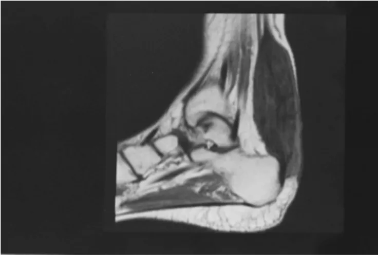
Magnetic resonance imaging (MRI) of the patient’s skull revealed mild cerebellar atrophy (Figure 1). MRI of the left ankle showed fusiform thickening of the Achilles tendon with heterogeneous signals on all sequences, T2-hyperin-tense foci due to the presence of fat, and a slight increase in the uptake of paramagnetic contrast. These results were suggestive of a xanthoma of the Achilles tendon (Figure 2). Laboratory tests (complete blood count, biochemistry, liver and kidney function, lipid panel, and coagulation tests) were normal. The patient’s serum cholestanol level was high (28.3mg/mL, normal value,6mg/mL).
Based on these findings, the patient was diagnosed with CTX, and treatment with CDCA (750 mg/day) was imme-diately initiated. The drug was imported from Germany. A significant improvement in neurological symptoms was observed after one year of follow-up, especially with respect to ataxia and coordination. The patient retains a slightly ataxic gait but presents normal finger-to-nose and fast finger movements, and his serum cholestanol level has been reduced to 4.1mg/mL. In addition, the patient has
under-gone eye surgery to treat his cataracts. The patient authorized the publication of this case report by signing a consent form.
DISCUSSION
Van Bogaert et al. described the first CTX phenotype in 1937.2 Subsequent work established several additional symptoms of CTX, including cholestanol accumulation in
Copyrightß2010CLINICS– This is an Open Access article distributed under the terms of the Creative Commons Attribution Non-Commercial License (http:// creativecommons.org/licenses/by-nc/3.0/) which permits unrestricted non-commercial use, distribution, and reproduction in any medium, provided the original work is properly cited.
Figure 1 - Brain MRI sagittal section in T1, showing mild
cerebellar atrophy in a patient with CTX.
CLINICS 2010;65(11):1217-1218 DOI:10.1590/S1807-59322010001100028
several tissues and the absence of CDCA in the bile. These symptoms were determined to be the result of a disorder of hepatic conversion of cholesterol to cholic acid and CDCA.3
In 1975, Salen et al. reported that administration of CDCA dramatically reduced cholestanol synthesis in CTX patients.4 In 1984, Berginer et al. demonstrated that one
year of CDCA oral supplementation treatment at 750 mg/ day was sufficient to produce a significant improvement in neurological symptoms, normalization of EEG readings, and a reduction in serum cholestanol in CTX patients.5 In 1991, Cali et al. identified a defect in the gene encoding the 27-hydroxylase enzyme in CTX patients.6 More than fifty different mutations of this gene have since been reported worldwide, and molecular analysis has enabled diagnosis during the pre-symptomatic period.7
The unexplained presence of bilateral cataracts and chronic diarrhea in children suggests a diagnosis of CTX. Tendon xanthomas usually appear in the second or third decade of life, especially in the Achilles tendons. Neuro-logical symptoms usually begin in the second decade of life and include ataxia, pyramidal and extrapyramidal signs, peripheral sensory-motor neuropathy, epilepsy, and dementia.7,8Psychiatric manifestations such as depression, suicidal thoughts, catatonia, and psychotic symptoms may also present with CTX and usually appear at early stages.9 Other clinical manifestations include osteoporosis, bone fractures, premature arteriosclerosis, and coronary and lung disease.1,8
The mechanism underlying the progressive neurological dysfunction in CTX remains unknown. Some authors support the hypothesis that increased concentrations of apolipoprotein B in the cerebrospinal fluid (CSF) of CTX patients facilitates cholestanol and cholesterol transport across the blood-brain barrier. Presumably, this accumula-tion of cholestanol activates apoptotic pathways, resulting in neuronal death. However, CDCA treatment restores the selective permeability of the blood-brain barrier, normal-izing the concentration of sterols and lipoproteins in the CSF and promoting reversal of the neurological symptoms.1,10
In a recent study, Berginer et al. demonstrated that early diagnosis and initiation of CDCA treatment during the pre-clinical and initial phases of CTX may prevent the development of clinical manifestations of CTX. These
authors suggest that the following three steps are funda-mental to prevent irreversible damage in patients with CTX: 1) recognition of early symptoms, including chronic diarrhea and juvenile cataracts, by pediatricians, 2) con-firmation of the diagnosis through biochemical and genetic analysis, and 3) immediate CDCA treatment to prevent the CTX phenotype.7
This report describes the case of a patient with clinical, biochemical, and radiological characteristics indicative of CTX. In support of this diagnosis, biochemical analysis demonstrated elevated cholestanol levels. Tendon xantho-mas can occur in other lipidoses, such as familial hyperch-olesterolemia and sitosterolemia, but juvenile cataracts and progressive neurological symptoms are only seen in CTX.1 We were unable to perform molecular genetic analysis in our patient, but MRI analysis of the skull revealed cerebellar atrophy. A similar finding has been reported by other authors, as have bilateral and symmetric hyperintense lesions surrounding the white matter of the dentate nuclei, cerebral atrophy, and demyelinating lesions.11 These changes were not demonstrated in our case due to techno-logical limitations. CDCA treatment was immediately initiated in our patient, and a significant improvement in neurological symptoms was observed after one year of treatment. Furthermore, the clinical improvement correlated with a reduction in cholestanol serum levels. Unfortunately, because treatment was initiated at a late stage in this case, it did not result in a functional cure of CTX. However, the treatment did promote a significant improvement in quality of life and, more importantly, prevented progression of the disease.
REFERENCES
1 . M o gha d a s i an M H , S a l en G , F r o h l i ch J, S cu d a mo r e C H . Cerebrotendinous Xanthomatosis: A Rare Disease with Diverse Manifestations. Arch Neurol. 2002;59:527-9, doi: 10.1001/archneur.59.4. 527.
2. Van-Bogaert L, Scherer HJ, Epstein E. A Cerebral Form of Generalized Cholesterinose [in French]. Paris, , France: Masson et Cie; 1937. 3. Menkes JH, Schimschock JR, Swanson PD. Cerebrotendinous
xantho-matosis: the storage of cholestanol within the nervous System. Arch Neurol. 1968;19:47–53.
4. Salen G, Meriwether TW, Nicolau G.Chenodesoxycholic acid inhibits increased cholesterol and cholestanol synthesis in patients with cerebrotendinous xanthomatosis. Biochem Med. 1975;14:57-74, doi: 10. 1016/0006-2944(75)90020-4.
5. Berginer VM, Salen G, Shefer S. Long-term treatment of cerebrotendi-nous xanthomatosis with chenodeoxycholic acid.N Engl J Med. 1984;311:1649–52, doi: 10.1056/NEJM198412273112601.
6. Cali JJ, Hsieh CL, Francke U, Russell DW. Mutations in the bile acid biosynthetic enzyme sterol 27-hydroxylase underlie cerebrotendinous xanthomatosis. J Biol Chem. 1991;266:7779–83.
7. Berginer VM, Gross B, Morad K, Kfir N, Morkos S, Aaref S, et al. Chronic Diarrhea and Juvenile Cataracts: Think Cerebrotendinous Xanthomatosis and Treat. Pediatrics. 2009;123:143–7, doi: 10.1542/peds.2008-0192. 8. Frederico A, Dotti MT. Cerebrotendinous Xanthomatosis: Clinical
Manifestations, Diagnostic Criteria, Pathogenesis, and Therapy. J Child Neurol.2003;18:633-8, doi: 10.1177/08830738030180091001.
9. Lee Y, Lin PY, Chiu NM, Chang WN, Wen JK. Cerebrotendinous xanthomatosis with psychiatric disorders: report of three siblings and literature review. Chang Gung Med J. 2002;25:334-40.
10. Salen G, Berginer V, Shore V, Horak I, Horak E, Tint GS, et al. Increased concentrations of cholestanol and apolipoprotein B in cerebrospinal fluid of patients with cerebrotendinous xanthomatosis: effect of chenodeoxy-cholic acid. N Engl J Med. 1987;316:1233-8, doi: 10.1056/ NEJM198705143162002.
11. De Stefano N, Dotti M, Mortilla M, Frederico A. Magnetic resonance Imaging and spectroscopic changes in brains of patients with cerebro-tendinous xanthomatosis. Brain. 2000;124:121-31, doi: 10.1093/brain/124. 1.121.
Figure 2 -Ankle MRI sagittal section in T1 showing Xanthoma of
the Achilles in a patient with CTX. Cerebrotendinous Xanthomatosis
de Cerqueira ACR et al. CLINICS 2010;65(11):1217-1218

