Neutrophil function and metabolism in
individuals with diabetes mellitus
1Departamento de Fisiologia e Biofísica, 2Departamento de Farmacologia, 3Departamento de Imunologia, Instituto de Ciências Biomédicas,
Universidade de São Paulo, São Paulo, SP, Brasil
4Divisão de Experimentação, Instituto do Coração, LIM-11,
Faculdade de Medicina, Universidade de São Paulo, São Paulo, SP, Brasil T.C. Alba-Loureiro1,
C.D. Munhoz2, J.O. Martins3,
G.A. Cerchiaro4, C. Scavone2,
R. Curi1 and P. Sannomiya4
Abstract
Neutrophils act as first-line-of-defense cells and the reduction of their functional activity contributes to the high susceptibilityto and severity of infections in diabetes mellitus. Clinical investigations in diabetic patients and experimental studies in diabetic rats and mice clearly demonstrated consistent defects of neutrophil chemotactic, phagocyt-ic and mphagocyt-icrobphagocyt-icidal activities. Other alterations that have been re-ported to occur during inflammation in diabetes mellitus include: decreased microvascular responses to inflammatory mediators such as histamine and bradykinin, reduced protein leakage and edema forma-tion, reduced mast cell degranulaforma-tion, impairment of neutrophil adhe-sionto the endothelium and migration to the site of inflammation, production of reactive oxygen species and reduced release of cyto-kines and prostaglandin by neutrophils, increased leukocyte apopto-sis, and reduction in lymph node retention capacity. Since neutrophil function requires energy, metabolic changes (i.e., glycolytic and glutaminolytic pathways) may be involved in the reduction of neutro-phil function observed in diabetic states. Metabolic routes by which hyperglycemia is linked to neutrophil dysfunction include the ad-vanced protein glycosylation reaction, the polyol pathway, oxygen-free radical formation, the nitric oxide-cyclic guanosine-3'-5’mono-phosphate pathway, and the glycolytic and glutaminolytic pathways. Lowering of blood glucose levels by insulin treatment of diabetic patients or experimental animals has been reported to have significant correlation with improvement of neutrophil functional activity. There-fore, changes might be primarily linked to a continuing insulin deficiency or to secondary hyperglycemia occurring in the diabetic individual. Accordingly, effective control with insulin treatment is likely to be relevant during infection in diabetic patients.
Correspondence
P. Sannomiya
Divisão de Experimentação InCor, LIM-11
Av. Dr. Enéas C. Aguiar, 44 05403-000 São Paulo, SP Brasil
Fax: +55-11-3061-7178 E-mail: [email protected] This study is dedicated to the memory of Professor João Garcia-Leme.
Research supported by FAPESP. R. Curi and C. Scavone are recipients of CNPq research fellowships.
Received March 9, 2007 Accepted May 21, 2007
Key words
•Neutrophils •Inflammation •Diabetes mellitus •Insulin
•Metabolism and cellular
function
Diabetes mellitus and inflammation
There is evidence that hormones are in-volved in the development of the inflamma-tory response. Inflammation evokes
an important part in the control of inflamma-tion (1,2).
Several aspects have been shown to be impaired during inflammation in diabetes mellitus. These include decreased microvas-cular responses to inflammatory mediators such as histamine and bradykinin (3,4), de-creased protein leakage and edema forma-tion (5-7), reduced mast cell degranulaforma-tion (8), decreased leukocyte-endothelial cell in-teractions and reduced number of leuko-cytes in inflammatory lesions (9-15), low-ered airway inflammatory response to anti-gen challenge (16,17), low superoxide anti- gen-eration, and reduced release of tumor necro-sis factor-α (TNF-α), interleukin-1ß and prostaglandin E2 by leukocytes upon
expo-sure to lipopolysaccharide (LPS) (18-20); low content of arachidonic acid in neutro-phils (20), and a reduction in lymph node retention capacity (21). These abnormalities might contribute to the increased suscepti-bility and severity of infections in diabetic patients.
In a series of experiments conducted in the early 1970’s, Garcia-Leme and co-work-ers (5,6) clearly demonstrated that insulin exerts direct regulatory effects on several stages of inflammation. Rats rendered dia-betic by the administration of alloxan or by subtotal pancreatectomy fail to present the edema that follows the injection of chemical irritants or application of physical stimuli. This inhibition is abolished by previous in-jection of insulin and is not associated with increased blood glucose concentrations. In addition, diabetic rats exhibit decreased re-sponses to permeability factors such as his-tamine, bradykinin, or serotonin injected into the skin compared to normal rats (5,6). Light and electron microscopy studies have re-vealed that microvessels of alloxan diabetic rats challenged with histamine or serotonin exhibit less labeling by intravenously in-jected colloidal carbon particles than do ves-sels of normal animals (7). The integrity of the microcirculatory responses to noxious
stimuli may consequently depend on the availability of insulin.
Neutrophil function in diabetes mellitus
Neutrophils play an essential role in the host inflammatory response against infec-tion. Mowat and Baum (22) showed for the first time that the chemotactic activity of neutrophils from diabetic patients is signifi-cantly lower than in cells from healthy con-trols. Studies of the phagocytic and microbi-cidal activities of diabetic patients reveal, with few exceptions, an impairment of these functions. Decreased bactericidal activity (23), impairment of phagocytosis and de-creased release of lysosomal enzymes (24), and reduced production of reactive oxygen species (25) by neutrophils of diabetic pa-tients have been described. Furthermore, re-duction in leukocyte phagocytosis and bac-tericidal activity showed a significant corre-lation with increases in blood glucose levels (26). It should be emphasized that investiga-tion of well-controlled diabetic patients may fail to demonstrate any consistent defect that might predispose the patient to infection. However, in poorly controlled diabetic pa-tients abnormalities in granulocyte chemo-taxis, phagocytosis and microbicidal activ-ity have been described by several groups.
Studies with diabetic rats and mice also showed a decreased neutrophil migration (9-12), phagocytosis capacity (27) and hydro-gen peroxide production (28). Furthermore, the reduction of blood glucose levels by insulin treatment of diabetic patients (26) or rats (28) has been reported to be significant-ly correlated with improvement of neutro-phil phagocytosis capacity.
of leukocytes and endothelial cells play a relevant role in the accumulation of leuko-cytes in the inflammatory lesion (29,30). Members of the selectin family of cell adhe-sion molecules are thought to mediate leu-kocyte rolling along the walls of the mi-crovasculature (29). Glycoproteins of the CD11/CD18 complex (ß2integrins) ex-pressed on leukocytes interact with ligands such as intercellular adhesion molecule-1 (ICAM-1) on endothelial cells to mediate leukocyte adhesion and migration (30). Monoclonal antibodies against cell adhe-sion molecules either on leukocytes or endo-thelial cells, or both can effectively inhibit inflammation (31). Accordingly, one pos-sible explanation for the abnormal leuko-cyte function in diabetes mellitus might be a down-regulation of adhesion molecules that regulate leukocyte recruitment during the course of inflammatory processes.
The impaired local exudative cellular re-action in alloxan-induced diabetic rats is a consequence of defective leukocyte-endo-thelial interactions (11,12,15). Intravital mi-croscopic examination of the internal sper-matic fascia microcirculatory network showed that a reduced number of leukocytes rolling along the venular endothelium is ob-served from the early stages of diabetes. Under the influence of an inflammatory stimulus, only few leukocytes accumulate in the perivascular tissue of diabetic rats in contrast to the massive number of leuko-cytes that normally emerge into the tissue around a vessel (12). These abnormalities are not related to changes in mean arterial pressure and heart rate values, blood flow velocity, venular shear rate, or blood glu-cose levels (15). Treatment of diabetic ani-mals with insulin completely reverses defec-tive leukocyte-endothelial interactions. Quan-titation of the immune staining for ICAM-1 in postcapillary venules showed that, rela-tive to naive rats, there is a significant in-crease in ICAM-1 protein levels in control rats under the influence of recombinant rat
TNF-α. A similar effect is observed in ICAM-1 mRNA levels, suggesting that TNF-α in-duces transcription and synthesis of ICAM-1 in the microcirculation. However, in allox-an-induced diabetic rats increased levels of ICAM-1 mRNA are not accompanied by a similar increase in ICAM-1 protein levels. Furthermore, treatment of diabetic rats with insulin restores the expression of ICAM-1 on microvascular endothelium to values at-tained in control rats, without changes in the levels of mRNA for ICAM-1. The sugges-tion is that up-regulasugges-tion of ICAM-1 in-duced by recombinant rat TNF-α depends on the availability of insulin (15). On the other hand, in LPS-induced lung inflamma-tion, despite no significant differences in lung ICAM-1 and E-selectin immune stain-ing between LPS-instilled diabetic rats and LPS-instilled controls, treatment of diabetic rats with insulin potentiates the expression of both adhesion molecules (19), suggesting that upregulation of both might be associ-ated with the circulating levels of this hor-mone. Therefore, the number of leukocytes migrating to the lungs remarkably increases after treatment of diabetic rats with insulin when compared to values observed in LPS controls (19,20).
High glucose levels and neutrophil function in diabetes mellitus
products like glycated hemoglobin (HbA1c)
that is elevated in diabetic patients. A small proportion of these products undergo further slow and irreversible chemical rearrange-ments to form AGEs, which accumulate in the vasculature under conditions that are accelerated during hyperglycemia and when protein turnover is delayed (32). By interact-ing with several different receptors on endo-thelial cells, smooth muscle cells and infil-trating mononuclear phagocytes, AGEs modify cell structure and function leading to microvascular and macrovascular compli-cations of diabetes (33). Non-enzymatic gly-cation of proteins may also interfere with leukocyte function. Masuda et al. (34) dem-onstrated that glycosylated proteins sepa-rated from the serum of diabetic rats de-crease the membrane fluidity of leukocytes from control animals and may affect leuko-cyte migration. Corroborating these obser-vations, aminoguanidine, an inhibitor of AGE formation, was shown to prevent the de-creased leukocyte rolling behavior, as well as the reduced adhesion and migration of leukocytes in response to noxious stimuli observed in alloxan-diabetic rats (11). In-deed, the presence of a functional receptor for AGEs at the mRNA and protein level in human neutrophils is linked to a rise in intra-cellular Ca2+ and to actin polymerization
(35). In addition, these investigators demon-strated impaired chemotactic peptide formyl-Met-Leu-Phe-induced migration of neutro-phils through endothelial cell monolayers, suggesting that sustained stimulation of neu-trophils with AGEs might reduce their abil-ity to respond to physiological chemotactic stimuli (35).
The polyol pathway is another metabolic route by which hyperglycemia is linked to leukocyte dysfunction. Under physiological conditions, glucose is converted to glucose-6-phosphate by hexokinase. When in ex-cess, because the hexokinase pathway is saturated, glucose is converted to sorbitol by aldose reductase, a rate-limiting enzyme of
the polyol pathway. Sorbitol is then con-verted to fructose by sorbitol dehydrogen-ase, and to fructose-3-phosphate by the ac-tion of 3-phosphokinase (36). Abnormali-ties in neutrophil functions have been shown to be associated with the polyol pathway. The impaired killing of Escherichia coli in
diabetic patients is improved by treatment with ponalrestat, an aldose reductase inhibi-tor (37). During the course of an inflamma-tory response, the reduced number of ad-hered and migrated leukocytes presented by diabetic rats is not observed when the ani-mals are treated with tolrestat, an aldose reductase inhibitor (13). These observations demonstrate a positive association between polyol pathway activation and leukocyte dysfunction in experimental diabetes melli-tus. The hypothesis is that the accelerated formation of sorbitol in diabetic animals may increase the intracellular osmolarity or decrease the availability of the enzyme co-factor NADPH, leading to a disturbance of endothelial cell functions that might alter leukocyte-endothelial cell interactions. In fact, inhibition of the polyol pathway cor-rects the defective leukocyte-endothelial in-teraction found in experimental diabetes and may have a similar effect in diabetic patients (13).
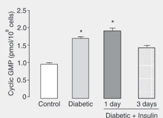
rendered diabetic by alloxan injection. In addition, treatment of these animals with NPH insulin (2 IU/day, for 3 days) reduces both inducible NO synthase activity and ex-pression to normal levels (38). Cyclic gua-nosine-3'-5'-monophosphate (cGMP) con-tent was determined in these cells for the investigation of the immediate second mes-senger effector of NO. Ten days after alloxan injection, basal levels of cGMP were increased in neutrophils from diabetic rats compared to controls. cGMP levels were reduced after treat-ment of diabetic animals with NPH insulin for at least 3 days. This information suggests that insulin modulates cGMP levels in neu-trophils by NO production and that an in-crease in the NO-cGMP pathway may con-tribute to the impaired leukocyte function in diabetes mellitus (Figure 1).
Neutrophil metabolism and diabetes mellitus
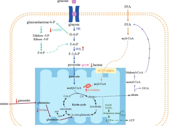
The maximal activities of several en-zymes of neutrophil metabolism such as hex-okinase (glycolytic pathway) and citrate syn-thase (Krebs cycle) have been investigated by our group (39,40). Also, the rates of metabolite production and utilization have been measured in incubated neutrophils (28,39,40). The ATP required for neutrophil functions (22) is mainly produced by the metabolization of glucose to lactate (41). In fact, only 2-3% of glucose is oxidized through the Krebs cycle in neutrophils (42). The stored glycogen can be utilized by neutro-phils to obtain energy (43) mainly during the phagocytosis process. When the intracellu-lar level of glucose is established, the re-synthesis of glycogen is then initiated (44). In addition to glucose, however, neutro-phils also utilize glutamine at high rates (39). Glutamine is the most abundant amino acid in plasma and skeletal muscle, being produced in muscle, lung and liver (45). Glutamine utilized by neutrophils is mainly converted to glutamate, aspartate, lactate,
and CO2 (39,46). The partial oxidation of
glucose to lactate and the glutaminolytic pathway are the main source of ATP and of the production of intermediate metabolites for the synthesis of macromolecules such as fatty acids and phospholipids (40).
Many studies have shown metabolic al-terations in neutrophils from diabetic pa-tients. High levels of glucose and ketone bodies seem to influence neutrophil func-tion by the producfunc-tion of polyols (47). Esmann (48) and Munroe and Shipp (49) did not observe differences in glucose utiliza-tion by neutrophils from healthy and dia-betic patients, whereas decreased utilization of glucose by neutrophils was verified by others (50,51). Munroe and Shipp (49) did not observe alterations, whereas Martin et al. (50) and Esmann (51) found decreased production of lactate by neutrophils from diabetic patients. In our study (28), there was no alteration in glucose consumption or oxidation by neutrophils from streptozoto-cin-induced diabetic rats, but decreased lac-tate production and increased phosphofruc-tokinase (PFK) maximal activity were ob-served (Figure 2).Similar results were ob-served in mesenteric lymph nodes and thy-mus lymphocytes from diabetic rats (52,53). The activity of PFK is stimulatedby fructose 2,6-biphosphate (54) andinhibited by ATP at low fructose 6-phosphate content, but not at high fructose 6-phosphate concentration (55). Therefore, under these conditions, the contentof intermediates of glycolysis, such
Figure 1. Cyclic GMP levels in neutrophils from diabetic rats, diabetic rats treated with insulin, and matching controls. Rats were rendered diabetic by the injection of alloxan (42 mg/kg,
iv) 10 days before. Insulin (NPH, 2 IU/day, sc) was given for 1 or 3 days before testing. Glycogen-elicited peritoneal neutrophils from 3 to 4 rats were pooled, each animal yielding approxi-mately 1 x 10⁸ cells. Data are
reported as pmol cyclic GMP per 108 cells, as mean ± SEM of 3
as fructose 6-phosphateand fructose 2,6-biphosphate, is expected to be elevated in neutrophils.This may partially explain the increase in PFK activity.In agreement with this proposition, Moreno-Aurioles et al. (52) found increased fructose 2,6-biphosphate content and PFK activityin neutrophils from streptozotocin-induced diabetic rat.
Neutrophils from diabetic rats showed no significant change in glucose oxidation, citrate synthase, or NAD+- and NADP+
-linked isocitrate dehydrogenase activities, suggesting that the flux of substrates through the Krebs cycle was unchanged (28) (Figure 2). The pentose-phosphate pathway oxidizes glucose-6-phosphate to intermediates of the glycolytic pathway, generating NADPH and ribose-5-phosphate for fatty acid and nucleo-tide synthesis, respectively (56). NADPH is important for NADPH oxidase activity and for glutathione reductase to recycle oxidized glutathione in neutrophils (57).
Glutamine oxidation and glutaminase
activity are significantly decreased in neu-trophils from diabetic rats (28) (Figure 2). Glutamine plays an important role in protein (as an amino acid source), lipid (by NAD(P)H production) and nucleotide synthesis (by purine and pyrimidine production), and in NADPH oxidase activity (58). Glutamine raises the in vitro bacterial killing activity and the rate of reactive oxygen species pro-duction by neutrophils (57,59). In addition, Pithon-Curi et al. (60) showed that gluta-mine has a protective effect on neutrophil apoptosis. Therefore, decreased glutamine utilization may contribute to the impaired inflammatory response in diabetic patients by increasing the occurrence of apoptosis in neutrophils.
These data, taken as a whole, show that neutrophils from diabetic rats present im-paired metabolism of glucose and glutamine. On the other hand, palmitic acid oxidation is increased, and this may compensate for the reduction in glucose and glutamine
tion to maintain the ATP supply for these cells (28).
Concluding remarks
Survival depends on the ability of the host to respond appropriately to pathogenic challenges. It has long been recognized that many diabetic patients have a worse progno-sis once infection is established. Abnormali-ties in neutrophil chemotaxis, phagocytosis and microbicidal mechanisms are responsi-ble, at least in part, for the increased suscep-tibility to and severity of infections in dia-betic patients. Considerable support to these clinical investigations was given by experi-mental studies on diabetic rats and mice. In addition, metabolic routes linked to leuko-cyte dysfunction, including advanced pro-tein glycosylation, the polyol pathway,
oxy-gen free radical formation, and the NO-cGMP pathway, have been described. Insulin re-stores the appropriate response to injury through a direct or indirect action on endo-thelial cells and leukocytes. For future stud-ies the challenge remains to better under-stand the integration of both innate and adaptive immune systems with the endo-crine and nervous systems, providing new insights into how inflammation is regulated.
Acknowledgments
We apologize to the many researchers whose work we have not been able to dis-cuss in this limited review. The authors are indebted to E.P. Portiolli, G. de Souza, J.R. Mendonça, and L. de Sá Lima for constant assistance.
References
1. Garcia-Leme J. Hormones and inflammation. Boca Raton: CRC Press; 1989.
2. Garcia-Leme J, Farsky SP. Hormonal control of inflammatory re-sponses. Mediat Inflamm 1993; 2: 181-198.
3. Fortes ZB, Garcia LJ, Scivoletto R. Vascular reactivity in diabetes mellitus: role of the endothelial cell. Br J Pharmacol 1983; 79: 771-781.
4. Fortes ZB, Garcia LJ, Scivoletto R. Vascular reactivity in diabetes mellitus: possible role of insulin on the endothelial cell. Br J Pharma-col 1984; 83: 635-643.
5. Garcia LJ, Hamamura L, Leite MP, Rocha e Silva. Pharmacological analysis of the acute inflammatory process induced in the rat’s paw by local injection of carrageenin and by heating. Br J Pharmacol
1973; 48: 88-96.
6. Garcia-Leme J, Bohm GM, Migliorini RH, de Souza MZ. Possible participation of insulin in the control of vascular permeability. Eur J Pharmacol 1974; 29: 298-306.
7. Llorach MA, Bohm GM, Leme JG. Decreased vascular reactions to permeability factors in experimental diabetes. Br J Exp Pathol 1976; 57: 747-754.
8. Cavalher-Machado SC, de Lima WT, Damazo AS, de Frias Carvalho V, Martins MA, Silva PM, et al. Down-regulation of mast cell activa-tion and airway reactivity in diabetic rats: role of insulin. Eur Respir J
2004; 24: 552-558.
9. Pereira MA, Sannomiya P, Leme JG. Inhibition of leukocyte chemo-taxis by factor in alloxan-induced diabetic rat plasma. Diabetes
1987; 36: 1307-1314.
10. Sannomiya P, Pereira MA, Garcia-Leme J. Inhibition of leukocyte chemotaxis by serum factor in diabetes mellitus: selective
depres-sion of cell responses mediated by complement-derived chemoat-tractants. Agents Actions 1990; 30: 369-376.
11. Sannomiya P, Oliveira MA, Fortes ZB. Aminoguanidine and the prevention of leukocyte dysfunction in diabetes mellitus: a direct vital microscopic study. Br J Pharmacol 1997; 122: 894-898. 12. Fortes ZB, Farsky SP, Oliveira MA, Garcia-Leme J. Direct vital
microscopic study of defective leukocyte-endothelial interaction in diabetes mellitus. Diabetes 1991; 40: 1267-1273.
13. Cruz JW, Oliveira MA, Hohman TC, Fortes ZB. Influence of tolrestat on the defective leukocyte-endothelial interaction in experimental diabetes. Eur J Pharmacol 2000; 391: 163-174.
14. Zanardo RC, Cruz JW, Martinez LL, de Oliveira MA, Fortes ZB. Probucol restores the defective leukocyte-endothelial interaction in experimental diabetes. Eur J Pharmacol 2003; 478: 211-219. 15. Anjos-Valotta EA, Martins JO, Oliveira MA, Casolari DA, Britto LR,
Tostes RC, et al. Inhibition of tumor necrosis factor-alpha-induced intercellular adhesion molecule-1 expression in diabetic rats: role of insulin. Inflamm Res 2006; 55: 16-22.
16. Vianna EO, Garcia-Leme J. Allergen-induced airway inflammation in rats. Role of insulin. Am J Respir Crit Care Med 1995; 151: 809-814.
17. Belmonte KE, Fryer AD, Costello RW. Role of insulin in antigen-induced airway eosinophilia and neuronal M2 muscarinic receptor dysfunction. J Appl Physiol 1998; 85: 1708-1718.
18. Boichot E, Sannomiya P, Escofier N, Germain N, Fortes ZB, Lagente V. Endotoxin-induced acute lung injury in rats. Role of insulin. Pulm Pharmacol Ther 1999; 12: 285-290.
lipopolysaccha-ride-induced acute lung inflammation: Role of insulin. Shock 2006; 25: 260-266.
20. Alba-Loureiro TC, Martins EF, Landgraf RG, Jancar S, Curi R, Sannomiya P. Role of insulin on PGE2 generation during LPS-induced lung inflammation in rats. Life Sci 2006; 78: 578-585. 21. Moriguchi P, Sannomiya P, Lara PF, Oliveira-Filho RM, Greco KV,
Sudo-Hayashi LS. Lymphatic system changes in diabetes mellitus: role of insulin and hyperglycemia. Diabetes Metab Res Rev 2005; 21: 150-157.
22. Mowat A, Baum J. Chemotaxis of polymorphonuclear leukocytes from patients with diabetes mellitus. N Engl J Med 1971; 284: 621-627.
23. Tan JS, Anderson JL, Watanakunakorn C, Phair JP. Neutrophil dysfunction in diabetes mellitus. J Lab Clin Med 1975; 85: 26-33. 24. Bagdade JD, Nielson KL, Bulger RJ. Reversible abnormalities in
phagocytic function in poorly controlled diabetic patients. Am J Med Sci 1972; 263: 451-456.
25. Nielson CP, Hindson DA. Inhibition of polymorphonuclear leukocyte respiratory burst by elevated glucose concentrations in vitro. Diabe-tes 1989; 38: 1031-1035.
26. Jakelic J, Kokic S, Hozo I, Maras J, Fabijanic D. Nonspecific immu-nity in diabetes: hyperglycemia decreases phagocytic activity of leukocytes in diabetic patients. Med Arh 1995; 49: 9-12.
27. Panneerselvam S, Govindasamy S. Sodium molybdate improves the phagocytic function in alloxan-induced diabetic rats. Chem Biol Interact 2003; 145: 159-163.
28. Alba-Loureiro TC, Hirabara SM, Mendonca JR, Curi R, Pithon-Curi TC. Diabetes causes marked changes in function and metabolism of rat neutrophils. J Endocrinol 2006; 188: 295-303.
29. Rosen SD. Ligands for L-selectin: homing, inflammation, and be-yond. Annu Rev Immunol 2004; 22: 129-156.
30. Mayadas TN, Cullere X. Neutrophil beta2 integrins: moderators of life or death decisions. Trends Immunol 2005; 26: 388-395. 31. Argenbright LW, Letts LG, Rothlein R. Monoclonal antibodies to the
leukocyte membrane CD18 glycoprotein complex and to intercellu-lar adhesion molecule-1 inhibit leukocyte-endothelial adhesion in rabbits. J Leukoc Biol 1991; 49: 253-257.
32. Brownlee M. Biochemistry and molecular cell biology of diabetic complications. Nature 2001; 414: 813-820.
33. Goldin A, Beckman JA, Schmidt AM, Creager MA. Advanced glycation end products: sparking the development of diabetic vascu-lar injury. Circulation 2006; 114: 597-605.
34. Masuda M, Murakami T, Egawa H, Murata K. Decreased fluidity of polymorphonuclear leukocyte membrane in streptozocin-induced diabetic rats. Diabetes 1990; 39: 466-470.
35. Collison KS, Parhar RS, Saleh SS, Meyer BF, Kwaasi AA, Hammami MM, et al. RAGE-mediated neutrophil dysfunction is evoked by advanced glycation end products (AGEs). J Leukoc Biol 2002; 71: 433-444.
36. Hotta N. New approaches for treatment in diabetes: aldose reduc-tase inhibitors. Biomed Pharmacother 1995; 49: 232-243. 37. Boland OM, Blackwell CC, Clarke BF, Ewing DJ. Effects of
ponalrestat, an aldose reductase inhibitor, on neutrophil killing of
Escherichia coli and autonomic function in patients with diabetes mellitus. Diabetes 1993; 42: 336-340.
38. Cerchiaro GA, Scavone C, Texeira S, Sannomiya P. Inducible nitric oxide synthase in rat neutrophils: role of insulin. Biochem Pharma-col 2001; 62: 357-362.
39. Curi TC, De Melo MP, De Azevedo RB, Zorn TM, Curi R. Glutamine utilization by rat neutrophils: presence of phosphate-dependent glutaminase. Am J Physiol 1997; 273: C1124-C1129.
40. Curi R, Newsholme P, Pithon-Curi TC, Pires-de-Melo M, Garcia C, Homem-de-Bittencourt Junior PI, et al. Metabolic fate of glutamine in lymphocytes, macrophages and neutrophils. Braz J Med Biol Res
1999; 32: 15-21.
41. Beck WS, Valentine WN. The aerobic carbohydrate metabolism of leukocytes in health and leukemia. I. Glycolysis and respiration.
Cancer Res 1952; 12: 818-822.
42. Beck WS. Occurrence and control of the phosphogluconate oxida-tion pathway in normal and leukemic leukocytes. J Biol Chem 1958; 232: 271-283.
43. Stjernholm RL, Burns CP, Hohnadel JH. Carbohydrate metabolism by leukocytes. Enzyme 1972; 13: 7-31.
44. Scott RB. Glycogen in human peripheral blood leukocytes. I. Char-acteristics of the synthesis and turnover of glycogen in vitro. J Clin Invest 1968; 47: 344-352.
45. Curi R, Newsholme EA. The effect of adenine nucleotides on the rate and fate of glutamine utilization by incubated mitochondria isolated from rat mesenteric lymph nodes. Mol Cell Biochem 1989; 86: 71-76.
46. Ardawi MS, Newsholme EA. Glutamine metabolism in lymphocytes of the rat. Biochem J 1983; 212: 835-842.
47. Wilson RM, Tomlinson DR, Reeves WG. Neutrophil sorbitol produc-tion impairs oxidative killing in diabetes. Diabet Med 1987; 4: 37-40. 48. Esmann V. The diabetic leukocyte. Enzyme 1972; 13: 32-55. 49. Munroe JF, Shipp JC. Glucose metabolism in leucocytes from
pa-tients with diabetes mellitus, with and without hypercholesteremia.
Diabetes 1965; 14: 584-590.
50. Martin SP, Chaudhuri SN, Green R, McKinney GR. The effect of adrenal steroids on aerobic lactic acid formation in human leuko-cytes. J Clin Invest 1954; 33: 358-360.
51. Esmann V. The polymorphonuclear leukocyte in diabetes mellitus. J Clin Chem Clin Biochem 1983; 21: 561-567.
52. Moreno-Aurioles VR, Montano R, Conde M, Bustos R, Sobrino F. Streptozotocin-induced diabetes increases fructose 2,6-biphosphate levels and glucose metabolism in thymus lymphocytes. Life Sci
1996; 58: 477-484.
53. Otton R, Mendonca JR, Curi R. Diabetes causes marked changes in lymphocyte metabolism. J Endocrinol 2002; 174: 55-61.
54. Wegener G, Krause U. Different modes of activating phosphofructo-kinase, a key regulatory enzyme of glycolysis, in working vertebrate muscle. Biochem Soc Trans 2002; 30: 264-270.
55. Mansour TE. Studies on heart phosphofructokinase: purification, inhibition and activation. J Biol Chem 1963; 238: 2285-2292. 56. Casazza JP, Veech RL. The interdependence of glycolytic and
pentose cycle intermediates in ad libitum fed rats. J Biol Chem 1986; 261: 690-698.
57. Curi TC, De Melo MP, Palanch AC, Miyasaka CK, Curi R. Percent-age of phagocytosis, production of O2.-, H2O2 and NO, and
antioxi-dant enzyme activities of rat neutrophils in culture. Cell Biochem Funct 1998; 16: 43-49.
58. Newsholme P, Lima MM, Procopio J, Pithon-Curi TC, Doi SQ, Bazotte RB, et al. Glutamine and glutamate as vital metabolites.
Braz J Med Biol Res 2003; 36: 153-163.
59. Ogle CK, Ogle JD, Mao JX, Simon J, Noel JG, Li BG, et al. Effect of glutamine on phagocytosis and bacterial killing by normal and pedi-atric burn patient neutrophils. JPEN J Parenter Enteral Nutr 1994; 18: 128-133.